Bot. Bull. Acad. Sin. (1999) 40: 275-281

Pan et al. Over-expression of rice CuZnSOD in E. coli

Over-expression and characterization of copper/

zinc-superoxide dismutase from rice in Escherichia coli

Shu-Mei Pan1, Guan-Bor Hwang, and Hung-Chi Liu

Department of Botany, National Taiwan University, Taipei, Taiwan, Republic of China

(Received August 10, 1998; Accepted March 3, 1999)

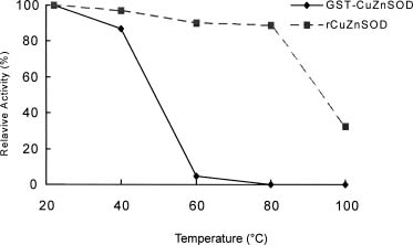
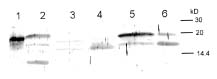
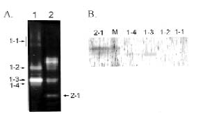
Abstract. The over-expression and characterization of cytosolic Cu/Zn-superoxide dismutase (CuZnSOD) from rice in Escherichia coli have been achieved. The full cDNA sequence coding for the rice cytosolic CuZnSOD was made by PCR and inserted into a pGEX-2T expression vector. The recombinant DNA was transformed to E. coli XL1 blue. Transformed E. coli expressed GST-CuZnSOD at levels greater than 20% of soluble protein under optimized conditions, and 10-30 mg of fusion protein can be purified from 1L bacterial culture by affinity gel. The purified fusion protein was cleaved to remove GST and produced recombinant CuZnSOD (rCuZnSOD). The MWs of a subunit of the fusion protein and rCuZnSOD were 43 kDa and 18 kDa, respectively, as predicted. The SOD activity was retained as the dimer for both forms. The fusion protein and rCuZnSOD were characterized for thermostability and the effects of pH and SDS on its activity by 10% nondenaturing gel. They showed resistance to the inhibition of hydrogen peroxide, in contrast to the native form of the plant CuZnSOD. Antiserum prepared from the GST-CuZnSOD fusion protein showed cross-reactivity to the subunits of rice cytosolic and plastidic CuZnSOD, and to those of other plants.

Keywords: CuZnSOD; Escherichia coli; Oryza sativa; Superoxide dismutase.

Abbreviations: CuZnSOD, copper/zinc superoxide dismutase; GST, glutathione transferase; PVDF, polyvinylidene difluoride; rCuZnSOD, recombinant CuZnSOD; SOD, superoxide dismutase.

Introduction

Reactive oxygen species—including superoxide, hydrogen peroxide, and hydroxyl radicals—are generated by a number of pathways (Fridovich, 1978). Accumulation of these active oxygen species in aerobic organisms may cause peroxidation of membrane lipids, DNA strand breakage, and inactivation of enzymes (Imlay and Linn, 1988). Defense systems derived from enzymatic and nonenzymatic antioxidants in living organisms can minimize the deleterious effects of active oxygen free radicals (Pell and Steffen, 1991). Superoxide dismutases (SOD), catalase, and ascorbate peroxidase are among the important antioxidant enzymes in plants. SODs are a group of metal-containing enzymes that catalyze the dismutation of superoxide radical to molecular oxygen and hydrogen peroxide (Beyer et al., 1991). These enzymes may be classified into three types according to their metal cofactor requirements: copper/zinc (CuZnSOD), manganese (MnSOD), and iron (FeSOD) forms. Isoforms of CuZnSOD, the most abundant SOD in higher plants, are located both in cytosol and in chloroplasts (Kanematsu and Asada, 1989). SOD activity in plants increases differentially in re

sponse to various environmental stresses (Bowler et al., 1989; Tsang et al., 1991). SOD activity often correlates with the severity of stress (Bowler et al., 1992). It has been reported that transgenic plants having high SOD activity enhance the tolerance towards oxidative stress (Bowler et al., 1991; Perl et al., 1993; Sen Gupta et al., 1993) and other stresses (Mckersie et al., 1993; Van Camp et al., 1994), although not all highly expressing plants are oxidative stress tolerant (Tepperman and Dunsmuir, 1990). Other enzyme activities may be needed at the same time (Foyer et al., 1994).

The cDNA encoding cytosolic CuZnSODs from several plants and rice (Sakamoto et al., 1992), as well as the gene structure and expression of CuZnSODs from rice were reported (Sakamoto et al., 1995a; Sakamoto et al., 1995b). In our laboratory, we are studying oxidative stress effect on the antioxidant enzymes of rice (Wei et al., 1995; Chen, 1994). In order to understand the regulation of rice SOD in response to a stress, cDNA clones of CuZnSOD and MnSOD were isolated and characterized from rice cDNA library (Pan et al., 1995; Chen et al., 1997). In this report, we describe the over-expression and characterization of CuZnSOD from rice in Escherichia coli, using the fusion protein to prepare antiserum. The antiserum that recognizes CuZnSOD was used to examine the SOD protein from different plant species.

1Corresponding author. Tel: 886-2-3630231 ext. 2366; Fax: 886-2-3918940; E-mail: pan@ccms.ntu.edu.tw


Botanical Bulletin of Academia Sinica, Vol. 40, 1999

Materials and Methods

Construction of Recombinant Expression Vector and Induction of CuZnSOD

A cDNA clone for cytosolic CuZnSOD isolated from a rice cDNA library constructed from an etiolated seedling was described previously (Pan et al., 1995). The expression vector used for the construction of the fused gene was the glutathione transferase gene Fusion System (GST Gene Fusion System, Pharmacia). Plasmid expression vectors have been constructed that direct the synthesis of foreign polypeptides in E. coli as fusions with the C terminus of Sj26, a 26-kDa glutathione S-transferase (GST; EC 2.5.1.18) encoded by the parasitic helminth Schistosoma japonicum (Smith and Johnson, 1988). The full length of CuZnSOD cDNA sequence having the BamHI and EcoRI sites as the ends was made by PCR, and ligated with the vector, which was derived from pGEX-2T cut with BamHI and EcoRI. The recombinant plasmid was transformed into competent XL1-blue cells by the CaCl2 method (Sambrook et al., 1989). More than ten transformed clones were picked and cultured in LB broth for mini-scale preparation. The transformed cell line used for biochemical study in this experiment was called cu101. The insert DNA and the fusion protein expression were checked using the protocol described by the manufacturer (Pharmacia Biotech).

Induction and Purification of the Fusion Protein

The culture of cell line cu101 and the conditions for induction were described by Smith and Johnson (1988). Overnight cultured cells were harvested and cell pellets were obtained by centrifugation (5,000 g, 10 min at 4°C). Pellets were resuspended in PBS (140 mM NaCl, 2.7 mM KCl, 10 mM Na2HPO4, and 1.8 mM KH2PO4, pH 7.3), and cells were sonicated for 15 min on ice. After centrifugation (12,000 g, 15 min at 4°C) the supernatant was collected for further purification. The fusion protein was batch-purified in a single step using bulk glutathione Sepharose 4B (Pharmacia Biotech).

Electrophoresis and SOD Activity Stain

Analytical polyacrylamide gel electrophoresis (PAGE) was performed according to a modified procedure of Gabriel (1971). Electrophoresis was conducted with a 1.5 mm 10% native acrylamide slab gel in a standard Tris-glycine buffer, pH 8.3. An appropriate amount of rice extract was applied and run at 80 V through the stacking gel for 15 min and 120 V through the separating gel for 60 min. After electrophoresis, a photochemical method modified from Beauchamp and Fridovich (1971) was used to visualize SOD activities. The gel was first soaked in 1.225 mM nitroblue tetrazolium solution for 15 min, briefly washed, then soaked in a 100 mM potassium phosphate buffer, pH 7.0 containing 28 µM riboflavin and 28 mM TEMED (N,N,N',N'-tetramethyl-ethylenediamine) for another 15 min. After the gel was briefly washed, it was illuminated on a light box with an intensity of 30 µE s-1m-2 for 15 min to initiate the photochemical reaction. To identify CuZnSOD,

8 mM KCN was included in the riboflavin solution for activity staining. For the inhibition of hydrogen peroxide towards CuZnSOD, the gels were first soaked in 8 mM hydrogen peroxide in a 100 mM potassium phosphate buffer, pH 7.0, for 30 min, followed by SOD activity staining. All procedures were carried out at room temperature, and the reaction mixture containing gels was shaken at 75 rpm. The tested gel was scanned with a laser densitometer (Molecular Dynamics) after the gel was stained, and relative activity was determined (Chen and Pan, 1996).

The purified fusion protein from cu101 cells was isolated and cleaved by the overnight incubation with thrombin (16 µg per 80 µg fusion protein). The reaction mixture was separated by a denaturing 12.5% SDS-PAGE to detect the purity, or separated by electrophoresis on 10% native-PAGE to detect the SOD activity of the GST-CuZnSOD and rCuZnSOD protein.

N-Terminal Sequencing

The rCuZnSOD protein was separated from the thrombin-digested fusion protein mixture by electrophoresis on a 12.5% SDS-PAGE, then blotted onto a transfer membrane (Immobilon-P, Millipore, Bedford, MA, USA) in a Hoefer mini-transblot cell at 0.4A for 80 min in the presence of 10 mM CAPS (3-[cyclohexylamino]-1-propanesulfonic acid), 10% methanol, pH 11, and submitted for N-terminal sequencing on an automatic amino acid sequencer (model 476A; Applied Biosystems Inc., San Jose, CA, USA). The sequence data revealed two extra amino acids residues, glycine and serine (derived from the thrombin-recognition sequences) before the ATG initiation codon.

Assay for Stability of rCuZnSOD Enzyme

Ten µg of the fusion protein and the rCuZnSOD protein after the thrombin-digestion were used for the following tests. For thermal stability, the enzyme samples were heated at room temperature (22°C), 40, 60, 80 and 100°C for 20 min. The enzyme samples were taken out and tested for pH stability, SDS effect, and proteolytic susceptibility to chymotrypsin, trypsin and pepsin (Liu, 1997). After treatment, every sample was electrophoresed on a 10% native PAGE to detect the SOD activity.

Preparation of Antisera

Purified rCuZnSOD or fusion protein was cut from the SDS-PAGE blots (6.0 mg protein obtained from each). This was taken to prepare the antiserum of SOD in rabbits. Sera collected from these rabbits were taken at week 7, or 9 or 11 and were used directly without any purification. The antiserum prepared from the fusion protein showed low nonspecific cross-reactivity and was therefore used for the immunoblotting analysis in this experiment.

Western Blotting

Antiserum was used to test cross-reactivity with CuZnSOD from different plant species. These tested species included spinach leaves, tobacco leaves, corn


Pan et al. Over-expression of rice CuZnSOD in E. coli

seedlings, rice roots, and rice leaves. Crude extracts from different species were boiled in SDS-PAGE buffer containing b-mercaptoethanol and then separated by 12.5% SDS-PAGE. Following electrophoresis, the proteins were transferred to a transfer membrane (Millipore, Immobilon-P) by using the protocol provided by the manufacturer (Genescreen). The blots were developed using a 1:5000 dilution of the primary antibody, a 1: 2000 dilution of horseradish peroxidase conjugated goat anti-rabbit IgG (Zymed), and DAB (3,3'-diaminobenzidine tetrahydro chloride dihydrate) as substrate.

Results

The full-length cDNA sequence coding for the rice cytosolic CuZnSOD was made by PCR and inserted into a pGEX-2T expression vector (Hwang, 1993). Total cell lysates of transformed E. coli cu101 containing recombinant plasmid were prepared at various times after the start of inoculation and analyzed by SDS-PAGE. Escherichia coli cells transformed with pcu101 showed maximum levels of expression at 3 h after the addition of 0.1 mM IPTG to the culture medium (Figure 1, lane 2-8). The molecular weight of this fusion protein was estimated to be 43 kDa, as expected (Figure 1). A high level of GST-CuZnSOD protein was induced in 1 L of E. coli cu101 cells, and 10-30 mg of fusion protein was purified. The purified GST-CuZnSOD protein gave a single protein band. The catalytic activity and the molecular size of the native fusion protein were analyzed by gradient PAGE to show that the SOD activity was present as the dimer form of the fusion protein (Figure 2). The dimer property of the fusion protein was confirmed by gel filtration analysis (Liu, 1997). The SOD activities of fusion protein and rCuZnSOD were inhibited by H2O2 and KCN, but they were not so sensitive to H2O2 inhibition as the endogenous CuZnSOD in rice leaf extracts (Figure 3, lane 5, 6). However, the endogenous CuZnSOD of rice leaf extract appeared as the fast-moving, major SOD band in the gel. Its activity was inhibited drastically by 8

Figure 2. SOD activity analysis of GST-CuZnSOD at various stages of purification by 5 to 20% acrylamide gradient PAGE. Lane 1: total cell extract of E. coli XL1-blue not containing pGEX-2T; lane 2: total cell extract from uninduced cells; lane 3: total cell extract and soluble fraction of the induced cells; lane 4: the flow through cell extract from glutathione Sepharose 4B gel; lane 5: the purified GST-CuZnSOD. The molecular markers are indicated, and the position of the native form of GST-CuZnSOD is indicated by the arrowhead. Cells were grown in shake flasks and harvested 3 h after induction with 0.1 mM IPTG.

mM H2O2 and KCN (Figure 3). Therefore, 10-30 mg of active rice rCuZnSOD can be obtained from 1 L of E. coli cu101 cells by such a cloning expression and purification procedure.

After the purified GST-CuZnSOD was digested overnight with thrombin, three protein bands, separated on an SDS-PAGE, were estimated to be 27, 43 and 18 kDa, respectively (Figure 4A). They correspond to the predicted molecular mass of GST, GST-CuZnSOD, and rCuZnSOD. If the same sample was separated on a native gel after PAGE, three SOD-activity bands appeared (Figure 4B). Presumably, the fast-moving one was rCuZnSOD, and the slow-moving two were dimer of GST-CuZnSOD and (GST)1-(CuZnSOD)2. This suggestion was confirmed by molecular size analysis and western blot analysis (Liu, 1997). The homogenous rCuZnSOD was used to determine its pI at pH 5.2 by the isoelectric focusing and activity staining on the gel (Liu, 1997). The first 15 N-terminus amino acids of the rCuZnSOD were sequenced (Table 1) and compared with those of a purified form from rice leaves (Kanematsu and Asada, 1989). We found the extra two amino acids glycine and serine, which belonged to the pGEX-2T vector, remained in the rCuZnSOD (Table 1). Moreover, the derived amino acid sequence of a cDNA sequence of rice CuZnSOD indicated that threonine at amino acid residue 31 replaced arginine, and lysine at amino acid residue 38 replaced threonine of the purified CuZnSOD protein from rice (Pan et al., 1995; Kanematsu and Asada, 1989). This discrepancy may be due to the different strains of rice used.

The thrombin-treated GST-CuZnSOD mixture was heated at various temperatures. The SOD activity of GST-

Figure 1. Expression of GST-CuZnSOD fusion protein in E. coli cells transformed with pcu101 plasmid. Lane 1: the cell extract of E. coli, lane 2-8: cells transformed with plasmid harvested at hourly intervals at to 6 h and 8 h after 0.1 mM IPTG induction of fusion protein; lane 9: low molecular weight standards (94, 67, 43, 30, 24, 14 kDa, respectively), lane 10: the purified fusion protein.


Botanical Bulletin of Academia Sinica, Vol. 40, 1999

Figure 3. The effect of inhibitors on the SOD activity of GST-CuZnSOD. Lane 1-3: as the control, no treatment; lane 4-6: 8 mM H2O2 as inhibitor; lane 7-9: 8 mM KCN as inhibitor; lane 1,4,7: bovine liver CuZnSOD, 3U; lane 2,5,8: crude extract of rice leaves, 70 µg; lane 3,6,9: 20 µg of GST-CuZnSOD after 2-day digestion with thrombin.

Figure 5. The effect of temperature on GST-CuZnSOD and rCuZnSOD. The thrombin-digested GST-CuZnSOD was heated at the various temperatures for 20 min. Then 80 µg of each sample were loaded onto a 10% native PAGE. The effects of temperature were determined by activity staining.

A. B.

CuZnSOD disappeared at 60°C, but the SOD activity of the rCuZnSOD protein remained complete at 80°C, and 70% was lost at 100°C (Figure 5).

The antiserum prepared from the GST-CuZnSOD showed better immunological properties than that from rCuZnSOD in terms of specificity and titer. Therefore, antiserum against fusion protein was used in all western blot experiments. Two nonspecific binding bands having the apparent molecular masses of 22 and 12 kDa were recognized by the preimmune antiserum in the spinach extract. Two protein bands with apparent molecular masses of 16 and 18 kDa were detected in all species (Figure 6), indicating the presence of two different molecular sizes of CuZnSOD. Presumably, the major CuZnSOD activity in the leaf extract but much lower activity in the root extract indicate what can be considered a plastidic form of the enzyme, with the major CuZnSOD activity in the root extract indicating a cytosolic form. Therefore, rice extracts of leaves or roots were separated on a native PAGE and stained for CuZnSOD activities (Figure 7A). The active bands corresponding to the MnSOD and plastidic and cytosolic CuZnSOD were cut separately, dissolved to run

Figure 4. Analysis of the product of GST-CuZnSOD digestion with thrombin. (A), protein analysis and (B), SOD activity. The GST-CuZnSOD was thrombin-digested at 22°C for the various periods. Then 80 µg of each sample was loaded onto a 12.5% SDS-PAGE followed by protein staining. Or the thrombin-digested GST-CuZnSOD were loaded on a 10% native PAGE for SOD activity analysis. Lane 1: control, not thrombin-treated; lane 2 to 4: thrombin-digested for 24, 48 and 72 h, respectively.

Table 1. Comparison of rCuZnSOD and protein deduced from a cDNA clone of rice CuZnSOD.

Characteristics rCuZnSODa Deduced rice CuZnSODb

N-terminal sequence GSMVKAVVVLGSSEIVKGT MVKAVVVLGSSEIVKGT

Amino acids residues 154 152

MW 18.5 kDa (deduced 15.5 kDa) 15.2 kDa

pI 5.2 ~ 5.6 5.98

arCuZnSOD was electrophoresis on a SDS-PAGE, blotted on to a PVDF membrane, and submitted for automatic N-terminal amino acid sequencing. MW or pI was determined by the electrophoretic method, respectively. Underlining indicates the identity of the N-terminal amino acids of rCuZnSOD and the deduced ones of a cDNA clone.

bThe data of cytosol CuZnSOD were derived from a cDNA clone isolated from a rice cDNA library (Pan et al., 1995).


Pan et al. Over-expression of rice CuZnSOD in E. coli

difference may be due to the different cultivars used (Tainung and Nipponbare) or the fact that they are encoded from the different genes (Pan et al., 1995). In this report, we have transformed our cDNA clone of rice CuZnSOD in the bacterium E. coli and expressed highly recombinant rice CuZnSOD protein in the transformed bacteria. A high level of GST-CuZnSOD protein was induced in 1 L of E. coli cu101 cells, and 10-30 mg of fusion protein was purified, but only 1-3 mg of GST-MnSOD was obtained in a similar procedure (Liu, 1997; Tzeng, 1997). The high yield of active rice CuZnSOD obtained from transformed E. coli facilitates its antibody preparation. The SOD activity of the purified rCuZnSOD displays some characteristics similar to the native enzyme, including the response to pH, temperature but more resistance to H2O2 inhibition. The thrombin-digested GST-CuZnSOD mixture was treated with various concentrations of SDS, and we found that the GST-CuZnSOD and the rCuZnSOD proteins were relatively stable under SDS treatment, remaining 40 and 60% active, respectively (Liu, 1997). A similar stability to pH in the range of 5 to 10.4 was observed for the GST-CuZnSOD and rCuZnSOD protein. But, when pH is below 4, the rCuZnSOD protein is more stable than the GST-CuZnSOD (data not shown). The catalytic rate constants for CuZnSOD were reported to be pH-independent (Ellerby et al., 1996). Although SOD activity of purified CuZnSOD from rice etiolated seedlings was not stable at higher temperature, it was inactivated 50% after 30 min at 60°C (Padiglia et al., 1996). The high thermostability of rCuZnSOD seems to be unique since the recombinant MnSOD of rice was heat labile (Tzeng, 1997). The thermostable property of rCuZnSOD protein can be useful for pharmaceutical applications if the SOD product needs to be processed at a higher temperature, such as 60°C during manufacturing. The thrombin-digested GST-CuZnSOD mixture was treated with chymotrypsin, trypsin, and pepsin for various periods, and we found that GST-CuZnSOD, but not the rCuZnSOD, was resistant to chymotrypsin (data not shown). Both proteins are labile to pepsin and trypsin digestion, and this behavior is different from that of sweet potato (Lin et al., 1995).

The specific activity of rCuZnSOD was estimated to be much lower than that of native forms isolated from various plant tissues (Liu, 1997; Kono et al., 1979; Baum et al., 1983; Kanematsu and Asada, 1989). Since copper and zinc ions added to the medium of cu101 bacterial culture did not bring up the specificity activity of rCuZnSOD, this rules out the amount of copper or zinc ions in the bacterial cell as a limiting factor to rCuZnSOD protein. However, we can not rule out the availability of copper and zinc ions to incorporate in rCuZnSOD apoprotein as the reason for the lower specific activity of rCuZnSOD observed in the transformed bacteria. Lately, several Cu chaperone proteins have been found in yeast (Lin and Culotta, 1995) and humans. These proteins are required for incorporating Cu into SOD in vivo (Culotta et al., 1997). Possibly, E. coli might not have the correct molecular machinery, such as metal chaperone proteins, to correctly incorporate the metals in the recombinant rice apoSOD. Therefore, the lower

Figure 6. Western blot analysis of CuZnSOD proteins from various plant species. Lane 1: rCuZnSOD; lane 2-6: the crude extract of spinach leaves, tobacco leaves, maize seedlings, rice roots, and rice leaves, 50 µg of soluble protein from each sample was loaded onto 12.5% SDS-PAGE. The molecular markers are indicated.

Figure 7. Western blot analysis of various -SODs of rice extract. (A), SOD activity. Lane 1: the crude extract of rice leaves, lane 2: the crude extract of rice roots; 50 µg of soluble protein from each was loaded onto 10% native-PAGE; bands corresponding to 1-1, and 1-2 were identified to be MnSODs, 1-3, and 1-4 were plastidic CnZnSODs, 2-1 was cytosolic CuZnSODs; the types of SOD were identified by the differential inhibition of 8 mM H2O2 and KCN. (B), Western analysis. The active bands of the MnSODs and plastidic and cytosolic CuZnSODs showed in Figure 7A were separately cut, dissolved, then loaded onto a 12.5% SDS- PAGE. Western blot analysis followed.

in SDS-PAGE, and analyzed by western blotting. Plastidic CuZnSOD was recognized and corresponds to the protein band of 16 kDa. The cytosolic form corresponds to the band of 18 kDa, but the MnSOD of rice can not be recognized (Figure 7B). Accordingly, the antiserum against GST-CuZnSOD showed cross-reactivity towards two different CuZnSODs, one of which was plastidic and one of which was cytosolic.

Discussion

We have isolated and characterized one cDNA clone of cytosolic CuZnSOD from a cDNA library of etiolated, 10-day-old seedlings of rice (Pan et al., 1995). Compared with the RSODA clone (Sakamoto et al., 1992), this clone showed a 99% identity in the coding region and 3' end untranslated region. Our clone has isoleucine at amino acid residue 57 while RSODA has methionine. This minor


Botanical Bulletin of Academia Sinica, Vol. 40, 1999

specific activity of rCuZnSOD in the transformed bacteria was observed. Alternatively, the two extra amino acids glycine and serine, added as an artifact of the fusion, may have some effects on the catalytic activity of rCuZnSOD. A similar inhibition of enzyme activity has been reported in the study of S-glutathione transferases in plants (Marr, 1996). The antiserum prepared from the GST-CuZnSOD showed the immunoreaction towards the different types of CuZnSODs. This property makes it possible to locate and measure the changes of both types of CuZnSOD proteins in the same tissues.

Acknowledgments. This work was supported in part by the Council of Agriculture, Republic of China under Grant AC85-1.1-F-49 (11).

Literature Cited

Baum, J.A., J.M. Chandlee, and J.G. Scandalios. 1983. Purification and partial characterization of a genetically-defined superoxide dismutases (SOD-1) associated with maize chloroplasts. Plant Physiol. 73: 31-35.

Beauchamp, C. and I. Fridovich. 1971. Superoxide dismutase: Improved assay and an assay applicable to acrylamide gels. Anal. Biochem. 44: 276-287.

Beyer, W., J. Imlay, and I. Fridovich. 1991. Superoxide dismutases. Prog. Nuc. Acid Res. Mol. Biol. 40: 221-248.

Bowler, C., T. Alliotte, M.V.D. Bulcke, G. Bauw, J. Vandekerckhove, M. Van Montagu, and D. Inze. 1989. A plant manganese superoxide dismutase is efficiently imported and correctly processed by yeast mitochondria. Proc. Natl. Acad. Sci. USA 86: 3237-3241.

Bowler, C., L. Slooten, S. Vandenbranden, R. De Rycke, J. Botterman, C. Sybesma, M. Van Montagu, and D. Inze. 1991. Manganese superoxide dismutase can reduce cellular damage mediated by oxygen radicals in transgenic plants. EMBO J. 10: 1723-1732.

Bowler, C., M. Van Montagu, and D. Inze. 1992. Superoxide dismutase and stress tolerance. Ann. Rev. Plant Physiol. Plant Mol. Biol. 43: 83-116.

Chen, C.N. and S.M. Pan. 1996. Assay of superoxide dismutase activity by combining electrophoresis and densitometry. Bot. Bull. Acad. Sin. 37: 107-111.

Chen, H.B. 1994. The Effect of Ozone on the Activity of Antioxidant Enzyme in Rice. Master thesis. National Taiwan University, Taiwan.

Chen, J.C., D.S. Wei, and S.M. Pan. 1997. Assay of superoxide dismutase activity by combining electrophoresis and densitometry. Taiwania 42: 53-62.

Culotta, V.C., L.W.J. Klomp, J. Strain, R.L.B. Casareno, B. Krems, and J. Gitlin. 1997. The copper chaperone for superoxide dismutase. J. Biol. Chem. 272: 23469-23472.

Ellerby, L.M., D.E. Cabelli, J.A. Graden, and J.S. Valentine. 1996. Copper-zinc superoxide dismutase: why not pH-dependent? J. Am. Chem. Soc. 118: 6556-6561.

Foyer, C.H., P. Descourvieres, and K.J. Kunert. 1994. Protection against oxygen radicals: an important defence mechanism studied in transgenic plants. Plant Cell Environ. 17: 507-523.

Fridovich, I. 1978. The biology of oxygen radicals. Science 201: 875-880.

Gabriel, O. 1971. Analytical disc gel electrophoresis. In W.B. Jakoby (ed.), Methods in Enzymology. vol. 22. Academic Press, New York, pp. 565-577.

Hwang, G.B. 1993. cDNA Cloning and Characterization of Cu/Zn-Superoxide Dismutase of Rice. Masters Thesis. National Taiwan University, Taiwan.

Imlay, J.A. and S. Linn. 1988. DNA damage and oxygen radical toxicity. Science 240: 1302-1308.

Kanematsu, S. and K. Asada. 1989. CuZn-superoxide dismutase in rice: occurrence of an active, monomeric enzyme and two types of isozyme in leaf and non-photosynthetic tissues. Plant Cell Physiol. 30: 381-391.

Kono, Y., M. Takahashi, and K. Asada. 1979. Superoxide dismutase from kidney bean leaves. Plant Cell Physiol. 20: 1229-1235.

Lin, C.T., M.T. Lin, T. Chen, and J.F. Shaw. 1995. Subunit interaction enhances enzyme activity and stability of sweet potato cytosolic Cu/Zn-superoxide dismutase purified by a His-tagged recombinant protein method. Plant Mol. Biol. 28: 303-311.

Liu, H.C. 1997. Over-Expression, Purification and Characterization of Rice CuZn-Superoxide Dismutase in E. coli. Masters thesis. National Taiwan University, Taiwan.

Lin, S.J. and V.C. Culotta. 1995. The ATX1 gene of Saccharomyces cerevisiae encodes a small metal homeostasis factor that protects cells against reactive oxygen toxicity. Proc. Natl. Acad. Sci. USA 92: 3784-3788.

Marr, K.A. 1996. The functions and regulation of glutathione S-transferases in plants. Ann. Rev. Plant Physiol. Plant Mol. Biol. 47: 127-158.

Mckersie, B.D., Y. Chen, M. De Beus, S. R. Bowley, C. Bowler, D. Inze, K. D'Halluin, and J. Botterman. 1993. Superoxide dismutase enhances tolerance of freezing stress in transgenic alfalfa (Medicago sativa L.). Plant Physiol. 103: 1155-1163.

Padiglia, A., R. Medda, E. Cruciani, A. Lorrai, and G. Floris. 1996. Purification and properties of Oryza sativa Cu-Zn superoxide dismutase. Prep. Biochem. Biotechnol. 26: 135-142.

Pan, S.M., G.B. Huang, D.S. Wei, and K.W. Yeh. 1995. Isolation and characterization of a cDNA for CuZn-superoxide dismutase of rice. Taiwania 40: 1-12.

Pell, E.J. and K.L. Steffen. 1991. Active Oxygen/oxidative Stress and Plant Metabolism. An American Society of Plant Physiologists Press, Rockville, MD.

Perl, A., R. Perl-Treves, S. Galili, D. Aviv, E. Shalgi, S. Malkin, and E. Galun. 1993. Enhanced oxidative-stress defense in transgenic potato expressing tomato Cu, Zn superoxide dismutases. Theor. Appl. Genet. 85: 568-576.

Sakamoto, A., H. Ohsuga, and K. Tanaka. 1992. Nucleotide sequences of two cDNA clones encoding different Cu/Zn-superoxide dismutase expressed in developing rice seed (Oryza sativa L.). Plant Mol. Biol. 19: 323-327.

Sakamoto, A., T. Okumura, H. Tkaminaka, and K. Tanaka. 1995a. Molecular cloning of the gene (SodCc1) that encodes a cytosolic copper/zinc-superoxide dismutase from rice (Oryza sativa L.). Plant Physiol. 107: 651-652.

Sakamoto, A., T. Okumura, H. Kaminaka, K. Sumi, and K. Tanaka. 1995b. Structure and differential response to ab


Pan et al. Over-expression of rice CuZnSOD in E. coli

scisic acid of two promoters for the cytosolic copper/zinc-superoxide dismutase genes, SodCc1 and SodCc2, in rice protoplasts. FEBS Letters 358: 62-66.

Sambrook, J., E.F. Fritsch, and T. Maniatis. 1989. Molecular Cloning: A Laboratory Manual. Cold Spring Harbor Laboratory Press, New York, USA.

Sen Gupta, A., R.P. Webb, A.S. Holaday, and R.D. Allen. 1993. Overexpression of superoxide dismutase protects plant from oxidative stress. Plant Physiol. 103: 1067-1073.

Smith, D.B. and K.S. Johnson. 1988. Single-step purification of polypeptides expressed in Escherichia coli as fusions with glutathione S-transferase. Gene 67: 31-40.

Tepperman, J.M. and P. Dunsmuir. 1990. Transformed plants with elevated levels of chloroplastic SOD are not more resistant to superoxide toxicity. Plant Mol. Biol. 14: 501-511.

Tsang, E.W.T., C. Bowler, D. Heriuart, W. Van Camp, R. Villarroel, C. Genetello, and D. Inze. 1991. Differential regulation of superoxide dismutases in plants exposed to environmental stress. Plant Cell 3: 783-792.

Tzeng, Y.C. 1997. Over-Expression and Characterization of Rice Manganese Superoxide Dismutase in E. coli. Master thesis. National Taiwan University, Taiwan.

Van Camp, W., H. Willekens, C. Bowler, M. Van Montagu, D. Inze, P. Reupold-Popp, Jr. H., Sandermann, and C. Langebartels. 1994. Elevated levels of superoxide dismutase protect transgenic plants against ozone damage. Biotechnology 12: 165-168.

Wei, D.S., C.P. Shen, and S.M. Pan. 1995. The effects of salt stress on the superoxide dismutase of rice. J. Chinese Agric. Chem. Soc. 33: 747-755.

¤ô½_»É¾N¶W®ñª[¤Æú@¦b¤j¸z±ìµß¤¤ªºªí¹F»P©Ê½è¬ã¨s

¼ï¯À¬ü¶À«a³Õ¼B§»°ò

°ê¥ß¥xÆW¤j¾Ç´Óª«¾Ç¬ã¨s©Ò

¥H PCR ¦X¦¨¤ô½_²Ó­M½è«¬»É¾N¶W®ñª[¤Æú@ (CuZnSOD) ªº cDNA §Ç¦C«á±µ¶i pGEX-2T ªí²{¸üÅé¡A ¥B°e¤J¤j¸z±ìµß XL1 blue «~¨t¤¤ªí¹F¡C¥H IPTG »¤¾É¤wÂ૬¤§¤j¸z±ìµß¨ä¿Ä¦X³J¥Õ½èªí²{¡A¨ä³J¥Õ½è²£ ¶q¬ù¥e¥i·»³J¥Õ½è¤§ 20%¡C¨Ã¸g¿Ë©M¼hªRªk¯Â¤Æ«á¡A¨C¤½¤Éµß²G¥i¦^¦¬ 10~30 mg ªº GST-CuZnSOD ¿Ä¦X³J¥Õ½è¡C GST-CuZnSOD ¿Ä¦X³J¥Õ½è¸g thrombin §@¥Î«á¡A¥i±N GST ¤Á¶}¦Ó±o¨ì­«²Õ«¬ CuZnSOD (rCuZnSOD)¡CGST-CuZnSOD ©M rCuZnSOD ¤§³æ¤¸Åé¤À¤l¶q¤À§O¬° 43 kDa ¤Î 18 kDa¡A¨âªÌ¤§­ìºA¬Ò¥H ¤G¤¸Å骺§Î¦¡¦s¦b¡F¥B³£«O¦³¶W®ñª[¤Æú@ªº¬¡©Ê¡C¤ñ¸û¿Ä¦X³J¥Õ½è©M rCuZnSOD ªº¬¡©Ê¹ï¼ö¡B pH ­È©M SDS ³B²z¤§®ÄÀ³¡C¤GªÌ¨ä»Ã¯À¬¡©Ê·|¨ü¹L®ñ¤Æ²B¤ÎÙæ¤Æ¹[ªº§í¨î¡A¦ý¨äªí²{¹ï¹L®ñ¤Æ²B§í¨îªº§Ü©Ê¡C¦¹ ¯S©Ê»P´Óª«­ìºA CuZnSOD ³J¥Õ½èªº¯S©Ê¤£¦P¡C¥H¯Â¤Æ¤§¿Ä¦X³J¥Õ½è»s³Æ¤§§ÜÅé¡A·|¿ëÃѤô½_²Ó­M½è«¬©M ¸­ºñÅ髬¤§ CuZnSOD ªº³æ¤¸Åé¡C¨Ã¹ï¥É¦Ì¡B·Ï¯ó¤ÎµÔµæ¤§µÑ¨ú²G¥çªí²{¥æ¤e¤ÏÀ³¡C

ÃöÁäµü¡G »É¾N¶W®ñª[¤Æ üÄ¡F¤j¸z±ìµß¡F¤ô½_¡F¶W®ñª[¤Æú@¡C