Bot. Bull. Acad. Sin. (1999) 40: 305-310

Hou et al. — Fatty acid esterases from yam tuber

Purification and properties of fatty acid esterases from yam (Dioscorea batatas Decne) tuber

Wen-Chi Hou, Hsien-Jung Chen, Chung-Fang Chang and Yaw-Huei Lin1

Institute of Botany, Academia Sinica, Nankang, Taipei 115, Taiwan

(Received February 5, 1999; Accepted March 5, 1999)

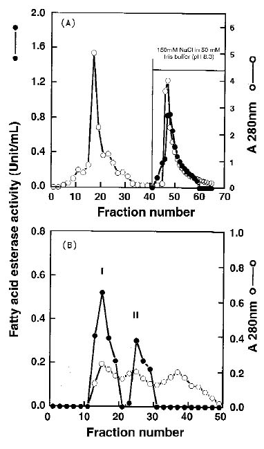
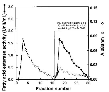
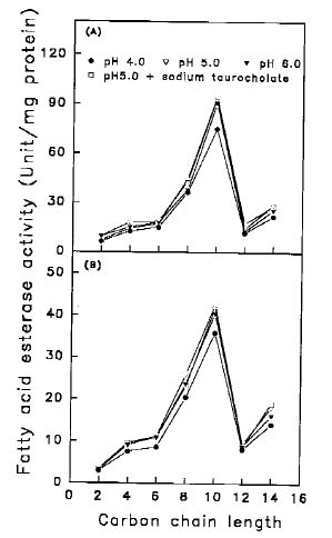
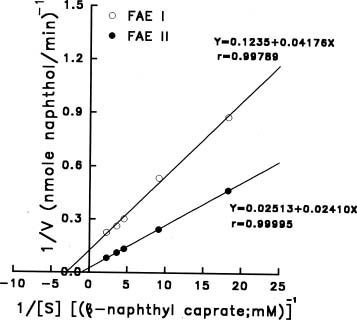
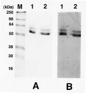
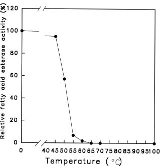
Abstract. Using b-naphthyl myristate (C14 fatty acid ester) as a screening substrate, we purified fatty acid esterases (FAEs) from yam (Dioscorea batatas Decne) tuber. Two FAE fractions (named FAE I and FAE II) were obtained after DE-52 ion exchange chromatography and Sephadex G-75 gel filtration purification steps, and then further purified by Con A Sepharose 4B affinity chromatography. FAE I and II fractions contained the same three protein bands about 50-64 kDa corresponding to esterase activity bands on SDS-PAGE gels. Among b-naphthyl esters determined at pH 4.0, 5.0 and 6.0 the best substrate for both FAE fractions was a C10-containing one with a maximum pH at 5.0. The Km and Vmax for b-naphthyl caprate (C10 fatty acid ester) of FAE I and II at 37°C, pH 5.0 were 0.338 and 0.959 mM; 0.405 and 0.585 nmole b-naphthol/min µg protein, respectively. FAE activity was stable below 50°C and lost completely above 65°C.

Keywords: Dioscorea batatas Decne; Fatty acid esterase (FAE); b-naphthyl caprate; b-naphthyl myristate; purification.

Abbreviations: APS, ammonium persulfate; Bis, N,N'-methylene-bis-acrylamide; CBB G-250, coomassie brilliant blue G-250; FAE, fatty acid esterase; 2-ME, 2-mercaptoethanol; SDS-PAGE, sodium dodecylsulfate-polyacrylamide gel electrophoresis; TCA, trichloroacetic acid; TEMED, N,N,N',N'-tetramethyl-ethylene-diamine.

Introduction

Multiple forms of esterase catalyzing the hydrolysis of carboxylic esters of short-chain fatty acids have been demonstrated in several plant tissues including leaves (Rudolph and Stahmann, 1966) and seeds (Schwartz et al., 1964; Veerabhadrappa and Montgomery, 1971) of Phaseolus species, carrot roots (Carino and Montgomery, 1968) and tubers of potato (Desborough and Peloquin, 1966; 1967; Galliard and Dennis, 1974). However, few works concerning long-chain fatty acid esters from plant sources have appeared except for studies of patatin, the potato tuber storage protein, which was demonstrated to have fatty acid esterase (Racusen, 1984), lipid acyl hydrolase, and acyltransferase activities involved wound responses of the tuber (Andrews et al., 1988; Jefferson et al., 1990).

Yam (Dioscorea species) is a member of the monocotyledonous family Dioscoreaceae and is a major tuber crop in West Africa, Southeast Asia and the Caribbean (Akoruda, 1984). We found yam (Dioscorea batatas Decne) tubers contained high fatty acid esterase (FAE) activities toward b -naphthyl esters of long-chain fatty acids in a preliminary work. In this report, we purified FAEs from yam tuber using b -naphthyl myristate as a screening substrate and characterized their properties.

Materials and Methods

Materials

Fresh tubers of yam (Dioscorea batatas Decne), imported from Japan, were purchased from a local market. Electrophoresis grade acrylamide and Bis, TEMED and APS were from E. Merck Inc. (Darmstadt, Germany). b -naphthyl esters of C2, C4, C6, C8, C10, C12 and C14 fatty acid were purchased from Sigma Chemical Company (St. Louis, MO, USA). A See BlueTM pre-stained standard kit for SDS-PAGE was obtained from NovexTM (San Diego, CA, USA). It contained myosin (250 kDa), BSA (98 kDa), glutamate dehydrogenase (64 kDa), alcohol dehydrogenase (50 kDa), carbonic anhydrase (36 kDa), myoglobin (30 kDa), and lysozyme (16 kDa). Con A Sepharose 4B and Sephadex G-75 gels were obtained from Pharmacia (Uppsala, Sweden).

Purification of FAEs from Yam Tuber

Extraction and purification processes were performed at 4°C. After being cleaned with water, yam tubers were peeled and cut into strips immediately for FAE extractions. Samples were homogenized with four volumes (W/V) of 50 mM Tris-HCl buffer (pH 8.3). After centrifugation at 14000 g for 30 min, the supernatants were saved and loaded onto a DE-52 ion exchange column (2.0 × 20 cm). The column was washed with 50 mM Tris-HCl buffer (pH 8.3) of three column volumes and then eluted batchwise with 150 mM NaCl in 50 mM Tris-HCl buffer (pH 8.3). Flow rate

1Corresponding author. Fax: 886-2-2782-7954; Tel: 886-2-2789-9590 ext. 320; E-mail: boyhlin@ccvax.sinica.edu.tw


Botanical Bulletin of Academia Sinica, Vol. 40, 1999

was 50 mL/h, and each fraction contained 5 mL. The eluted fraction containing FAE activity was collected and concentrated with Centriprep 10 (Amicon, USA) and purified by a Sephadex G-75 column (1.6 × 70 cm). The column was eluted with 100 mM Tris-HCl buffer (pH 7.9) containing 100 mM NaCl. Flow rate was 27 mL/h, and each fraction contained 3.6 mL. Two FAE activity fractions (FAE I and FAE II) were separated, and each of them was further purified by a Con A Sepharose 4B column (1.0 × 10 cm). The column was washed with 20 mM Tris buffer (pH 7.4) containing 200 mM NaCl and then eluted batchwise with 200 mM methylglucoside in the same buffer. Flow rate was 50 mL/h, and each fraction contained 5 mL. The purified FAE I and FAE II were collected and concentrated with Centriprep 10 for further use.

Determination of FAE Activity from Yam Tuber

FAE activity was determined according to the method of Andrews et al. (1988) with some modifications. b-naphthyl myristate was dissolved in acetone to make a stock solution of 3 mM and stored at 4°C. Assay mixtures contained 400 µL of 100 mM acetate buffer (pH 5.0), 400 µL of SDS mix (one hundred mL SDS mix contained 1 mL Triton X-100, 1.7 mL of 1% SDS, and 97.3 ml water), 50 µL b-naphthyl myristate, and 100 µL enzyme solution. The enzyme reaction was carried out at 37°C for 30 min and ended by adding 200 µL of 50% acetic acid. The assay mixture was centrifuged at 14,500 rpm for 15 min. The supernatants were saved, and their absorbance was determined at 322 nm. One unit of FAE was defined as the amount of enzyme preparation capable of releasing 1 µmole b-naphthol at pH 5.0 in one hour. The b-naphthol (7-400 nmole) was used to plot the standard curve.

Protein and Activity Stainings of FAEs from Yam Tubers on 15% SDS-PAGE Gels

A discontinuous SDS slab gel electrophoresis of 15% acrylamide was performed using a vertical mini-gel system (Bio-Rad Inc., USA) with a 0.75 mm thickness. The gel was prepared mainly according to Laemmli (1970). A one hundred µL sample was mixed with 25 µL of 60 mM Tris buffer (pH 6.8) containing 2% SDS, 25% glycerol, 14.4 mM 2-ME and 0.1% bromophenol blue and then incubated at room temperature overnight. When SDS-PAGE was finished, gels were cut into two parts. One was fixed with 12.5% TCA and stained with CBB G-250 dye (Neuhoff et al., 1985). The other was immersed and shaken twice in 25% isopropanol in 10 mM Tris buffer (pH 7.9) for 10 min to remove SDS (Hou and Lin, 1998) and then equilibrated in 10 mM Tris buffer (pH 7.9) for 15 min before activity staining. The gel was incubated in the dark at 37°C for at least 20 min with 160 mL of the substrate-dye solution immediately prepared before use. The substrate-dye solution consisted of 40 mg b-naphthyl acetate in 16 mL of N,N-dimethylformamide that was brought to 160 mL with 144 mL of 10 mM Tris buffer (pH 7.9), in which 80 mg tetrazotized o-dianisidine was dissolved. The gel was destained with 10% acetic acid for 30 min.

Hydrolysis of b-Naphthyl Esters Containing Various Carbon Chain Lengths by FAEs

The b-naphthyl esters of C2, C4, C6, C8, C10, C12 and C14 fatty acids were used to determine the best substrate of FAE I and FAE II. These b-naphthyl esters were dissolved in acetone to a final concentration of 4 mM, and the assay method was described above. 100 mM acetate buffer (pH 4.0, 5.0), 100 mM phosphate buffer (pH 6.0) and 0.5 mM sodium taurocholate in 100 mM acetate buffer (pH 5.0) were used in testing pH effect on the enzyme activity.

Figure 1. The chromatograms of fatty acid esterases on (A) DE-52 ion exchange column (2.0 × 20 cm) and the active portions were further purified by (B) Sephadex G-75 column (1.6 × 70 cm). For DE-52 ion exchange column, the column was washed with 50 mM Tris-HCl buffer (pH 8.3) of three column volumes and then eluted batchwise with 150 mM NaCl in 50 mM Tris-HCl buffer (pH 8.3). Flow rate was 50 mL/h, and each fraction contained 5 mL; for Sephadex G-75 column, the column was eluted with 100 mM Tris-HCl buffer (pH 7.9) containing 100 mM NaCl. Flow rate was 27 mL/h, and each fraction contained 3.6 mL.


Hou et al. — Fatty acid esterases from yam tuber

The Enzyme Kinetic Parameters of FAE I and FAE II Hydrolyzing b-Naphthyl Caprate

The enzyme kinetic parameters (Km and Vmax) of FAE I and FAE II were determined at pH 5.0, 37°C using b-naphthyl caprate as a substrate. The proteins added were 20 µg and 68 µg, respectively, for FAE I and FAE II. The final concentrations of b-naphthyl caprate included 0.0547, 0.1094, 0.2187, 0.2734 and 0.4374 mM.

Temperature Stability

FAEs were pre-incubated at 45, 50, 55, 60, 65, 70 and 100°C for 5 min and then immediately cooled in an ice bath. The assay method was described above using b-naphthyl caprate as a substrate. The relative esterase activity of unheated FAEs was set at 100%.

Results

Purification of FAEs from Yam Tuber

In a preliminary study, we found yam tubers contained high FAE activities toward b-naphthyl esters of long-chain fatty acids. Therefore, we used b-naphthyl myristate (C14 fatty acid ester) as a screening substrate to purify FAEs from yam tubers. Figure 1 showed the chromatograms of FAEs on a DE-52 ion exchange column (Figure 1A), and the active portions toward b-naphthyl myristate were further purified by a Sephadex G-75 column (Figure 1B). Two FAE fractions were separated by the Sephadex G-75 column and named, in order, FAE I and FAE II (Figure 1B). Each of these two FAE fractions was purified by a Con A Sepharose 4B column and eluted batchwise with methylglucosides. Figure 2 showed the chromatogram of FAE II on the Con A Sepharose 4B column. The FAE II bound onto the Con A affinity column and was eluted batchwise with 200 mM methylglucoside. FAE I had the same chromatographic pattern on the Con A column (data not showed). The purification results were summarized in Table 1. Starting with crude extracts of yam tubers, 66- and 37-fold purification with 46% and 27% recoveries, respectively, of FAE I and FAE II were obtained.

Protein and Activity Stainings of FAEs from Yam Tubers on 15% SDS-PAGE Gels

Figure 3 showed protein (Figure 3A) and activity (Figure 3B) stainings of FAE I and II on 15% SDS-PAGE gels from yam tubers after Con A affinity column purification. Lane 1 and lane 2 were FAE I and FAE II, respectively. The b-naphthyl acetate was used as FAE`s substrate for esterase activity staining. From Figure 3A, it was found that the purified FAE I and FAE II contained the same three protein bands about 50-64 kDa corresponding to esterase activity bands (Figure 3B) on SDS-PAGE gels.

Figure 2. The chromatogram of fatty acid esterase II on Con A Sepharose 4B column (1.0 × 10 cm) after Sephadex G-75 gel filtration purification. The column was washed with 20 mM Tris buffer (pH 7.4) containing 200 mM NaCl and then eluted batchwise with 200 mM methylglucoside in the same buffer. Flow rate was 50 mL/h, and each fraction contained 5 mL.

Table 1. Purification of fatty acid esterases from yam tubers.

Activity Protein Specific activity Fold Recovery

(Unit)a (mg) (Unit/mg) (%)

Crude extracts 61.82 140.42 0.44 1.00 100.00

DE-52 column 31.43 39.48 0.80 1.81 50.84

Sepharose G-75

FAE I 7.03 1.41 4.99 11.33 11.37

FAE II 5.22 3.73 1.40 3.18 8.44

Con A-Sepharose 4B

FAE I 28.40 0.98 28.98 65.83 45.94

FAE II 16.41 1.01 16.25 36.91 26.54

aThe b-naphthyl myristate was used as a substrate. One unit was defined as the amount of enzyme preparation that catalyzed the formation of 1 µmole b-naphthol per hour at 37°C, pH 5.0.


Botanical Bulletin of Academia Sinica, Vol. 40, 1999

Hydrolysis of b-Naphthyl Esters Containing Various Carbon Chain Lengths by FAEs

Figure 4 showed the hydrolysis of b-naphthyl esters containing C2, C4, C6, C8, C10, C12 and C14 fatty acid by FAE I (Figure 4A) and FAE II (Figure 4B). The best one was b-naphthyl caprate (C10 fatty acid ester) at all three pHs (4.0, 5.0 and 6.0) with a maximum pH at 5.0. The result was the same as patatin purified from potato (Andrews et al., 1988) in that the p-nitrophenyl C10 fatty acid ester was the best substrate. The added 0.5 mM sodium taurocholate in 100 mM acetate buffer (pH 5.0) had the same effects as that of acetate buffer only (pH 5.0) on FAEs for b-naphthyl ester hydrolysis.

The Enzyme Kinetic Parameters of FAE I and FAE II Hydrolyzing b-Naphthyl Caprate

The double reciprocal plots for both FAE I and FAE II were determined using b-naphthyl caprate as a substrate at 37°C, pH 5.0 (Figure 5). Linear regressions were obtained for both FAE activities within substrate concentrations 0.0547, 0.1094, 0.2187, 0.2734 and 0.4374 mM. The Km and Vmax of FAE I and II were calculated to be 0.338 and 0.959 mM; 0.405 and 0.585 nmole b-naphthol/min µg protein, respectively. The FAE I had higher affinity toward b-naphthyl caprate than FAE II did.

Temperature Stability

Figure 6 showed the temperature stability of FAEs which were pre-incubated at 45, 50, 55, 60, 65, 70 and 100°C for 5 min and then immediately cooled in an ice bath. FAE activity was stable below 50°C and lost completely above 65°C.

Figure 4. The optimal hydrolysis of carbon chain length of fatty acid esterase I (A) and fatty acid esterase II (B) purified from yam tuber at 37°C, pH 4.0, 5.0 and 6.0 using b-naphthyl esters of C2, C4, C6, C8, C10, C12 and C14 fatty acid.

Figure 3. The protein (A) and activity (B) stainings of fatty acid esterase I and II on 15% SDS-PAGE gels from yam tubers after Con A affinity column purification. Lane 1 and lane 2 were FAE I and FAE II, respectively. The b-naphthyl acetate was used for esterase activity staining. 10 µg protein was added on each well. "M" indicated the Seeblue prestained electrophoretic markers.

Figure 5. The double reciprocal plot of fatty acid esterase I and fatty acid esterase II using b-naphthyl caprate at pH 5.0, 37°C. The proteins were 20 µg and 68 µg, respectively, for fatty acid esterase I and II. The final concentrations of b-naphthyl caprate included 0.0547, 0.1094, 0.2187, 0.2734 and 0.4374 mM.


Hou et al. — Fatty acid esterases from yam tuber

was found that the total FAE activity was changed during purification steps. It is possible that some inhibitors against FAEs are removed during Con A affinity chromatography and need further investigation.

b-naphthyl ester of C10 fatty acid was the best substrate for both FAE fractions at all three pHs tested (Figure 4). The result was the same as patatin purified from potato (Andrews et al., 1988) in that the p-nitrophenyl C10 fatty acid ester was the best one but at pH 8.2. The presence of C10 fatty acid esters in yam tuber was not known before. However, the low specificity toward fatty acid esters and board hydrolysis pH ranges in yam tuber will be beneficial if FAEs play an important role in hydrolyzing carboxylic esters as sources of tuber sprouting. Although 0.5 mM sodium taurocholate activates the fatty acid esterase activities of patatin (Andrews et al., 1988) and pancreatic lipase (Ravin and Seligman, 1953), it had no activation effects on FAEs for b-naphthyl esters hydrolysis. It might be that SDS and Triton X-100 in our reaction mixtures were enough for substrate emulsion.

The physiological roles of FAEs in yam tubers are still not clear. This enzyme family may be involved in lipid metabolism during sprouting. The reactions of lipid hydrolysis and transacylation reaction of FAEs purified from yam tuber need further investigation.

Acknowledgment. The authors want to acknowledge the National Science Council, Republic of China (ROC) for financial support (NSC88-2311-B001-43).

Literature Cited

Akoruda, M.O. 1984. Genetic improvement of vegetable crops: yam (Dioscorea spp.). In Kasloo et al. (eds.), Genetic Improvement of Vegetable Crops. Pergamon Press, pp. 717-733.

Andrews, D.L., B. Beames, M.D. Summers, and W.D. Park. 1988. Characterization of the lipid acyl hydrolase activity of the major potato (Solanum tuberosum) tuber protein, patatin, by cloning and abundant expression in a baculovirus vector. Biochem. J. 252: 199-206.

Carino, L.A. and M.W. Montgomery. 1968. Identification of some soluble esterases of the carrot (Daucus carota L.). Phytochemistry 7: 1483-1490.

Desborough, S. and S.J. Peloquin. 1966. Disc electrophoresis of tuber proteins from Solanum species and interspecific hybrids. Phytochemistry 5: 727-733.

Desborough, S. and S.J. Peloquin. 1967. Esterase isozymes from Solanum tuber. Phytochemistry 6: 989-994.

Galliard, T. and S. Dennis. 1974. Isoenzymes of lipolytic acyl hydrolase and esterase in potato tuber. Phytochemistry 13: 2463-2468.

Hou, W.C. and Y.H. Lin. 1998. Activity staining on polyacrylamide gels of trypsin inhibitors from leaves of sweet potato (Ipomoea batatas L. Lam) varieties. Electrophoresis 19: 212-214.

Jefferson, R., A. Goldbrough, and M. Bevan. 1990. Transcriptional regulation of a patatain-1 gene in potato. Plant Mol. Biol. 14: 995-1006.

Figure 6. Temperature stability of fatty acid esterases. Enzymes were pre-incubated at 45, 50, 55, 60, 65, 70 and 100°C for 5 min and then immediately cooled in an ice bath. The relative esterase activity of unheated fatty acid esterases was expressed as 100%.

Discussion

Few works concerning long-chain fatty acid esters from plant sources have been published except for studies of patatin, the potato tuber storage protein, which was demonstrated to have fatty acid esterase (Racusen, 1984), lipid acyl hydrolase and acyltransferase activities involved in wound responses of the tuber (Andrews et al., 1988; Jefferson et al., 1990). We found yam tubers contained high fatty acid esterase (FAE) activities toward b-naphthyl esters of long-chain fatty acids in a preliminary work. In this report, we purified FAEs for the first time from yam tuber using b-naphthyl myristate as a screening substrate and characterized their properties.

Two FAE fractions were separated by a Sephadex G-75 column (Figure 1B), and each of them was further purified by a Con A Sepharose 4B column and eluted batchwise with methylglucosides (Figure 2). It is possible that both FAE I and FAE II were glycoproteins in nature. From Figure 3A, it was found that the purified FAE I and FAE II contained the same three protein bands about 50-64 kDa corresponding to esterase activity bands (Figure 3B) on SDS-PAGE gels. The glycoproteins of patatin, the storage proteins of potato tuber, contained at least six ionic forms in isoelectric focusing gels, all of which exhibited esterase activities (Racusen, 1984). Although FAE 1 and FAE II showed different molecular sizes in the Sephadex G-75 column (Figure 1B), both fractions contained the same constitutes as shown on SDS-PAGE gels (Figure 3). Some impurities might have been removed from FAE fractions during Con A affinity chromatography. From Table 1, it


Botanical Bulletin of Academia Sinica, Vol. 40, 1999

Laemmli, U.K. 1970. Cleavage of structural proteins during the assembly of the head of bacteriophage T4. Nature 277: 680-685.

Neuhoff, V., R. Stamm, and H. Eibl. 1985. Clear background and highly sensitive protein staining with coomassie blue dyes in polyacrylamide gels: a systematic analysis. Electrophoresis 6: 427-448.

Racusen, D. 1984. Lipid acyl hydrolase of patatin. Can. J. Bot. 62: 1640-1644.

Ravin, H.A. and A.M. Seligman. 1953. Determinants for the specificity of pancreatic lipase. Arch. Biochem. Biophys. 42: 337-354.

Rudolph, K. and M.A. Stahmann. 1966. Multiple hydrolases in bean leaves (Phaseolus vulgaris L.) and the effect of the halo blight disease caused by Pseudomonas phaseolicola (Burkh.) Dowson. Plant Physiol. 41: 389-394.

Schwartz, H.M., S.I. Biedron, M.M. Von Holdt, and S. Rehm. 1964. A study of some plant esterases. Phytochemistry 3: 189-200.

Veerabhadrappa, P.S. and M.W. Montgomery. 1971. Polyacrylamide-gel electrophoresis of pea and garden bean carboxyesterases. Phytochemistry 10: 1171-1174.

¯Â¤Æ»P©w©Ê¤sÃĶô²ô¤¤ªº¯×ªÕ»Äà­ú@

«J¤åµX³¯Åãºa ±i¬îªÚ ªLÄ£½÷

¤¤¥¡¬ã¨s°|´Óª«¬ã¨s©Ò

¥H b-naphthyl myristate (¤Q¥|ºÒ¯×ªÕ»Äà­) ¬°°ò½è¡A§Ú­Ì¯Â¤Æ¤sÃÄ (Dioscorea batatas Decne) ¶ô²ô¤¤ªº¯×ªÕ»Äà­ú@ (FAEs)¡C¸g¹L DE-52 Â÷¤l¥æ´«ºÞ¬W¼hªR»P Sephadex G-75 ½¦Âo¼hªR¡A¨â­Ó FAE °Ï¤À³Q¤ÀÂ÷¶} (©R¦W¬° FAE I ©M FAE II)¡A¨â­Ó°Ï¤À¦U§O¶i¤@¨B¥H Con A Sepharose 4B ¿Ë©MºÞ¬W¼hªR¯Â¤Æ¡C FAE I ©M FAE II ¦b SDS-PAGE ½¦¤ù¤W¡A§t¦³¬Û¦Pªº¤T±ø³J¥Õ½è±a¨ä¤À¤l¶qªº¤À¥¬¬° 50-64 kDa¡A¨Ã¥B¬Ò¬Û¹ïÀ³ ©ó¨ä¯×ªÕ»Äà­ú@ªº¬¡©Ê¡C¥H b-naphthyl esters ¬°°ò½è¡A¦b pH 4.0¡A5.0 ©M 6.0 ¤U¡A¨â­Ó FAE °Ï¤À³Ì¾A¤ô ¸Ñªº¯×ªÕ»Ä୺ҼƬҬ°¤QºÒªº¯×ªÕ»Ä¡A³Ì¤j­È¦b pH 5.0¡C ¥H b-naphthyl caprate (¤QºÒ¯×ªÕ»Äà­) ¬°°ò½è¦b 37 °C¡ApH 5.0 ¤U¡AFAE I ©M FAE II ªº Km »P Vmax ¤À§O¬° 0.338 »P 0.959 mM¡F0.405 ©M 0.585 nmole b-naphthol/min µg protein¡CFAE ¦b 50°C ¤U¦w©w¡A¶W¹L 65°C «h¥¢¥h©Ò¦³ªº¬¡©Ê¡C

ÃöÁäµü¡G ¤sÃÄ¡F¯×ªÕ»Äà­ú@¡F¯Â¤Æ¡C