Bot. Bull. Acad. Sin. (1999) 40: 333-336

Chen et al. — A new species of Aphanocladium from Taiwan

Aphanocladium macrosporum sp. nov. from Taiwan

Jin-Liang Chen1,3, W.S. Lin1, and S.S. Tzean2

1Department of Hospital and Health Care Administration, Chia-Nan College of Pharmacy and Science, Tainan 717, Taiwan

2Department of Plant Pathology, National Taiwan University, Taipei 106, Taiwan

(Received October 7, 1998; Accepted March 12, 1999)

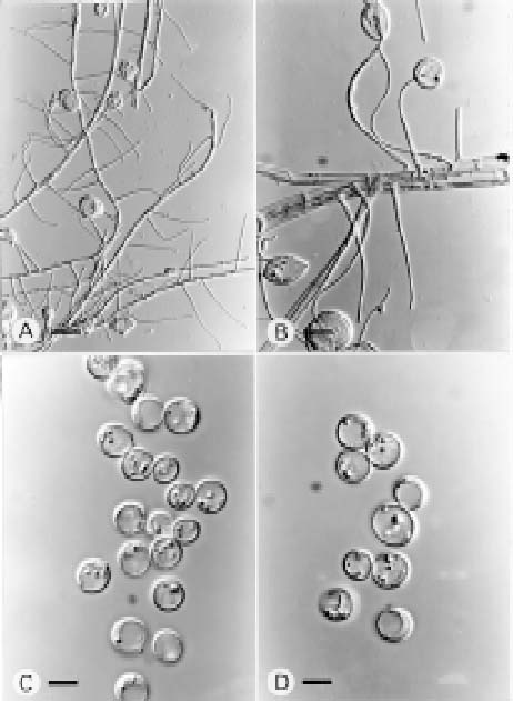
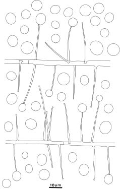
Abstract. A species of Aphanocladium was isolated from rotten bark in Kuohsing, Nantou, Taiwan. Its distinguishing characteristics are slender, acicular phialide-like conidiogenous cells and solitary, large, smooth, hyaline conidia. As these morphological characteristics differ from all other known species in the genus, this isolate can be recognized as a new species, Aphanocladium macrosporum J.L. Chen, W.S. Lin and S.S. Tzean.

Keywords: Aphanocladium macrosporum sp. nov.; Hyphomycetes; Taxonomy; Taiwan.

Introduction

Gams (1971) erected the genus Aphanocladium to include three species, A. album (Preuss) W. Gams (Basionym: Acremonium album Preuss), A. aranearum (Petch) W. Gams (Basionym: Acremonium aranearum Petch) and A. meliolae (Hansf.) W. Gams (Basionym: Oospora meliolae Hansf.). In 1973, the generic conception of Aphanocladium was revised as having solitary conidia borne on phialide-like conidiogenous cells, and A. spectabile W. Gams was named as a new species (Gams, 1973). Aphanocladium album (Preuss) W. Gams was designated the type species. Later, a further three species, A. tomentosum Arambarri, A. aranearum (Petch) W. Gams var. sinense J.D. Chen and A. dimorphum J.D. Chen were added to the genus (Arambarri, 1981; Chen et al., 1984; 1985; Petch, 1932) bringing the total number of species in Aphanocladium to seven. Three species—A. album, A. aranearum var. sinense and A. dimorphum—are parasitic on Agaricus bisporus (Lange) Sing and cause disease in the mushroom. Aphanocladium aranearum var. sinense is capable of infecting other mushroom species including Hericium erinaceus (Bull.) Pers., Lentinus edodes (Berk.) Sing. and Pleurotus ostreatus (Jacq. ex Fr.) Quél. (Chen et al., 1984). Aphanocladium dimorphum has two distinct types of conidial morphs, but conidial size was not described or measured (Chen et al., 1985).

During a taxonomic study of hyphomycetes, Duteromycotina, from rotten leaf litter in Taiwan, an interesting fungus was isolated from rotten bark in Kuohsing, Nantou County. The general morphological

3Corresponding author. Tel: 06-266-4911 ext. 284; Fax: 06-266-7322; E-mail: ccl51911@ms29.hinet.net

Figure 1. Aphanocladium macrosporum. Characteristics of its conidiophores and conidia on oat meal agar.


Botanical Bulletin of Academia Sinica, Vol. 40, 1999

characteristics of this isolate fit the generic concept of Aphanocladium. Phialide, conidial morphology and dimensions easily distinguish this isolate from other known species in the genus. Thus, a new species, Aphanocladium macrosporum J.L. Chen, W.S. Lin and S.S. Tzean is proposed.

Materials and Methods

Samples were collected from rotten bark in Kuohsing, Nantou County during October, 1996 and incubated in moist chambers (plastic boxes, 30 × 20 × 12 cm, with three layers of moistened papers) to encourage fungal sporulation. Pure culture was established by isolating a single spore or spores with a sterile glass microneedle on 3% agar. A piece of agar containing isolated spores

was cut out and transferred to oat meal agar (OMA) slants or plates under a stereomicroscope. Details of fungal morphology and conidiogenesis were studied and recorded. The fungus was illustrated using a drawing tube and photographed using an Olympus light microscope (BX50). The taxonomic systems of Barron (1968), Hughes (1953), Tubaki (1963), Ellis (1971) and Saccardo (1882-1931) were used for identification. Both live cultures and dried specimens were deposited in the Herbarium of the Chen-Fungi-Collection (Herb. CFC).

Species Descriptions

Aphanocladium macrosporum J.L. Chen, W.S. Lin, and S.S. Tzean sp. nov. (Figures 1-2)

Figure 2. Aphanocladium macrosproum. A-B, phialide-like conidiogenous cells. A. Bar = 50 µm; B. Bar = 20 µm. C-D, globose, subglobose or ellipsoidial conidia. Bars = 20 µm.


Chen et al. — A new species of Aphanocladium from Taiwan

Coloniae in OMA effusae, floccosae, albae; reversae albae; Mycelium fere superficiale, ex hyphis ramosis, septatis, lenibus, hyalinis, 1.6-8.0 µm latum compositum; Cellulae conidiogenae terminales vel laterales, monoblastae, directae, simplices vel fasciculatae, aciculares, laeves, hyalinae, 20.0-67.2 × 0.6-1.6 µm, cum collum ad apicem; Conidia solitaria, globosa ad subglobosa vel ellipsoidea, lenia, hyalina, 5.2-14.1 µm longus, 4.8-13.6 µm latus.

In Cortice putrido, Kuohsing, Nantou, 13-X-96; Holotypus, Herb. CFC-2.

Colonies on Oat meal Agar effuse, floccose, white; reverse white; Mycelium mostly superficial, composed of branched, septate, smooth, hyaline, 1.6-8.0 µm wide hyphae; Conidiogenous cells terminal or lateral, monoblastic, simple or fasciculate, acicular, with a collar at the apex, smooth, hyaline, 20.0-67.2 × 0.6-1.6 µm; Conidia solitary, globose to subglobose or ellipsoidal, smooth, hyaline, 5.2-14.1 µm long, 4.8-13.6 µm wide.

Isolated from: rotten bark, Kuohsing, Nantou, Oct. 13 1996. CTN-69.

Chen et al. (1984, 1985) have made two comparative tables detailing the morphological characteristics of six species of Aphanocladium. The six species were compared and discussed including A. aranearum var. sinense, A. dimorphum, A. album, A. aranearum, A. meliolae and A. spectabile. The remaining species not included in the tables is A. tomentosum Arambarri, which is the only synnematous species of Aphanocladium and was collected from fallen leaves of Nothofagus pumilio in Argentina. The conidiogenous cells of A. tomentosum differ from those of A. macrosporum as they are shorter and broader (11.0-13.5 × 2.4-3.0 µm) and produce smaller conidia (8.0-9.6 × 1.8-4.5 µm). Conidiogenous cells of A. macrosporum are more slender and longer (20.0-67.2 × 0.6-1.6 µm) than those of all other known species in the genus, and the conidia, which are globose or subglobose, are the largest (5.2-14.1 × 4.8-13.6 µm) among the species. The conidial morphology of A. album

is similar to that of A. macrosporum, but the conidia of A. album are smaller (2.8-5.5 µm long, 1.7-3.7 µm wide). Conidial shape in other species of Aphanocladium is described as obovoid, ellipsoidal, short-ellipsoidal or ovoid. The unique characteristics of A. macrosperum clearly distinguish it from all other members of Aphanocladium.

Acknowledgments. We thank the National Science Council (NSC-86-2314-B-041-008) for funding this research.

Literature Cited

Arambarri, A.M. 1981. Micoflora de La hojarasca de Nothofagus obliqua de Y N. pumilio. I. Boln. Soc. Argent. Bot. 20(1-2): 19-30.

Barron, G.L. 1968. The Genera of Hyphomycetes from Soil. The Williams and Wilkins Co., Baltimore.

Chen, J.D., J.G. Liu, S.S. Chen, F.J. Cai, and Z.C. Zhang. 1984. Studies on Aphanocladium aranearum (Petch) Gams var. sinense var. nov. — A fungus parasitic on Agaricus bisporus (Lange) Sing. Acta Mycol. Sin. 3(2): 96-101

Chen, J.D., J.G. Liu, S.S. Chen, F.J. Cai, and Z.C. Zhang. 1985. A new species of Aphanocladium on Agaricus bisporus. Acta Mycol. Sin. 4(4): 227-233.

Ellis, M.B. 1971. Dematiaceous Hyphomycetes. Commonwealth Mycological Institute. Kew. Surrey, England.

Gams, W. 1971. Cephalosproiumartige Schimmelpilze (Hyphomycetes). Gustar Fischer Verlag, Stuttgart, 262 pp.

Gams, W. 1973. Phialides with solitary conidia? Remarks on conidium ontogeny in some hyphomycetes. Persoonia 7(2): 161-169.

Hughes, S.J. 1953. Conidiophores, conidia and classification. Can. J. Bot. 31: 577-659.

Petch, T. 1932. Notes on Entomogenous Fungi. Trans. Br. Mycol. Soc. 16: 242-243.

Saccardo, P.A. 1882-1931. Sylloge Fungorum Omnium Cognitorum. 25 volumes. Pavia, Italy.

Tubaki, K. 1963. Taxonomic study of Hyphomycetes. Annu. Rep. Inst. Ferment. Osaka 1: 25-54.


Botanical Bulletin of Academia Sinica, Vol. 40, 1999

¥xÆW²£¤£§¹¥þµß·sºØ Aphanocladium macrosporum sp. nov.

³¯«°ãS1 ªL¬°´Ë1 ´¿Å㶯2

1 ¨p¥ß¹Å«nÃIJz¾Ç°|Âå°ÈºÞ²z¨t

2 °ê¥ß¥xÆW¤j¾Ç´Óª«¯f²z¾Ç¨t
¥»¤å´y­z¤@ºØ¤ÀÂ÷¦Û«n§ë¿¤¡B°ê©m¶m¡A»G¦´¾ð¥Ö¤W¤§µ·ÌUºõ¤£§¹¥þµß·sºØ Aphanocldium macrosporum J.L. Chen, W.S. Lin and S.S. Tzean¡C¦¹·sºØ¨ä³Ì¥D­nªº§ÎºA¯S¼x»P²£ÌU¤è¦¡¬°¡G²£ÌU²Ó­M (conidiogenous cells; phialide-like) ¤W¡A¯à²£¥Í³æ¤@ (solitary)¡A¥¨¤j¡A¥­·Æ¡AµL¦â¡A¶ê§Î¡B¦¸¶ê§Î©Î¾ò¶ê§Î¤§¤À¥ÍÌU¤l¡C­Y »PÄݤº¤§¤wª¾ºØ¤¬¬Û¤ñ¸û§ÎºA¯S¼x¡A«h¥i»´©ö¦a°Ï§O¦¹¯uµß¡A¨Ò¦p¡GAphanocladium macrosporum ¨ã¦³²Óªøªº²£ÌU²Ó­M¥H¤Î¤j«¬ªº¤À¥ÍÌU¤l¡C¦¹µ¥¿W¤@µL¤Gªº¯S¼x¡A¤D½T¥ß¦¹¤@·sºØ¤§³Ð³]¡C

ÃöÁäµü¡G ¤£§¹¥þµß¡Fµ·ÌUºõ¡F·sºØ (Aphanocladium macrosporum sp. nov.)¡F¥xÆW¡C