Bot. Bull. Acad. Sin. (2000) 41: 19-25

Lee — Acid phosphatase in Ulva

Phosphate starvation induction of acid phosphatase in Ulva lactuca L. (Ulvales, Chlorophyta)

Tse-Min Lee1

Institute of Marine Biology, National Sun Yat-sen University, Kaohsiung 80424, Taiwan

(Received November 23, 1998; Accepted March 5, 1999)

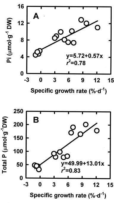
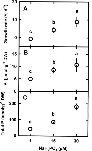
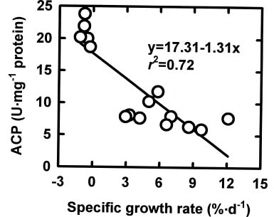
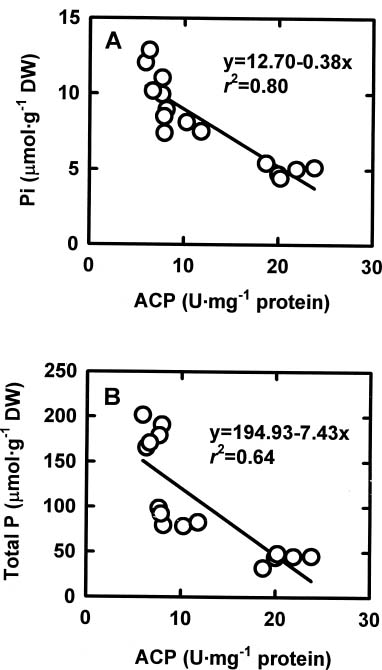
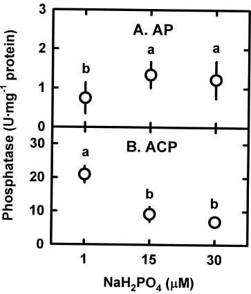
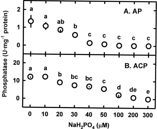
Abstract. The relationships among specific growth rate, intracellular inorganic phosphate (Pi) concentrations, total phosphorus (P) concentrations and specific activities of acid phosphatase (ACP; EC 3.1.3.2) and alkaline phosphatase (AP; EC 3.1.3.1) were studied in a green marine macroalga Ulva lactuca L. after 4 days of incubation at 1, 15 or 30 µM NaH2PO4. Both the specific growth rate and the intracellular Pi and total P concentrations increased with increasing NaH2PO4 levels; a linear correlation with specific growth rate was found for both intracellular Pi concentrations (r2=0.78, P<0.05) and the total P concentrations (r2=0.83, P<0.05). The growth rate and the Pi and total P concentrations were relatively low at 1 µM NaH2PO4, indicating U. lactuca L. could be P-deficient at 1 µM NaH2PO4. The specific activity of ACP was 4 to 10-fold greater than that of AP. Exposure to 1 µM NaH2PO4 increased the specific activity of ACP but slightly decreased that of AP. The ACP specific activity was inversely correlated with specific growth rate (r2=0.72, P<0.05) and also intracellular Pi concentrations (r2=0.80, P<0.05) and the total P concentrations (r2=0.64, P<0.05). The AP specific activity had no correlation with specific growth rate, intracellular Pi concentrations, or total P concentrations. The addition of NaH2PO4 concentrations higher than 20 µM to the assay mixture inhibited the specific activities of both ACP and AP. Overall, the P deficiency induction of ACP activity is related to a decrease in P availability in U. lactuca L.

Keywords: Acid phosphatase; Algae; Alkaline phosphatase; Phosphate starvation; Ulva lactuca.

Abbreviations: DW, dry weight; AP, alkaline phosphatase; ACP, acid phosphatase; P, phosphorus; Pi, inorganic oxyanion phosphate; p-NPP, p-nitrophenol phosphate.

Introduction

In algae, phosphorus (P) is an important and often limiting nutrient (Davies, 1988). P deficiency leads to metabolic disorders such as a decrease in photosynthesis and respiration, a retardation of growth, and even the death of algae cells (Davies, 1988; Gárciá-Sánchez et al., 1996; Lapointe, 1987; Theodorou et al., 1991). In general, algae take up P in the form of inorganic oxyanion phosphate (Pi) from the surrounding media via an active transport. However, most soluble Pi in seawater is esterified to organic compounds such as dissolved organic phosphomonoesters, rendering it is impossible for algae to uptake directly. It is known that most marine algae can synthesize extracellular alkaline phosphatase (AP; EC 3.1.3.1) to liberate Pi from dissolved organic phosphomonoesters, especially under P-deficient conditions (Fitzgerald and Nelson, 1966; Hernández et al., 1993; Weich and Graneli, 1989). In the case of the marine chlorophyte macroalga Ulva lactuca L., the activity of extracellular AP increased during cultivation in a P-deficient medium (Weich and Graneli, 1989). A similar trend was

also found in the freshwater microalgae Chlamydomonas reinhardtii grown in the absence of Pi (Quisel et al., 1996).

Algae can store P intracellularly as Pi or polyphosphate, which is utilized during the subsequent growth process or in P-deficient conditions (Cole and Huges, 1965; Kulaev and Vagabov, 1983; Lundberg et al., 1989; Watanabe et al., 1987). The concentrations of intracellular polyphosphates in U. lactuca L. decrease after transfer to P-deficient conditions, indicating that the intracellular polyphosphates could be hydrolyzed to meet P requirements (Weich and Graneli, 1989). Several intracellular phosphatases have been suggested to have a role in the hydrolysis of intracellular polyphosphates (Huber and Hamel, 1985; Marco and Orús, 1988). In microalgae, P-deficiency increases the activity of intracellular acid phosphatase (ACP; EC 3.1.3.2) (Blum, 1965; Price, 1962), which is localized in the cytoplasm (Moher et al., 1975; Price, 1962; Sommer and Blum, 1965). There are two kinds of ACP in microalgae: constitutive and inducible enzymes (Blum, 1965). In macroalgae, the responses of phosphatase to P-deficiency have been mainly studied on AP. Although the role of ACP in phosphorus metabolism has been extensively studied in terrestrial plants (Duff et al., 1994), we still have little knowledge about the relationships between intracellular soluble ACP and P availability in macroalgae.

1Tel: 886-7-5252000 ext. 5110; Fax: 886-7-5255100; E-mail: tmlee@mail.nsysu.edu.tw


Botanical Bulletin of Academia Sinica, Vol. 41, 2000

In this study, the relationships among specific growth rate, intracellular Pi concentrations, total P concentrations, and intracellular soluble ACP specific activity were determined in U. lactuca L. in response to varying external NaH2PO4 levels (1, 15 and 30 µM). The changes in intracellular soluble AP specific activity at different external NaH2PO4 levels were also determined. The in vitro effects of NaH2PO4 on ACP and AP activities were also determined.

Materials and Methods

Algal Culture and Treatment

Ulva lactuca L. was collected in August from the intertidal regions of Kenting, Taiwan, Republic of China (ROC) and was immediately transferred to the laboratory and used for experiments. The average total P concentration in seawater in August is 26.63 µM (17.95~31.40 µM) and the average Pi concentration is 12.47 µM (8.25~15.07 µM). After washing with sterilized seawater, three thallus discs (5 cm in diameter) of approximately 1.2 g fresh weight were cultured for 4 days at 25°C in a polycarbonate vessel (Magenta GA-7 vessel, Sigma, St. Louis, MO., USA). Each vessel contained 300 ml of 35‰ artificial seawater (405 mM NaCl, 10 mM KCl, 10 mM CaCl2, 30 mM MgSO4 and 10 mM Tris-HCl, pH 8.0) enriched with P-free Provasoli nutrient solution (Provasoli, 1968). This seawater was changed every day. The levels of Pi in the artificial seawater were adjusted by adding NaH2PO4 to a final concentration of 1, 15 or 30 µM. The photoperiod was 12 h and the photon irradiance was 250 to 300 µmol photon m-2 s-1, achieved by ten 60W cool-fluorescent lamps (FL60D, China Electric Apparatus Ltd., Tao-yuan, Taiwan, ROC). After treatments, thallus discs were washed with MQ H2O three times within 1 min, fixed in liquid nitrogen, lyophilized at -60°C and then stored at -70°C for further analysis. In this study, a vessel is a replicate, and each treatment had at least 5 replicates. Each experiment was performed three times. Since they showed a similar trend, only one of them was shown in this paper.

Determination of Specific Growth Rate

Fresh weight of discs was determined at the beginning (W0) and after 4 days of incubation (W4). The specific growth rate was expressed as the percentage of fresh weight increase (expressed as the natural log value) per day, %•d-1 = ln(W4-W0)/4d ×100 (Lee and Chen, 1998). The specific growth rate was the average of five replicates.

Determination of Intracellular Pi and Total P Concentrations

For the intracellular Pi determination, lyophilized material was mixed with H2O in a ratio of plant material : H2O = 1 g dry weight (DW) : 50 mL. The mixture was incubated at 80°C in a water bath for 30 min and then centrifuged at 15,000 g for 10 min at 4°C. For the total P determination, 0.1 g DW of lyophilized material was digested in an acid solution (concentrated HNO3 : HClO4 = 2 : 1, v/v) until the

mixture became clear. The amount of intracellular Pi or total P was determined by the Mo blue method (Murphy and Riley, 1962).

Determination of ACP and AP Specific Activities

Lyophilized material was homogenized into powder in liquid nitrogen and mixed with extraction buffer (100 mM Tris-HCl, pH 7.0) in a ratio of plant material : extraction buffer = 1 g dry weight : 10 mL. After centrifugation at 15,000 g for 10 min under 4°C, the supernatant was collected as the sample extract for the determination of activity of intracellular soluble enzymes. The ACP activity was assayed spectrophotometrically according to the method of Pan and Chen (1988). The reaction was started by adding p-nitrophenol phosphate (p-NPP) and then incubating at 40°C for 30 min. The reaction solution contained 100 mM sodium acetate buffer (pH 5.4), 5 mM p-NPP and 5 µL of sample extract in a total volume of 200 µL. Fifty µL of 1 M KOH was added to terminate the reaction. For the AP activity determination, the reaction mixture consisted of 100 mM Tris-HCl (pH 8.3), 10 mM p-NPP and 5 µL of sample extract in a total volume of 200 µL. The reaction was also initiated by adding p-NPP and then incubating at 40°C for 30 min. Then, 50 µL of 1 M KOH was added to terminate the reaction. We found that the enzyme activities of both ACP and AP were linear between 0 to 60 min and also proportionally increased as the amount of sample extract increased. The amount of released p-nitrophenol was detected by the absorbance at 405 nm. ACP activity was calculated using the molar extinction coefficient for p-nitrophenol of 3.8 × 10³ M-1 cm-1, and AP activity was calculated using the molar extinction coefficient for p-nitrophenol of 4.6 × 10³ M-1 cm-1. The extinction coefficient for p-nitrophenol for ACP (pH 5.4) or AP (pH 8.3) was obtained from our experiments using different concentrations of p-nitrophenol purchased from Sigma (St. Louis, MO, USA). One unit (U) of phosphatase activity is the amount of enzyme producing 1 µmol p-nitrophenol min-1. Since the polyphenols in sample extracts also have a significant absorbance at 405 nm, the blank of each sample was prepared by adding 50 µL of 1 M KOH to the reaction solution before the addition of p-NPP. Soluble proteins were determined according to the Bradford dye binding method (Bradford, 1976) using bovine serum albumin as a standard.

Statistical Analysis

Data of specific growth rate, intercellular Pi and total P concentrations, and ACP and AP activities among different NaH2PO4 levels were firstly analyzed by the ANOVA test at P<0.05 (SAS, SAS Ltd., NC, USA), and then the significance of differences between treatments was analyzed by using Duncan’s new multiple range test (SAS, SAS Ltd., NC, USA). The relationships among specific growth rate, intracellular Pi concentration, total P concentration, and enzyme activity were tested by using the association and regression analysis at P<0.05 (SAS, SAS Ltd., NC, USA).


Lee — Acid phosphatase in Ulva

Results

Specific Growth Rate, Intercellular Pi and Total P Concentrations

After being collected from the sea, the algae were extensively washed with autoclaved artificial seawater and then sampled to determine the initial values. The initial concentrations of intracellular Pi and total P were 8.7 ± 0.19 and 72.5 ± 3.6 µmol g-1 DW, respectively, which were similar to those incubated at 15 µM NaH2PO4 for 4 days (t-Test, P>0.05, n=10). The similarity between the initial and the 15 µM NaH2PO4 treatment may be due to the highly eutrophicated (the Pi levels were around 15 µM) seawater where the algae were collected.

As the external NaH2PO4 levels increased, the specific growth rate increased; exposure to 1 µM NaH2PO4 ceased growth (Figure 1A). Both the intracellular Pi (Figure 1B) and total P (Figure 1C) concentrations also increased with increasing external NaH2PO4 levels.

Figure 2. The relationships between specific growth rate and intracellular Pi concentration (A) and between specific growth rate and total P concentration (B) in Ulva lactuca L. exposed to varying external NaH2PO4 levels.

The changes in intracellular Pi concentrations showed a linear relation with the changes in specific growth rate (r2=0.78, P<0.05) (Figure 2A). The changes in total P concentrations were also positively related to the changes in specific growth rate (r2=0.83, P<0.05) (Figure 2B).

ACP and AP Specific Activities

The initial intracellular soluble ACP and AP activities were 11.7 ± 2.9 and 1.4 ± 0.3 U mg-1 protein, respectively. In the U. lactuca L. grown in the 15 µM NaH2PO4 artificial seawater for 4 days, the specific activities of both ACP and AP remained unchanged compared to the initial ACP and AP activities (t-Test, P>0.05, n=10), and the specific activity of intracellular soluble ACP was still 10-fold greater than that of intracellular soluble AP (Figure 3). As compared to the 15 and 30 µM NaH2PO4 treatments, the AP

Figure 1. Changes in specific growth rate (A), intracellular Pi concentration (B) and total P concentration (C) in Ulva lactuca L. exposed to varying external NaH2PO4 levels. Different symbols indicate significant differences at P<0.05. Vertical bars indicate the 95% confidence interval (n=5).


Botanical Bulletin of Academia Sinica, Vol. 41, 2000

specific activity was slightly lower in thalli grown at 1 µM NaH2PO4 (Figure 3A). At 1 µM NaH2PO4, the ACP specific activity was the highest, almost 2-fold higher than in the other treatments (Figure 3B).

Figure 4 shows that the changes in the specific activity of ACP were inversely related to the changes in the specific growth rate (r2=0.72, P<0.05). The changes in the specific activity of ACP were also inversely related to the changes in the intracellular Pi concentrations (r2=0.80, P< 0.05) (Figure 5A) and total P concentrations (r2=0.64, P< 0.05) (Figure 5B). However, the specific activity of AP had no correlation with the specific growth rate, the intracellular Pi concentrations, or the total P concentrations (data not shown).

Influence of NaH2PO4 on in vitro Phosphatase Activity

To test whether Pi regulates the ACP and AP activities in U. lactuca L., different NaH2PO4 concentrations (0, 10, 20, 30, 40, 50, 100, 200, 300 µM) were added to the assay mixture of phosphatase extracted from thalli grown at 30 µM NaH2PO4 and enzyme activity was determined. The addition of NaH2PO4 at concentrations higher than 20 µM inhibited the ACP activity, and 50% inhibition was reached around 50 µM (Figure 6A). The AP activity was inhibited by NaH2PO4, and 50% inhibition occurred at about 30 µM (Figure 6B). Thus AP was slightly more sensitive to Pi inhibition than was ACP.

Figure 4. Relationship between specific growth rate and ACP specific activity in Ulva lactuca L. exposed to varying external NaH2PO4 levels.

Figure 3. Specific activities of AP (A) and ACP (B) in Ulva lactuca L. exposed to varying external NaH2PO4 levels. Different symbols indicate significant differences at P<0.05. Vertical bars indicate the 95% confidence interval (n=5).

Figure 5. Relationship between ACP specific activity and intracellular Pi concentration (A) and between ACP specific activity and total P concentration (B) in Ulva lactuca L. exposed to varying external NaH2PO4 levels.


Lee — Acid phosphatase in Ulva

Exposure of U. lactuca L. to 1 µM NaH2PO4 markedly increased the specific activity of ACP, but not of AP (Figure 3). This P deficiency-induced increase in ACP activity has also been found in microalgae (Knutsen, 1968; Lien and Knutsen, 1973; Lubián et al., 1992; Price, 1962; Theodorou et al., 1991). In higher plants, the ACP activity also increases after transfer to P-deficient conditions (Barrett-Lennard et al., 1982; Dracup et al., 1984; Duff et al., 1994). ACP activity has been used in higher plants as a biochemical marker of P limitation (Barrett-Lennard et al., 1982; Duff et al., 1994). Our results demonstrating the linear correlation between ACP activity and intracellular Pi and total P concentrations show that in U. lactuca L. ACP can be an indicator of P deficiency. The increment on ACP activity in Pi-limited conditions suggests that ACP could be involved in polyphosphate degradation in U. lactuca, as has been suggested for some higher plants (Dewald et al., 1992; Duff et al., 1994).

In this study, the in vitro experiments show that NaH2PO4 is a potent inhibitor of the activities of ACP and AP in U. lactuca L.; the intracellular soluble ACP and AP activities are inhibited by the addition of NaH2PO4. A high correlation with intracellular Pi concentrations was found for ACP activity. The induction of ACP activity at 1 µM NaH2PO4 is, therefore, possibly due to a lower intracellular Pi concentration. However, our data show no correlation between intracellular Pi and AP activity.

In conclusion, to our knowledge, this is the first paper showing P deficiency can induce ACP activity in the marine macroalga U. lactuca L. The results of this investigation using thallus discs grown under different NaH2PO4 levels suggest that the P deficiency induction of ACP activity may be correlated to P availability in U. lactuca L.

Acknowledgments. We are grateful to Dr. W. C. Plaxton and one anonymous reviewer in critical reading the manuscript, and to Dr. Yaw-Huei Lin from the Institute of Botany, Academia Sinica, Taipei, Taiwan, Republic of China, for providing the facilities. Thanks also to Fan-Pao Lee, Min-Chiao Shih and May-Yu Kuo for manuscript preparation and technical assistance. The financial assistance from the National Science Council, Executive Yuan, Republic of China to Tse-Min Lee is acknowledged.

Literature Cited

Barrett-Lennard, E.G., E.G. Robin, and H. Greenway. 1982. Effect of phosphorus and water deficit on phosphatase activities from wheat leaves. J. Exp. Bot. 33: 682-693.

Blum, J.J. 1965. Observations on the acid phosphatases of Euglena gracilis. J. Cell Biol. 24: 223-234.

Bradford, M.M. 1976. A rapid and sensitive method for quantitative of microgram quantities of protein using the principle of protein-dye binding. Anal. Biochem. 72: 248-254.

Cole, J.A. and D.E. Huges. 1965. The metabolism of polyphosphates in Chlorobium thiosulfatophikum. J. Gen. Microbiol. 38: 65-72.

Davies, A.G. 1988. Nutrient interactions in the marine environment. In D. Rogers and J. Gallon (eds.), Biochem

Figure 6. Changes in AP (A) or ACP (B) activities in response to in vitro application of NaH2PO4 (0, 10, 20, 30, 40, 50, 100, 200, 300 µM). NaH2PO4 was added to the enzyme assay solution. Different letters indicate significant differences at P< 0.05. Vertical bars indicate the 95% confidence interval (n=5).

Discussion

This study shows that the growth of U. lactuca L. depends on the external supply of NaH2PO4. There is a linear relationship between external NaH2PO4 level and specific growth rate; the growth of thallus discs ceased after 4 days of incubation in the 1 µM NaH2PO4 medium, so U. lactuca L. faces P deficiency when incubated at 1 µM NaH2PO4.

The external NaH2PO4 level affects the concentrations of intracellular Pi and total P; the lower the external NaH2PO4 level, the lower the intracellular Pi and total P concentrations (Figure 1). The initial intracellular Pi and total P concentrations were 8.7 and 72.5 µmol g-1 DW, respectively, which were similar to those found in thalli after 4 days of incubation in the 15 µM NaH2PO4 medium. It has also been shown in other algae that the levels of intracellular Pi decrease when exposed to P-deficient conditions (Lundberg et al., 1989; Tillberg and Rowley, 1989). The intracellular Pi and total P concentrations significantly decreased to 4.9 and 42.9 µmol g-1 DW, respectively, after incubation in the 1 µM NaH2PO4 medium, which indicated that at this concentration, U. lactuca L. could be P-limited. The specific growth rate of U. lactuca L. thallus discs showed a linear relationship with the intracellular Pi concentrations.

Besides, our data from Figure 2 show that the ratio Pi/total P in U. lactuca L. changed in response to different external NaH2PO4 levels, although most P in thalli exists in the bound form rather than free Pi whenever grew at all treated NaH2PO4 levels. Based on the total P amount, the percentage of Pi was approximately 9% in thallus discs grown at 1 or 15 µM NaH2PO4 but decreased to 5.5% when exposed to 30 µM NaH2PO4.


Botanical Bulletin of Academia Sinica, Vol. 41, 2000

istry of the Algae and Cyanobacteria. Proc. Phytochem. Soc. Europe, vol. 29. Oxford University Press. Oxford, pp. 241-256.

Dewald, D.B., H.S. Mason, and J.E. Mullet. 1992. The soybean vegetative storage proteins VSP alpha and VSP beta are acid phosphatases active in polyphosphates. J. Biol. Chem. 267: 15958-15964.

Dracup, M.N.H., E.G. Barrett-Lennard, H. Greenway, and A.D. Robson. 1984. Effect of phosphorus deficiency on phosphatase activity of cell walls from roots of subterranean clover. J. Exp. Bot. 35: 466-480.

Duff, S.M.G., G. Sarath, and W.C. Plaxton. 1994. The role of acid phosphatases in plant phosphorus metabolism. Physiol. Plant. 90: 791-800.

Fitzgerald, G.P. and T.C. Nelson. 1966. Extractive and enzymatic analyses for limiting or sulpus phosphorus in algae. J. Phycol. 2: 32-37.

Gárciá-Sánchez, M.J., J.A. Fernández, and F.X. Niell. 1996. Photosynthetic response of P-deficient Gracilaria tenuistipitata under two different phosphate treatments. Physiol. Plant. 96: 601-606.

Hernández, I., J.A. Fernández, and F.X. Niell. 1993. Influence of phosphorus status on the seasonal variation of alkaline phosphatase activity in Porphyra umbilicalis (L.) Kutzing. J. Exp. Mar. Biol. Ecol. 173: 181-196.

Huber, A.L. and K.S. Hamel. 1985. Phosphatase activities in relation to phosphorus nutrient in Nodularia spumigena (Cyanobactieaceae). Hydrobiologia 123: 81-88.

Kulaev, I.S. and U.M. Vagabov. 1983. Polyphosphate metabolism in micro-organisms. Adv. Microbiol. Physiol. 24: 83-171.

Knutsen, G. 1968. Repressed and derepressed synthesis of phosphatase during synchronous growth of Chlorella pyrenoidosa. Biochim. Biophys. Acta 161: 205-214.

Lapointe, B.E. 1987. Phosphorus- and nitrogen-limited photosynthesis and growth of Gracilaria tikvahiae (Rhodophyceae) in the Florida Keys: an experimental field study. Mar. Biol. 93: 561-568.

Lee, T.M. and M.H. Chen. 1998. Hyposaline effect on polyamine accumulation in Ulva fasciata (Ulvales, Chlorophyta). Bot. Bull. Acad. Sin. 39: 167-174.

Lien, T. and G. Knutsen. 1973. Synchronous cultures of Chlamydomonas reinhardti: properties and regulation of repressible phosphatases. Physiol. Plant. 28: 291-298.

Lubián, L.M., J. Blasco, and R. Establier. 1992. A comparative study of acid and alkaline phosphatase activities in several strains of Nannochloris (Chlorophyceae) and

Nannochloropsis (Eustigmatophyceae). Br. Phycol. J. 27: 119-130.

Lundberg, P., R.G. Weich, P. Jensen, and H.J. Vogel. 1989. Phosphorus-31 and nitrogen-14 NMR studies of the uptake of phosphorus and nitrogen compounds in the marine macroalgae Ulva lactuca. Plant Physiol. 89: 1380-1387.

Marco, E. and M.I. Orús. 1988. Variation in growth and metabolism with phosphorus nutrient in two cyanobacteria. J. Plant Physiol. 132: 339-344.

Moher, M., S. Myklestad, and A. Haugh. 1975. Alkaline and acid phosphatases of the marine diatoms Chaetoccros affinis var. Willei (Gran) Hustedi and Skeletonema costatum (Grev.) Cleve. J. Exp. Mar. Biol. Ecol. 19: 217-226.

Murphy, J. and J.P. Riley. 1962. A modified single solution method for the determination of phosphate in nature waters. Anal. Chim. Acta 27: 31-36.

Pan, S.M. and Y.R. Chen. 1988. The effects of salt stress on acid phosphatase activity of Zea mays seedlings. Bot. Bull. Acad. Sin. 29: 33-38.

Price, C.A. 1962. Repression of acid phosphatase synthesis in Euglena gracilis. Science 135: 46.

Provasoli, L. 1968. Media and prospects for the cultivation of marine algae. In A. Watanabe and A. Hattori (eds.), Cultures and Collections of Algae. Proceeding of the U.S., Japan Conference. Hakone. Japanese Society of Plant Physiology, pp. 63-75.

Quisel, J.D., D.D. Wykoff, and A.R. Grossman. 1996. Biochemical characterization of the extracellular phosphatase produced by phosphorus-deprived Chlamydomonas reinhartii. Plant Physiol. 111: 839-848.

Sommer, J.R. and J.J. Blum. 1965. Cytochemical localization of acid phosphatase in Euglena gracilis. J. Cell Biol. 24: 235-251.

Theodorou, M.E., I.R. Elrifi, D.H. Turpin, and W.C. Plaxton. 1991. Effects of phosphorus limitation on respiratory metabolism in the green alga Selenastrum minutum. Plant Physiol. 95: 1089-1095.

Tillberg, J.E. and J.R. Rowley. 1989. Physiological and structural effects of phosphorus starvation on the unicellular alga Scenedesmus. Physiol. Plant. 75: 315-332.

Watanabe, M., K. Kohata, and M. Kunugi. 1987. 31P Nuclear Magnetic Resonance study of intracellular phosphate pools and polyphosphate metabolism in Heterosigma akashiwo (hada) Hada (Raphidophyceae). J. Phycol. 23: 54-62.

Weich, R.G. and E. Graneli. 1989. Extracellular alkaline phosphatase activity in Ulva lactuca L. J. Exp. Mar. Biol. Ecol. 129: 33-44.


Lee — Acid phosphatase in Ulva

¯ÊÁC»¤¾É¥Ûç] (Ulva lactuca L.) »Ä©ÊÁC»Ä¤À¸Ñ»Ã¯À¬¡©Ê¼W°ª

§õ¿A¥Á

°ê¥ß¤¤¤s¤j¾Ç®ü¬v¥Íª«¬ã¨s©Ò

¥»¬ã¨s±´°Q¤j«¬®ü¬vºñĦ¥Ûç] (Ulva lactuca L.) ¸g 4 ¤Ñªº 1¡B15 ©Î 30 µM NaH2PO4 ³B²z«á¡A¤ñ¥Íªø³t²v¡B²Ó­M¤ºµL¾÷ÁC»Ä§t¶q¡BÁ`ÁC§t¶q¡B»Ä©ÊÁC»Ä¤À¸Ñ»Ã¯À (ACP; EC 3.1.3.2) ¤ñ¬¡©Ê¤ÎÆP©ÊÁC»Ä¤À¸Ñ»Ã¯À (AP; EC 3.1.3.1) ¤ñ¬¡©Ê¤§Ãö«Y¡C¤ñ¥Íªø³t²v¡B²Ó­M¤ºµL¾÷ÁC»Ä§t¶q¤ÎÁ`ÁC§t¶qÀH NaH2PO4 ¿@«×¼W°ª¦Ó¼W¥[¡C¤ñ¥Íªø³t²v»P²Ó­M¤ºµL¾÷ÁC»Ä§t¶q¦¨ª½½u¬ÛÃö (r2=0.78, P <0.05)¡A»PÁ`ÁC§t¶q¤]¦¨ª½½u¬ÛÃö (r2=0.83, P <0.05)¡C·t¥Ü¥Ûç]»PÁC»Äªº§Q¥Î©Ê¦³Ãö¡CACP ªº¤ñ¬¡©Ê¬O AP ¤ñ¬¡©Êªº 4~10 ­¿¡C¦b 1 µM NaH2PO4 ¤U¡A¼W¥[¦Ó AP ¤ñ¬¡©Ê­°§C¡CACP ¤ñ¬¡©Ê»P¤ñ¥Íªø³t²v¦¨ª½½u¬ÛÃö (r2=0.72, P <0.05)¡A»P²Ó­M¤ºµL¾÷ÁC»Ä§t¶q¤]¦¨ª½½u¬ÛÃö (r2=0.80, P<0.05)¡C¦ý¬O¡AAP ¤ñ¬¡©Ê»P¤ñ¥Íªø³t²v©Î²Ó­M¤ºµL¾÷ÁC»Ä§t¶qµLÃö¡C§Q¥Î¥~¥[©ó»Ã¯À¤ÏÀ³²Gªº in vitro ¤è¦¡¡ANaH2PO4 ¦b¿@«×°ª©ó 20 µM ¤U¹ï ACP ¤Î AP ¤ñ¬¡©Ê¦³©úÅ㪺§í¨î§@¥Î¡C¥»¬ã¨sªºµ²ªG«ü¥X¯ÊÁC©Ò»¤¾Éªº ACP ¬¡©Ê¼W°ª¥i¯à»P¤j«¬®ü¬vºñĦ¥Ûç]¦b¯ÊÁCª¬ªp¤U¥i§Q¥ÎÁCªº§t¶q­°§C¦³Ãö¡C

ÃöÁäµü¡G»Ä©ÊÁC»Ä¤À¸Ñ»Ã¯À¡FÆP©ÊÁC»Ä¤À¸Ñ»Ã¯À¡F¯ÊÁC¡F¥Ûç]¡C