Bot. Bull. Acad. Sin. (2000) 41: 57-60

Zheng and Ho — The sexual stage of Phytophthora polygoni

The sexual stage of Phytophthora polygoni Saw.

X.B. Zheng1 and H.H. Ho2,3

1Plant Pathogen Research Institute, Nanjing Agricultural University, Nanjing 210095, People’s Republic of China

2Department of Biology, State University of New York, New Paltz, New York 12561, USA

(Received March 1, 1999; Accepted May 20, 1999)

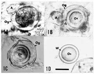
Abstract. The sexual stage of Phytophthora polygoni Saw.—which is distinguishable from other species of Phytophthora by its inability to grow on common agar media, its sporangial characteristics and its disease symptoms—has been successfully induced to form sexual organs in diseased leaves collected from the field or by artificial inoculation. The oogonia are smooth, spherical, thin-walled, (28.5-) 33 (-42) µm diam, with an aplerotic to markedly aplerotic, thick walled (3-4 µm) oospore, (24.7-) 28.1 (-32.3) µm diam, and a paragynous antheridium, 13-25 × 10-15 µm, attached laterally or apically to the oogonium. Interesting chlamydospore-like bodies were also observed.

Keywords: China; Phytophthora polygoni; Rumex dentatus; Sex organs.

Introduction

Phytophthora polygoni Saw., first described as a new species by Sawada (1922) causing leaf spot disease of Polygonium japonicum Meisn. in Taiwan was recently rediscovered on Rumex dendatus L. in Nanjing in Jiangsu Province of mainland China. It was identified based on similar disease symptoms and sporangial characteristics (Zheng and Ho, 1998). However, the fungus could not be isolated on culture media, and the sexual stage had not been found. Consequently, the species was excluded from treatment of the genus Phytophthora (Waterhouse, 1963) due to incomplete knowledge of its life cycle. Reported here are successful attempts to induce, under laboratory conditions, the formation of sex organs on diseased leaves of R. dentatus collected from the field and by artificial inoculation of healthy leaves with P. polygoni.

Materials and Methods

Diseased leaves of R. dentatus infected by P. polygoni were collected from Nanjing (late April to early July, 1998) and Yangzhou (early June, 1998) in China’s Jiangsu Province. Freshly collected leaves (2-3) were placed in 12-15 ml sterile tap water in a 9-cm petri dish. Three replicates were used for each collection from the same locality at a specific time. The dishes were incubated in darkness for 12-15 days at 25, 20 and 14°C. At the end of the incubation period, a small piece of rotten tissue was placed on a glass slide. The excess water was removed with filter paper. The leaf tissues were cut into smaller pieces with a scalpel. Lactophenol (2-3 drops) was added to the specimen, heated over an alcohol lamp until it just started to boil, then removed from the flame until the boiling

stopped before it was reheated again. The procedure was repeated 3-5 times so that the tissues became clarified. They were then covered with a cover glass for studies under a light microscope.

For artificial inoculation, freshly collected diseased leaves were first rinsed clean under running tap water. Small pieces of leaf lesions (ca. 15 × 15 mm) were cut out and placed in 10 ml sterile tap water in a 9-cm petri dish, with the leaf surface facing upwards. After incubation under light at 20°C, abundant zoospores were released into the water. The zoospore suspension was transferred to a misting gun and sprayed on the healthy leaves of potted R. dentatus from the greenhouse. Then the entire plant was wrapped in a plastic bag for 24 h to maintain saturated humidity. At the end, the treated plants were kept in a greenhouse (25-28°C) for 5-7 days. When leaf lesions developed fully, diseased leaves were submerged in water and heated with lactophenol as described above.

Results

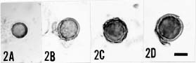
Sexual structures that were not found at any stage in the diseased leaves in the field, could be induced to produce in the laboratory by the methods described (Figure 1A-D). The sexual organs were frequently formed along and within the leaf veins and most abundantly in the midrib and petiole (adjacent to the lamina) of the diseased leaf. The mature oogoonia (Figure 1B-C) were smooth, spherical, thin-walled, straw-colored, measuring (28.5-) 33 (-42) µm diam with an aplerotic to markedly aplerotic spherical oospore (24.7-) 28.1 (-32.3) µm diam. The single-layered oospore wall was smooth and uniformly thick (3-4 µm) (Figure 1D). The paragynous antheridium 13-25 × 10-15 µm was attached laterally or apically to the side of the oogonium (Figure 1A-B). Presumably, fertilization occurred when the antheridial contents were discharged into the oogonium through a fertilization tube.

3Corresponding author. Tel: 914-257-3780; Fax: 914-257-3791; E-mail: hoh@matrix.newpaltz.edu


Botanical Bulletin of Academia Sinica, Vol. 41, 2000

Figure 1. Sexual structures of Phytophthora polygoni. A, Fertilization of young oogonium (Og) by a paragynous antheridium, (A); B, Mature oogonium with aplerotic oospore (Os) and paragynous antheridium. Note the remains of fertilization tube (ft); C, Mature oogonium with markedly aplerotic oospore; D, Isolated oospore released from oogonium with uniformly thick wall (W). Scale bar: 20 µm. All at the same magnification.

Figure 2. A-D, Small chlamydospore-like structures. Scale bar: 10 µm. All at the same magnification.

Figure 3. A-B, Large chlamydospore-like structures. Scale bar: 20 µm. Both at the same magnification.


Zheng and Ho — The sexual stage of Phytophthora polygoni

Leaf lesions, developed as a result of artificial inoculation and treated similarly as naturally infected leaves, produced identical sex organs, confirming that the sex organs found in diseased leaves collected from the field were indeed formed by the pathogen, P. polygoni. The induction of sexual reproduction in P. polygoni was favored at 25 and 20°C whereas at 14°C oogonia were fewer and often aborted. Instead, abundant small, spherical chlamydospore-like bodies (12.3-) 16 (-24.6) µm diam with one or two wall layers (0.5-1 µm) were produced in small chains, clusters, or singly (Figure 2A-D). Occasionally, similar but larger spherical structures (24.6-) 31(-36.9) µm diam with single walls (1 µm) were also found (Figure 3A-B).

Discussion

Waterhouse (1963) excluded P. polygoni from her key to Phytophthora species due to a lack of description of the sexual organs. It has now been demonstrated that at least under laboratory conditions P. polygoni is able to produce sex organs similar to other Phytophthora spp. with paragynous antheridia. Consequently, P. polygoni should be now accepted as a valid species within the genus Phytophthora.

The formation of abundant small chalmydospore-like bodies with one or two walls at 14°C is of special interest. They resemble the chlamydospores (4.2-11.5 µm diam) of P. drechsleri formed in lupin roots and cantaloupe hypocotyls when buried in moist soil (Cother and Griffin, 1973; Alavi and Strange, 1982). These structures could be found also at 25 and 20°C, but were not as numerous as at 14°C. It has been reported that low temperatures stimulated chlamydospore production in P. cactorum (Erwin and Ribeiro, 1996) and ‘P. palmivora’ MF4 (Alizadeh and Tsao, 1985). The larger, single-walled structures resemble the chlamydospore-like bodies (15.4-33.3 µm) produced by P. drechsleri in culture (Katsura, 1958). Sawada (1922) found no chlamydospores in his original description of polygoni. Unfortunately, the term “chlamydospore” has been used by various authors to describe different structures due to the lack of a precise definition (Griffiths, 1974; Hughes, 1985). In Phytophthora taxonomy, Blackwell (1949) described the chlamydospore as “a perennating walled spore which is slowly built up from a portion of the mycelium....... more or less swollen with reserves.........and with an extra inner, thickened wall layer..........” It is not clear if the chlamydospore-like bodies observed in the present study

are swollen with reserves. Whereas the small spherical structures have an extra inner-thickened wall which may be fused with the outer wall or separated by a small but detectable space, the larger ones have only one wall. The true nature of these chlamydospore-like bodies associated with P. polygoni cannot be ascertained until they can be germinated to infect the leaves of R. dentatus producing the same symptoms and the ontogeny of these structures can be studied.

Acknowledgements. The work was supported in part by a grant awarded to X.B. Zheng from the Trans-century Training Programme Foundation for the Talents by the State Education Commission and a grant from the National Geographic Society to H.H. Ho.

Literature Cited

Alavi, A. and R.N. Strange. 1982. The relative susceptibility of some cucurbits to an Iranian isolate of Phytophthora drechsleri. Pl. Pathol. 31: 221-227.

Alizadeh, A. and P.H. Tsao. 1985. Chlamydospore formation in ‘Phytophthora palmivora’ MF4. Trans. Br. Mycol. Soc. 85: 71-79.

Blackwell, E. 1949. Terminology in Phytophthora. Mycol. Pap. 30. C.M.I. Kent, Surrey, England, 24 pp.

Cother, E.T. and D.M. Griffin. 1973. Formation of chlamydospores by Phytophthora drechsleri. Trans. Br. Mycol. Soc. 61: 379-402.

Erwin, D.C. and O.K. Ribeiro. 1996. Phytophthora Diseases Worldwide. Am. Phytopathol. Soc., St. Paul, Minnesota.

Griffiths, D.A. 1974. The origin, structure and function of chlmydospores in fungi. Nova Hedwigia 25: 503-547.

Hughes, S.J. 1985. The term chlamydospore. In T. Kuga, K. Terao, M. Yamazaki, M. Miyaji and T. Unemotoa (eds.), Filamentous Microorganisms. Japan Scientific Soc. Press, Tokyo, pp. 1-19.

Katsura, K. 1958. A Phytophthora rot of watermelon caused by Phytophthora drechsleri. Sci. Rep. Saikyo Univ. Agric. 10: 77-85.

Sawada, K. 1922. Descriptive catalogue of the Formosan fungi II. Rept. Dept. Agr. Govt. Res. Inst. Formosa 2: 1-173.

Waterhouse, G.M. 1963. Key to the Species of Phytophthora de Bary. Mycol. Pap. 92. C.M.I., Kew, Surrey, England, 22 pp.

Zheng, X.B. and H.H. Ho. 1998. The rediscovery of Phytophthora polygoni Saw. Bot. Bull. Acad. Sin. 39: 209-212.


Botanical Bulletin of Academia Sinica, Vol. 41, 2000

çd¬Ì¾` (Phytophthora polygoni Saw.) ¤§¦³©Ê¶¥¬q

¾G¤pªi1 ¦óº~¿³2

1 ¤¤°ê«n¨Ê¹A·~¤j¾Ç´Óª««OÅ@¾Ç¨t

2 ¬ü°ê¯Ã¬ù¦{¥ß¤j¾Ç¥Íª«¾Ç¨t

çd¬Ì¾` (Phytophthora polygoni Saw.)¡A¦]¨ä¤£¯à¦b±`³W¤H¤u°ö¾i°ò¤W¥Íªø©MÌU¤lÅnªº¯S¼x¤Î¯f®`¯gª¬ ¦Ó¦³§O©ó¬Ì¾`Äݪº¨ä¥LºØ¡A¤w³Q¦¨¥\¦a»¤¾É¦b±Ä¶°¦Û¥Ð¶¡©M¤H¤u±µºØªº¾¦ªG»Ä¼Ò (Remex dentatus L.) ¯f¸­¤¤²£¥Í¦³©Ê¾¹©x¡C¸ÓºØªºÂçZ¾¹¾ÀÁ¡¡Aªí­±¥ú·Æ¡A²y§Î¡Aª½®| (28.5-) 33 (-42) ·L¦Ì¡A¤º§t¤@­Ó¤£º¡¾¹¦ÜÅãµÛ¤£º¡¾¹¡B¨ã«p¾À (3-4 ·L¦Ì)¡Bª½®| (24.7-) 28.1 (-32.3) ·L¦Ìªº§ZÌU¤l¡C¶¯¾¹°¼¥Í¡A¤j¤p 13-25 × 10-15 ·L¦Ì¡A¥H°¼­±©Î³»ºÝªþ¤_ÂçZ¾¹¤W¡C¦¹¥~ÁÙÆ[¹î¨ì³\¦hÃþ¦ü«p«®ÌU¤lªºµ²ºc¡C

ÃöÁäµü¡G¤¤°ê¡Fçd¬Ì¾`¡F¾¦ªG»Ä¼Ò¡F¦³©Ê¾¹©x¡C