Bot. Bull. Acad. Sin. (2000) 41: 81-84

Chen et al. — A new species of Lambdasporium from Taiwan

A new species of Lambdasporium from Taiwan

J.L. Chen1,3, W.S. Lin1, and S.S. Tzean2

1Department of Hospital and Health Care Administration, Chia-Nan College of Pharmacy & Science, Tainan 717, Taiwan

2Department of Plant Pathology, National Taiwan University, Taipei 106, Taiwan

(Received September 21, 1998; Accepted April 1, 1999)

Abstract. Lambdasporium lushanense sp. nov., an interesting hyphomycetes collected from rotten leaves of Taiwan, was described, illustrated, photographed, and briefly discussed with L. viridense and L. wauense. This fungus was also compared with similar speices Curucispora ponapensis, Tricladium angulatum, Varicosporium elodeae and Volucrispora graminea. This fungus can be recognized as a new species, characterized by micronematous, mononematous conidiophores and k-, rarely y-shaped, septate, smooth, hyaline or subhyaline conidia.

Keywords: Hyphomycetes; Lambdasporium lushanense sp. nov.; Taxonomy; Taiwan.

Introduction

Matsushima (1971b) established a new genus Lambdasporium with L. wanense Matsushima as the type species. The holotype (MFC-2989) of L. wanense deposited in Masushima Fungus Collection (MFC) was found from decaying leaves in Wau, Papua-New Guinea. Its main charcteristics include the absence of conidiophores and l-shaped and the production of pale brown conidia arising from a denticle of vegetative hyphae. The main axis of L. wanense conidia is subulate, smooth, constricted, and the branches are lateral, single, subulate, smooth, constricted. The second species, L. viridense Nawawi was obtained from river spume in W. Malaysia (Nawawi, 1985), and was also isolated by Marvanová and Bärlocher (1998) and from foam in England (Dickson and Leonard, 1996). The main characteristics of L. viridense are numerous, simple or sparsely branched conidiophores; single, apical, monoblastic, denticulate, proliferation-sympodial condiogenous cells; tri-radiate conidia; a septate, curved, attenuated main axis and single, attenuated, aseptate, lateral branches (Nawawi, 1985).

An interesting fungus was isolated from decaying leaves in Lushan, Nantou County while we studied the taxonomic hyphomycetes, Duteromycotina, from rotten litter of Taiwan. The present fungus not only fits in the generic circumscription of Lambdasporium but also is easily distinguished from other known species of this genus when the fungal conidiogenesis and other microcharacteristics are examined by light microscope. Therefore, a new species is proposed.

Materials and Methods

Samples were collected from various decaying stems and rotten leaves in Lushan, Nantou County during November, 1995. Collections were incubated in moist chambers (plastic boxes, 30×20×12 cm, with three layers of moistened papers) for fungal sporulation. Pure culture was established by isolating a single spore or spores on 3% agar with a sterile glass microneedle. A piece of agar containing isolated spores was transfered to oatmeal agar (OMA) slants or plates under a stereomicroscope. Details of fungal characteristics and conidiogenesis were studied, measured, described, illustrated, and photographed with an Olympus light microscope (BX50). The taxonomic systems of Barron (1968), Hughes (1953), Tubaki (1963), Ellis (1971) and Saccardo (1882-1931) were used for identification. Both live culture and dried specimens were deposited in the Herbarium of the Chen Fungus Collection (Herb. CFC).

Species Descriptions

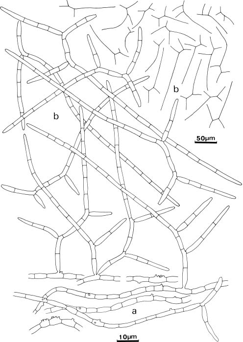
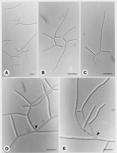
Lambdasporium lushanense J.L. Chen, W.S. Lin and S.S. Tzean sp. nov. (Figures 1-2)

Coloniae diametro in OMA 10 mm in 14 diebus ad 25°, velutinae ad planae griseolae brunneae; reversae griseolae brunneae ad atro-brunneas. Mycelium partim superficiale, partim immersum ex hyphis ramosis, frequenter catenulatis, septatis, lenibus, pallidis griseis brunneis ad griseis brunnea, 1.4-6.4 µm latum compositum. Conidiophora micronematoidea, mononematoidea. Cellulae conidiogenae intergrae, terminales, cylindricae, doliiformes, clavatae monoblastae vel ployblastae, denticulatae. Conidia lateralia vel terminalia fere k-, raro

3Corresponding author. Tel: 06-266-4911 ext. 284; Fax: 06-266-6411; E-mail: ccl51911@ms29.hinet.net


Botanical Bulletin of Academia Sinica, Vol. 41, 2000

y-formia, septata, laevigata, hyalina ad subhyalina vel pallida brunnea in massa; principalis axis truncatus ad basim, attenuatus, rotundatus ad apicem, usque ad 21 septatum, 61.8-164.6 µm longus, 1.6-2.4 µm latus, fere cum 1-2 divergens septatus ramus; ramus attenuatus, lateralis, usque ad 5-septatum, 12.0-54.0 µm longus, 1.4-2.2 µm latus.

In folio carioso, Lushan, Nantou, 5-XI-1995; Holotypus, Herb. CFC-1.

Colonies grew slowly on oatmeal agar (OMA), 10 mm in diameter in 14 days at 25°, velutinous to plane, greyish brown, margin irregular; reverse greyish brown to dark brown. Mycelium partly superficial, partly immersed composed of branched, septate, smooth, pale greyish brown to

greyish brown, 1.6-6.4 µm wide, often catenulate hyphae. Conidiophores micronematous, mononematous; conidiogenous cells intercalary, terminal, cylindrical, doliiform, clavate, monoblastic or polyblastic, with denticle scattered. Conidia lateral or terminal, mostly k-, rarely y-shaped, septate, smooth, hyaline to subhyaline or pale brown in mass; conidia composed of a longly main axis and two (rarely one) branched arm; main axis often truncate at the base, tapering and rounded at the apex, up to 21 septate, 61.8-164.6 µm long, 1.6-2.4 µm wide, occasionally slightly constricted at the septa; branches tapering, often lateral arising from the second and fourth cells (occasionally the third and fifth cells) of main axis, up to 5-septate, 12.0-54.0 µm long, 1.4-2.2 µm wide.

Figure 1. a-b. Lambdasporium lushanense. Characteristics of its conidiophores (a) and conidia (b) on oatmeal agar.


Chen et al. — A new species of Lambdasporium from Taiwan

Figure 2. A-C, Lambdasporium lushanense. k-, or y-shaped conidia, Bars = 50 µm. D-E, Conidia borne on the denticle of micronematous conidiophore (arrows). Bars = 10 µm.

Specimen examined. Decaying leaves, Lushan, Nantou county, Nov. 5, 1995.

The conidiogenesis and conidial morphology of L. lushanense are close to L. wauense Matsushima. However, L. lushanense has larger conidia, a longer main axis with more septa and two lateral branches arising from different locations. Lambdasporium wauense also has smaller conidia and only one branched arm arising from the second cell of the main axis (Matsushima, 1971a,b). Lambdasporium viridense Nowawi has simple or sparsely branched conidiophores, single, apical, monoblastic, denticulate, proliferation-sympodial conidiogenous cells, and tri-radiate (Y-shaped), attenuated conidia (Nawawi, 1985), while L. lushanense has micronematous conidiophores; monoblastic or polyblastic, denticulate conidiogenous cells and k-, and occasionally y-shaped

conidia. Therefore L. viridense and L. lushanense are different.

The microcharacteristics of L. lushanense are not similar to those of Curucispora ponapensis Matsushima, Tricladium angulatum Ingold, Varicosporium elodeae Kegel, or Volucrispora graminea Ingold, Mc Dougall & Dann (Matsushima, 1981, 1983, 1987; Ingold et al., 1968; Roldan, 1991). Curucispora ponapensis has X-shaped conidia, which is different from the conidial morphology of L. lushanense. Varicosporium elodeae has complex blastopore, but it has inconspicuously denticulate conidiogenous cells. Therefore, it can be easily distinguished from L. lushanense. Both T. angulatum and L. lushanense also have closely similar conidial morphology, but L. lushanense has denticulate, porliferation-sympodial conidiogenous cells, rostrate basal cell conidia, and longer


Botanical Bulletin of Academia Sinica, Vol. 41, 2000

branches. Volucrispora graminea is distributed over a wide range of climates. It is similar to L. lushanense in conidial morphology, but V. graminea has a short, slightly curved or somewhat sinuate main axis (22-75 × 1.5-2 µm, up to 3 septate), and has 1-(2) short branches which are pleurogenous, sequential, arranged in one order, and arise mostly immediately below the septa (Inglod et al., 1991).

Acknowledgments. Thanks to a grant from the National Science Council (NSC 85-0409-B-0231-011) in support of this project.

Literature Cited

Barron, G.L. 1968. The Genera of Hyphomycetes from Soil. The Williams and Wilkins Co., Baltimore.

Dickson, G and A. Leonard. (eds.) 1996. Fungi in the New Forest. A Mycota. Hampshire Wildlife Trust, British Mycological Society, England.

Ellis, M.B. 1971. Dematiaceous Hyphomycetes. Commonwealth Mycological Institute. Kew. Surrey, England.

Hughes, S.J. 1953. Conidiophores, conidia and classification. Canada J. Bot. 31: 577-659.

Ingold, C.T., P.J. Mc Dougall, and V. Dann. 1968. Volucrispora

graminea sp. Nov. Trans. Br. Mycol. Soc. 51: 325-328.

Marvanová L. and F. Bärlocher. 1998. Hyphomycetes from Canadian streams. IV. Spirosphaera dimorpha sp. nov. Mycotaxon 68: 33-40.

Matsushima, T. 1971a. Microfungi of the Solomon Islands and Papua-New Guinea. Matsushima: Kobe, Japan.

Matsushima, T. 1971b. Mycological reports from New Guinea and the Solomon Islands 7. Some interesting Fungi Imperfecti. Bull. Nat. Sc. Mus. Tokyo 14: 460-480.

Matsushima, T. 1981. Matsushima Mycological Memoires, 2. Matsushima: Kobe, Japan.

Matsushima, T. 1983. Matsushima Mycological Memoires, 3. Matsushima: Kobe, Japan.

Matsushima, T. 1987. Matsushima Mycological Memoires, 5. Matsushima: Kobe, Japan.

Nawawi, A. 1985. Some interesting hyphomycetes from water. Mycotaxon 24: 217-226.

Roldan, A. 1991. Pure culture studies on Volucrispora graminea and Isthmotricladia britannica (Hyphomycetes). Mycotaxon 42: 297-306.

Saccardo, P.A. 1882-1931. Sylloge Fungorum Omnium Cognitorum. 25 volumes. Pavia, Italy.

Tubaki, K. 1963. Taxonomic study of Hyphomycetes. Annual Report Institute Fermentation. Osaka 1: 25-54.

¤@ºØ¥xÆW²£ Lambdasporium Äݤ§¥@¬É©Ê·sºØ

³¯ãS1 ªL¬°´Ë1 ´¿Å㶯2

1 ¨p¥ß¹Å«nÃIJz¾Ç°|Âå°ÈºÞ²z¨t

2 °ê¥ß¥xÆW¤j¾Ç´Óª«¯f²z¾Ç¨t
¥»¤å°O­z¡A¨Ã´yø¤@ºØ¦Û¥xÆW¬\¸­¤W¤ÀÂ÷¨ì¤§·sªºµ·ÌUºõ¤£§¹¥þµß Lambdasporium lushanense J.L. Chen, W.S. Lin & S.S. Tzean¡C¦¹µß°£»P¦PÄݤº¤§¤wª¾ºØ¶i¦æ©¼¦¹¶¡²§¦P³B°Q½×¥~¡A¨Ã¥B¥ç»P§ÎºA ¯S¼x¬Ûªñ¦ü¤§¨ä¥Lµ·ÌUºõ¤£§¹¥þµß¥]¬A¡GCurucispora ponapensis¡BTricladium angulatum¡BVaricosporium elodeae ¤Î Volucrispora graminea §@¤ñ¸û¡C

ÃöÁäµü¡G¤£§¹¥þµß¡Fµ·ÌUºõ¡F·sºØ (Lambdasporium lushanense sp. nov.)¡F¥xÆW¡C