association with cellular pigment contents in transgenic tobacco suspension cultures

Bot. Bull. Acad. Sin. (2000) 41: 93-98

Chen et al. — RP3 promoter expression and pigment contents

Expression of rice prolamin RP3 promoter exhibits a positive association with cellular pigment contents in transgenic tobacco suspension cultures

Hsien-Jung Chen1, Chih-Yuan Yang1, Yean-Ching Chen1 and Ching-San Chen1,2

1Institute of Botany, Academia Sinica, Nankang, Taipei, Taiwan 11529, Republic of China

(Received March 3, 1999; Accepted September 23, 1999)

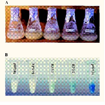
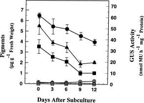
Abstract. Three transgenic tobacco regenerants (lines RP3/2, RP3/7 and RP3/18) harboring rice RP3/GUS chimeric gene inserts in their genomes were isolated previously and used to establish cell suspension cultures (Yang et al., 2000). A differential GUS expression pattern together with morphological, biochemical, and molecular variations were observed among these cell lines (Chen et al., 1999). In this report we used pigments such as carotenoids and chlorophylls a and b as markers to study the possible relationship between RP3 promoter activity and cellular pigment contents, and a positive association was found among these cell lines. RP3/2, which contained the highest cellular pigment contents, also exhibited the highest GUS expression level. The RP3 promoter activity in RP3/2 continuously decreased and was parallel with the reduction of pigment contents within a 12-day growth period after subculture. In RP3/18, both also showed a parallel association and remained relatively constant during cell growth. Based on these results, a positive association between RP3 promoter activity and cellular pigment content was found among and within cell lines. The meaning of the association and its possible explanation are discussed.

Keywords: Pigment; Prolamin; Rice; RP3 promoter; Tobacco.

Introduction

Rice is an important cereal crop in Asia and contains prolamins and glutelins as its major seed storage proteins (Bietz, 1982; Juliano, 1972). Prolamins form a small gene family (Kim and Okita, 1988a) and are comprised of heterogeneous proteins (Hibino et al., 1989) with molecular weights of 12 to 17 kDa (Mandac and Juliano, 1978; Padhye and Salunkhe, 1979). Prolamins constitute ca. 6 to 25% of total seed storage proteins on a weight basis, depending on the genotypes (Huebner et al., 1990), subcellular fractionation and pepsin digestion assay (Ogawa et al., 1987), and different extraction solvents (Sugimoto et al., 1986). Expression of rice prolamin is seed-specific and can be detected as early as 5 to 8 days after flowering by Northern blot (Kim et al., 1993; Shyur et al., 1992) or as early as 8 to 11 days using Western blot (Li and Okita, 1993; Shyur et al., 1994). Prolamins accumulate within protein body-I formed by direct dilation of the endoplasmic reticulum membrane (Krishnan et al., 1986; Yamagata and Tanaka, 1986).

Several rice prolamin genes have been isolated previously (Barbier and Ishihama, 1990; Feng et al., 1990; Kim and Okita, 1988a & b; Masumura et al., 1990; Shyur and Chen, 1993; Shyur et al., 1992; Wen et al., 1993; Yamagata

et al., 1992). However, factors involved in the prolamin gene expression and regulation are not yet clear. In our laboratory three genomic DNA clones (RP3, RP5 and RP6) of rice prolamins were isolated (Chen et al., 1996; Shyur et al., 1992; Wen et al., 1993). The RP3 promoter has been constructed with a bacterial GUS reporter gene and expressed in the embryo and endosperm tissues of transgenic tobacco seeds, and in the suspension-cultured cells established from transgenic tobacco regenerants (Yang et al., 2000). Characterization of these tobacco suspension cultures derived from transgenic regenerants showed that the line RP3/2 exhibited distinct sub-organelle structures of chloroplasts, and over-expressed the pigment contents and mRNA of a 23 kDa polypeptide of PSII oxygen-evolving complex (Chen et al., 1999). In this report a positive association between the GUS expression level and cellular pigment contents was observed. The indication and possible explanation for the association are discussed.

Materials and Methods

Plant Materials

The rice RP3 gene was isolated previously and encodes a putative seed prolamin storage protein (Chen et al., 1996). Transgenic tobacco regenerants (lines RP3/2, RP3/7 and RP3/18) harboring rice RP3/GUS chimeric gene construct in their genomes were isolated previously (Yang et al., 2000) and used to establish cell suspension cultures (Chen et al., 1999).

2Corresponding author. Tel: 886-2-27899590 ext. 251; Fax: 886-2-27858936; E-mail: bocschen@ccvax.sinica.edu.tw


Botanical Bulletin of Academia Sinica, Vol. 41, 2000

Tobacco cell suspension cultures were regularly maintained with a 3% sucrose MS medium (pH 5.6) supplemented with 1 ppm 2,4-D, 1 mg l-1 nicotinic acid, 10 mg l-1 thiamine hydrochloride, 1 mg l-1 pyridoxine hydrochloride, and 100 mg l-1 myo-inositol. Cell suspensions were subcultured every 7 days in a 1:2 (v:v) dilution ratio with fresh culture medium at 25°C in a 16 h light / 8 h dark cycle and used for the experiments in the meantime.

Analyses of Pigments

Tobacco cells were collected from suspensions every 3 days within a 12-day growth period after subculture, then extracted with an 2.5 mM sodium phosphate buffered 80% acetone (pH 7.8) in a 1:4 (w:v) ratio at room temperature on an orbital shaker for 5 h before being centrifuged at 1,000 g for 10 min. The supernatant was transferred to a new 50 ml centrifuge tube and measured with wavelengths of 663.2 nm, 646.8 nm and 470 nm, respectively, using a Beckman DU50 spectrophotometer. The quantitative values of pigments (carotenoids, chlorophylls a and b) were calculated from optical density data based on the equations reported by Porra et al. (1989).

GUS Assay

GUS assay was carried out basically as reported by Jefferson (1987) in transgenic suspension-cultured cells. Cells collected every 3 days after subculture as described earlier were ground into powder in liquid N2 with a mortar and pestle, then extracted with 0.8 ml of ice-cold GUS lysis buffer (50 mM sodium phosphate pH 7.0, 1 mM EDTA, 0.1% Triton X-100, 1 mM DTT and 0.1% laurylsarcosine). The supernatant after being centrifuged at 13,000 g at 4°C for 10 min was assayed for GUS activity with 4-methyl umbelliferyl glucuronide (MUG) substrate by a Hoffer TKO-100 minifluorometer at the excitation/emission wavelengths of 365 nm/455 nm. For histochemical analysis, cells 7 days after subculture were incubated in GUS staining buffer (1 mM 5-bromo-4-chloro-3-indolyl-b-D-glucuronide, 0.5 mM potassium ferricyanide, 0.5 mM potassium ferrocyanide and 0.1 M sodium phosphate pH 7.0) at 37°C for 12 to 16 h, then de-stained in 70% ethanol.

Results

A Positive Association of RP3 Promoter Activity with Cellular Pigment Contents Among Lines RP3/2, RP3/7 and RP3/18

Tobacco cell suspension cultures derived from transgenic regenerants (lines RP3/2, RP3/7 and RP3/18) harboring the rice prolamin RP3 promoter/GUS chimeric gene inserts in their genomes were established and used to study the possible factors affecting RP3 promoter activity. Morphological variations of cell suspension 7 days after subculture were observed among cell lines (Figure 1A). RP3/2 displayed a yellowish phenotype while the others (RP3/7, RP3/18, pBI101 and pBI121) were near whitish (Figure 1A). When these suspension-cultured

Figure 1. The morphologies and GUS activity staining of cell suspensions 7 days after subculture. A, Morphologies of cell suspension cultures; B, GUS activity staining of suspension-cultured cells with 5-bromo-4-chloro-3-indolyl-b-D-glucuronide substrate. RP3/2, RP3/7 and RP3/18 were transgenic cell lines harboring the rice prolamin RP3 promoter/GUS chimeric gene. pBI101 was a transgenic line containing the promoterless GUS structural gene, and pBI121 was a transformant with the CaMV 35S promoter/GUS chimeric gene.

cells were incubated with 5-bromo-4-chloro-3-indolyl-b-D-glucuronide substrate, differential GUS stainings were found (Figure 1B). RP3/2 exhibited a bluer stain than RP3/7 and RP3/18, and RP3/18 was almost the same as the negative promoterless control pBI101. PBI121 with a CaMV 35S promoter was the positive control (Figure 1B). Using these data, differential GUS expression levels and morphological variations were observed among cell lines RP3/2, RP3/7 and RP3/18 (Figure 1). This led us to study the possible association between the morphological variations using cellular pigment contents as markers and RP3 promoter activity among and within cell lines. A quantitative analysis of cellular pigment contents was accomplished with 80% acetone extracts. RP3/2 contained higher amounts of chlorophyll a (ca. 2 to 8 times), chlorophyll b (ca. 1.5 to 8 times), and carotenoids (ca. 2 to 3 times) than the others (Table 1). The pigment contents in RP3/7 were higher than in wild type control W38, and the smallest quantity was found in RP3/18 (Table 1). For GUS expression levels, RP3/2 showed ca. 6 and 25 times the expression of RP3/7 and RP3/18, respectively. RP3/18 had slightly higher GUS activity than the promoterless pBI101 and wild type controls. The GUS activity of pBI121 positive control was 840.46 ± 27.29 nmol MU h-1 mg-1 protein (Table 1). Thus, the results showed a positive association between cellular pigment contents and GUS expression level among transgenic cell lines RP3/2, RP3/7 and RP3/18.


Chen et al. — RP3 promoter expression and pigment contents

Table 1. A positive association of cellular pigment contents with GUS activities among transgenic tobacco cell suspension cultures. The cellular pigment contents and GUS activities were measured 7 days after subculture. RP3/2, RP3/7 and RP3/18 are transgenic cell lines harboring rice RP3 prolamin promoter/GUS chimeric genes. W38 is a wild type control. The data are the average from three independent experiments and are shown as mean ± SE. SE, standard error. The values of 840.46 ± 27.29 and 1.18 ± 0.71 (nmol MU h-1 mg-1 protein) were GUS activities for positive (pBI121) and negative (pBI101) controls, respectively.

Cell lines Chlorophyll a Chlorophyll b Carotenoids GUS activity (ng g-1 fresh weight) (nmol MU h-1 mg-1 protein)

W38 984 ± 120 175 ± 54 1183 ± 68 0

RP3/2 5976 ± 505 1411 ± 232 2472 ± 261 69.14 ± 4.37

RP3/7 2791 ± 143 944 ± 285 1283 ± 110 11.10 ± 0.88

RP3/18 726 ± 150 185 ± 50 801 ± 39 2.71 ± 0.69

A Parallel Reduction of Cellular Pigment Contents with RP3 Promoter Activity in Cell Line RP3/2 During Cell Growth

In order to study this association further, we measured the changes of GUS expression levels and cellular pigment contents during cell growth. In RP3/2, a gradual decrease of GUS expression level was found within a 12-day growth period, which was parallel to the reduction of cellular pigment contents (Figure 2). The GUS activity and the pigment content were both reduced to from one-half to one-third their original amounts 12 days after subculture (Figure 2). For RP3/18, the cellular pigment contents and GUS expression levels were relatively constant during the whole growth period (Figure 2). A similar cell growth rate was observed for RP3/2 and RP3/18 (Chen et al., 1999), and the result excludes the possibility that reduction of GUS activities and cellular pigments in RP3/2 were due to cell death after subculture. These data also exhibit a positive association between RP3 promoter activity and cellular pigment contents within cell lines.

Discussion

Rice prolamin is one of the two major seed storage proteins and has been studied intensively on protein and mRNA levels (Kim et al., 1993; Li and Okita, 1993; Mandac et al., 1978; Ogawa et al., 1987; Padhye et al., 1979; Shyur and Chen, 1993; Shyur et al., 1992; Shyur et al., 1994; Sugimoto et al., 1986; Yamagata and Tanaka, 1986). However, components involved in the induction of prolamin gene expression and regulation are not yet clear. In this study three transgenic tobacco suspension-cultured cells harboring rice prolamin RP3/GUS chimeric gene inserts in their genomes were used to study the possible factors influencing RP3 promoter activity. The GUS activity could be detected in suspension-cultured cells (Figures 1B and 2). Robert and Okita (1991) previously reported that nuclear extracts isolated from suspension-cultured cells of rice, wheat, and tobacco were capable of RNA polymerase II-dependent transcription of a 250 bp fragment of a wheat gliadin promoter. These data may support the suitability of tobacco suspension-cultured cells for RP3 promoter induction.

Differential GUS expression levels among cell lines RP3/2, RP3/7 and RP3/18 were observed and positively correlated to the pigment contents (Table 1). The reasons are not clear. Previous results showed that transgenic tobacco plants possibly contained one RP3/GUS chimeric gene insert for RP3/2 and RP3/18, and two for RP3/7 in their genomes using a X2 test. They also expressed similar GUS expression levels in T1 seeds (Yang et al., 2000). Thus, differential GUS expression levels among cell lines RP3/2, RP3/7 and RP3/18 derived from these transgenic regenerants could not be explained merely with the differences of copy numbers and/or position effects of RP3/GUS chimeric gene inserts in their genomes. In Figure 2, parallel changes of RP3 promoter activities with cellular pigment contents were also found for RP3/2 and RP3/18 during cell growth. These results provide further evidence to support the association and exclude the bias due to the influence of copy numbers and position effects.

How the pigments could directly or indirectly affect rice prolamin RP3 promoter induction in transgenic cell suspension culture is not clear. The carotenoids, chlorophylls a and b are pigments for photosynthesis. Variations of photosynthetic rates could influence intracellular metabo

Figure 2. Changes of GUS activities and cellular pigment contents during cell growth in transgenic cell lines RP3/2 and RP3/18. The GUS activities and cellular pigment contents were measured every 3 days within a 12-day growth period. The solid symbols (˜; p; ¢) were for RP3/2 and open symbols (™; r; £) were for RP3/18. ˜ & ™, GUS activities; p & r, carotenoids + chlorophyll a + chlorophyll b; ¢ & £, chlorophyll a. The data were from three independent experiments, and the vertical bars were the sizes of standard errors.


Botanical Bulletin of Academia Sinica, Vol. 41, 2000

lites such as sucrose, soluble sugars, and nitrogen compounds, and in turn alter plant physiological conditions and gene expression patterns. The influence of metabolites on gene expression has been demonstrated for potato tuber patatin storage protein (Grierson et al., 1994; Martin et al., 1997), vegetative storage proteins (vspB) of soybean (Mason et al., 1993), zein seed storage proteins of maize (Lee and Tsai, 1984), 23 kDa polypeptide of PSII oxygen evolving complex (Kochhar et al., 1996), and rbcS and cab proteins of tobacco (Herbers et al., 1996). We have studied the influence of culture medium components on RP3/GUS chimeric gene expression, and the preliminary results showed that RP3 promoter expression level was increased in RP3/2 when sucrose was removed from, or nitrogen base was added to, the culture medium (data not shown). We also demonstrated that the mRNA of a 23 kDa polypeptide of PSII oxygen-evolving complex was expressed exclusively in RP3/2, but not in RP3/18, using Northern blot hybridization (Chen et al., 1999). These data support and suggest the possibility of influence of metabolites such as sucrose, soluble sugars, and nitrogen compounds on rice prolamin RP3 promoter induction, and imply a possible indirect explanation for pigment effects.

RP3/2 with yellowish phenotype (Figure 1) showed morphological variations from the other cell lines. It contained more pigments, thylakoids, osmiophilic plastoglobuli, and electron-dense materials in the chloroplasts, and expressed the mRNA of a 23 kDa polypeptide of PSII oxygen-evolving complex (Chen et al., 1999). The causal agents for the morphological, biochemical, and molecular variations of RP3/2 from the other cell lines are not clear; however, they seem to be associated with mutations that occurred during the tissue culture procedure or T-DNA integration during agroinfection. Examples of mutation affecting seed prolamin contents have been reported in fluory-2 mutant of maize (Jones, 1978), high-lysine mutant of barley (Shewry et al., 1980), and low glutelin/high prolamin mutant of rice (Iida et al., 1993). The altered physiological conditions and/or gene expression patterns for RP3/2 suggest a possible interpretation for the positive association of pigment contents with RP3 promoter activity. Identification of the agents responsible for the variations of RP3/2 may provide directions in the future for studying the components of rice prolamin gene expression and regulation.

Literature Cited

Barbier, P. and A. Ishihama. 1990. Variation in the nucleotide sequence of a prolamin gene family in wild rice. Plant Mol. Biol. 15: 191-195.

Bietz, J.A. 1982. Cereal prolamin evolution and homology revealed by sequence analysis. Biochem. Genet. 20: 1039-1053.

Chen, H.J., C.Y. Yang, W.N. Jane, and C.S. Chen. 1999. Increase of pigments, plastoglobuli and the mRNA of a 23 kDa polypeptide of PSII oxygen-evolving complex in a transgenic tobacco cell line RP3/2. J. Plant Physiol. 155: 584-590.

Chen, J.M., J.J. Lin, S.T. Jhiang, and C.S. Chen. 1996. Characterization of rice prolamin genes. Czech-Taiwan Symposium on Biotechnology, Praque Czech Republic, pp. 1-6.

Feng, G., L. Wen, J.K. Huang, B.S. Shorrosh, S. Muthukrishnan, and R. Reeck. 1990. Nucleotide sequence of a cloned rice genomic DNA fragment that encodes a 10 kDa prolamin polypeptide. Nucl. Acids Res. 18: 683.

Grierson, C., J.S. Du, M. de-Torres-Zabala, K. Beggs, C. Smith, M. Holdsworth, and M. Bevan. 1994. Separate cis sequence and trans factors direct metabolic and developmental regulation of a potato tuber storage protein gene. Plant J. 5: 815-826.

Herbers, K., P. Meuwly, J.P. Metraux, and U. Sonnewald. 1996. Salicylic acid-independent induction of pathogenesis-related protein transcripts by sugars is dependent on leaf developmental stage. FEBS Lett. 397: 239-244.

Hibino, T., K. Kidzu, T. Masumura, K. Ohtsuki, K. Tanaka, M. Kawabata, and S. Fujii. 1989. Amino acid composition of rice prolamin polypeptide. Agric. Biol. Chem. 53: 512-518.

Huebner, F.R., J.A. Bietz, B.D. Webb, and B.O. Juliano. 1990. Rice cultivar identification by high-performance liquid chromatography of endosperm proteins. Cereal Chem. 67: 129-135.

Iida, S., E. Amano, and T. Nishio. 1993. A rice (Oryza sativa) mutant having a low content of glutelin and a high content of prolamin. Theor. Appl. Genet. 87: 374-378.

Jefferson, R.A. 1987. Assaying chimeric genes in plants: the GUS gene fusion system. Plant Mol. Biol. Rep. 5: 387-405.

Jones, R.A. 1978. Effects of fluory-2 locus on zein accumulation and RNA metabolism during maize endosperm development. Biochem. Genet. 16: 7-38.

Juliano, B.O. 1972. The rice caryopsis and its composition. In D.F. Houston (ed.), Rice: Chemistry and Technology. American Association of Cereal Chemist, Inc., St. Paul, Minnesota, pp. 16-74.

Kim, W.T. and T.W. Okita. 1988a. Structure, expression and heterogenicity of the rice seed prolamins. Plant Physiol. 88: 649-655.

Kim, W.T. and T.W. Okita. 1988b. Nucleotide and primary sequence of a major rice prolamin. FEBS Lett. 231: 308-310.

Kim, W.T., X. Li, and T.W. Okita. 1993. Expression of storage protein multigene families in developing rice endosperm. Plant Cell Physiol. 34: 595-603.

Kochhar, A., J.P. Khurana, and A.K. Tyagi. 1996. Nucleotide sequence of the psbP gene encoding precursor of 23-kDa polypeptide of oxygen-evolving complex in Arabidopsis thaliana and its expression in the wild-type and a constitutively photomorphogenic mutant. DNA Res. 3: 277-285.

Krishnan, H.B., V.R. Franceschi, and T.W. Okita. 1986. Immunochemical studies on the role of the Golgi complex in protein-body formation in rice seeds. Planta 169: 471-480.

Lee, L. and C.Y. Tsai. 1984. Zein synthesis in the embryo and endosperm of maize mutants. Biochem. Genet. 22: 729-737.

Li, X. and T.W. Okita. 1993. Accumulation of prolamins and glutelins during rice seed development: a quantitative evaluation. Plant Cell Physiol. 34: 385-390.

Mandac, B.E. and B.O. Juliano. 1978. Properties of prolamin


Chen et al. — RP3 promoter expression and pigment contents

in mature and developing rice grain. Phytochemistry 17: 611-614.

Martin, T., H. Hellmann, R. Schmidt, L. Willmitzer, and W.B. Frommer. 1997. Identification of mutants in metabolically regulated gene expression. Plant J. 11: 53-62.

Mason, H.S., D.B. Dewald, and J.E. Mullet. 1993. Identification of a methyl jasmonate-responsive domain in the soybean vspB promoter. Plant Cell 5: 241-251.

Masumura,T., T. Hibino, K. Kidzu, N. Mitsukawa, K. Tanaka, and S. Fujii. 1990. Cloning and characterization of a cDNA encoding a rice 13 kDa prolamin. Mol. Gen. Genet. 221: 1-7.

Ogawa, M., T. Kumamaru, H. Satoh, N. Iwata, T. Omura, Z. Kasai, and K. Tanaka. 1987. Purification of protein body-I of rice seed and its polypeptide composition. Plant Cell Physiol. 28: 1517-1527.

Padhye, V.W. and D.K. Salunkhe. 1979. Extraction and characterization of rice proteins. Cereal. Chem. 56: 389-393.

Porra, R.J., W.A. Thompson, and P.E. Kriedemann. 1989. Determination of accurate extinction coefficients and simultaneous equations for assaying chlorophylls a and b extracted with four different solvents: verification of the concentration of chlorophyll standards by atomic absorption spectroscopy. Biochim. Biophys. Acta 975: 384-394.

Robert, M.W. and T.W. Okita. 1991. Accurate in vitro transcription of plant promoters with nuclear extracts prepared from cultured plant cells. Plant Mol. Biol. 16: 771-786.

Shewry, P.R., A.J. Faulk, and B.J. Miflin. 1980. Effect of high-lysine mutations on the protein fractions of barley grain. Biochem. Genet. 18: 133-151.

Shyur, L.F. and C.S. Chen. 1993. Rice prolamins: heterogeneity of cDNAs and synthesis of precursors. Bot. Bull. Acad. Sin. 34: 143-154.

Shyur, L.F., T.N. Wen, and C.S. Chen. 1992. cDNA cloning and gene expression of the major prolamin of rice. Plant Mol. Biol. 20: 323-326.

Shyur, L.F., T.N. Wen, and C.S. Chen. 1994. Purification and characterization of rice prolamins. Bot. Bull. Acad. Sin. 35: 65-71.

Sugimoto, T., K. Tanaka, and Z. Kasai. 1986. Improved extraction of rice prolamin. Agri. Biol. Chem. 50: 2409-2411.

Wen, T.N., L.F. Shyur, J.C. Su, and S.C. Chen. 1993. Nucleotide sequence of a rice (Oryza sativa) prolamin storage protein gene, RP6. Plant Physiol. 101: 1115-1116.

Yamagata, H. and K. Tanaka. 1986. The site of synthesis and accumulation of rice storage proteins. Plant Cell Physiol. 27: 135-145.

Yamagata, H., T. Nomura, S. Arai, K. Tanaka, and T. Iwasaki. 1992. Nucleotide sequence of a cDNA that encodes a rice prolamin. Biosci. Biotech. Biochem. 56: 537.

Yang, C.Y., H.J. Chen, Y.F. Chen, M.H. Chen, and C.S. Chen. 2000. Seed-specific expression of a rice prolamin RP3/GUS chimeric gene in Nicotiana tabacum cv. Wisconsin 38. J. Plant Physiol. 156: 100-105.


Botanical Bulletin of Academia Sinica, Vol. 41, 2000

Âà´ÞµÒ¯óÄa¯B²Ó­M¤º¤ô½_¾J·»³J¥Õ RP3 °_°Ê¤lªºªí²{

»P¦â¯À§t¶q¦³¥¿¬ÛÃö©Ê

³¯Åãºa ·¨´¼¤¸ ³¯Ú|«C ³¯¼y¤T

¤¤¥¡¬ã¨s°|´Óª«¬ã¨s©Ò

¤T­Ó§t¦³¤ô½_¾J·»³J¥Õ RP3/GUS chimeric gene inserts ¤§Âà´ÓµÒ¯ó¡A´Ó®è¡]«~¨t RP3/2¡ARP3/7 ¤Î RP3/18¡^¤w³Q¤ÀÂ÷¤Î«Ø¥ßÄa¯B²Ó­M°ö¾i¡]Yang et al., 1999¡^¡A²Ó­M«~¨t¶¡®t²§©Êªº GUS ªí²{¤Î§ÎºA¡B¥Í¤Æ¡B¤Î¤À¤lªºÅܲ§¥ç³QÆ[¹î¨ì¡]Chen et al., 1999¡^¡C¦b³o½g³ø§i¤¤§Ú­Ì¨Ï¥Î¦â¯À¦pÃþ¸¬ÅÚ½³¯À¡B¸­ºñ¯À a ¤Î b §@¬°¼Ð»x±´°Q RP3 °_°Ê¤l¬¡©Ê»P²Ó­M¦â¯À§t¶qªº¥i¯àÃöÁp¡A¨Ã¥Bµo²{¦¹¤GªÌ©ó²Ó­M«~¨t¶¡¦³¥¿¬ÛÃö ©Ê¡CRP3/2 ªºÄa¯B²Ó­M¨ã¦³³Ì¦hªº¦â¯À§t¶q¤Î³Ì¤jªº GUS ªí²{¶q¡ARP3 °_°Ê¤l¬¡©Ê¦b¦¹Äa¯B²Ó­M¤º 12 ¤ÑªºÄ~¥N°ö¾i¹Lµ{¤¤³vº¥ªº´î¤Ö¡A¦Ó¥B»P²Ó­M¦â¯À§t¶q¤U­°¶q¥­¦æ¡C©ó RP3/18¡A©ó²Ó­M¥Íªø¹Lµ{¸Ó¤GªÌ¥çÅã¥Ü¥­¦æªºÃöÁp©Ê¥B«O«ù¬Û·íªº«ù¥­¡C¥Ñ³o¨Ç¹êÅçµ²ªGÅã¥Ü¤ô½_¾J·»³J¥Õ RP3 °_°Ê¤l¬¡©Ê¦bÂà´ÞµÒ¯óÄa¯B²Ó­M«~¨t¶¡¤Î«~¨t¤º»P¦â¯À§t¶q¦³¥¿¬ÛÃö©Ê¡C§Ú­Ì¹ï¸ÓÃöÁp¤§·N¸q¤Î¥i¯àªº¸ÑÄÀ´£¥X¤@¨Ç¬Ýªk¡C

ÃöÁäµü¡G¾J·»³J¥Õ½è¡F¤ô½_¡FRP3 °_°Ê¤l¡F¦â¯À¡FµÒ¯ó¡C