roots -- Involvement of the chaperonins in starch biosynthesis

Bot. Bull. Acad. Sin. (2000) 41: 105-111

Chang et al. — Chaperonin 60 from sweet potato roots

The isolation and characterization of Chaperonin 60 from sweet potato roots — Involvement of the chaperonins in starch

biosynthesis

Shih-Chung Chang, Pei-Chun Lin, Han-Min Chen, Jiann-Shing Wu, and Rong-Huay Juang1

Department of Agricultural Chemistry, National Taiwan University, Taipei 106, Taiwan

(Received August 17, 1999; Accepted September 9, 1999)

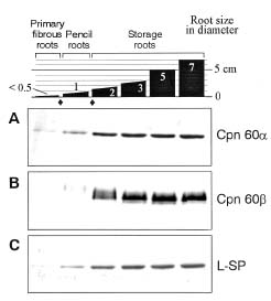
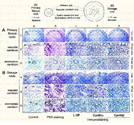
Abstract. Molecular chaperonins cpn60a and cpn60b were isolated from sweet potato roots and were characterized as homologous polymers. However, their immunochemical and biochemical properties were divergent. The two chaperonins as well as starch phosphorylase appeared concurrently during the starch-accumulating period and were localized in the starch granule by histochemical methods. Notably, these proteins increased substantially in cells around the anomalous cambium when sweet potato initiated starch biosynthesis. Observations in this study indicated that the chaperonins might be involved in the biosynthesis of starch in sweet potato roots.

Keywords: Amyloplast; Anomalous cambium; Chaperonins; Histochemical methods; Ipomoea batatas; Starch biosynthesis; Starch phosphorylase.

Introduction

Chaperonin 60 (cpn60) is a huge protein complex composed of fourteen subunits and shaped like a cylinder. The structure and function of chaperonins in protein folding have been reviewed by Sigler et al. (1998). In its physiological function, plastid cpn60 assists the assembly and folding of plastid proteins (Ellis, 1990; Ellis, 1994). Lubben et al. (1989) showed that several proteins are imported into pea chloroplasts by forming stable complexes with the GroEL-related chloroplast chaperone. Although heat shock treatment does not effect the expression of the cpn60b transcript in rye leaf, Schmitz et al. (1996) and Holland et al. (1998) observed the stress-induced accumulation of plastic chaperonin 60 during tobacco seedling development. Since the plastid is an active carbohydrate-metabolizing organelle and also contains a large amount of chaperonin molecules (Schlicher and Soll, 1996), we speculate that the chaperonins might play a role in regulating starch biosynthesis.

Starch phosphorylase (SP, EC 2.4.1.1) catalyzes the reversible phosphorolysis of a-glucan and produces Glc-1-P as one of the products (Hanes, 1940). In sweet potato roots, where the starch biosynthesis is active, only the low-starch-affinity isoform of starch phosphorylase (L-SP) was found (Chang et al., 1987). Although the biosynthesis of starch granules is conducted mainly by ADPglucose pyrophosphorylase, starch-branching enzyme, starch

synthase, and debranching enzyme (Ball et al., 1996; Smith et al., 1997), a possible role for starch phosphorylase in the primer synthesis has been suggested (Nelson and Pan, 1995). Brisson et al. (1989) indicated that potato starch phosphorylase is localized inside the stroma of the amyloplast in young tubers, whereas in mature tubers, potato starch phosphorylase is found within the cytoplasm in the immediate vicinity of the plastids. That observation aroused questions of how SP is transported into the amyloplasts and how this process is regulated.

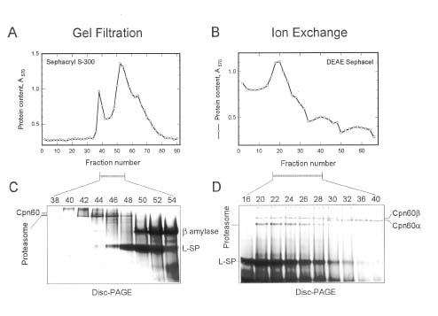
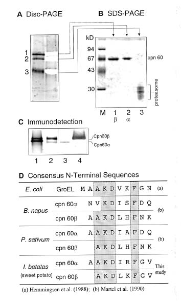
In this study, we explored the relationship between the chaperonins and the biosynthesis of starch by characterizing the chaperonin 60 using immunochemical and histochemical tools. We found that the accumulation of starch in the sweet potato roots correlated to the increase of both chaperonins and L-SP. This result was further sustained by histochemical observations.

Materials and Methods

Materials

Roots of sweet potato (Ipomoea batatas [L.] Lam. cv Tainong 57) were collected from the farm of the National Taiwan University. The roots were stored at room temperature and used for the analysis or purification procedures within two weeks.

Assays for Protein Content

Protein content was determined by the dye-binding method (Bradford, 1976) using the microassay system from Bio-Rad (Protein Assay Kit).

1Corresponding author. Tel and Fax: (02) 2363-1704; E-mail: juang@ccms.ntu.edu.tw


Botanical Bulletin of Academia Sinica, Vol. 41, 2000

Isolation and Characterization of Chaperonin 60

All purification procedures were carried out at 4°C using buffer A (50 mM Tris, pH 7.4) as the buffer system. Fresh sweet potato roots (400 g) were added to 400 mL of extraction buffer (buffer A containing 1% polyvinylpolypyrrolidone) and homogenized in a Waring blender. The crude extract was filtratedthrough four layers of gauze, and the filtrate was clarified by centrifugation (10,000 g, 40 min). Protein was fractionated by collecting the precipitate between a 20 to 80% saturation of ammonium sulfate. The pellet was resuspended in a minimal amount of buffer A. The brownish mixture was dialyzed overnight against buffer B (buffer A containing 0.15 M NaCl), and the precipitate subsequently formed was removed by centrifugation (10,000 g, 40 min) before applying 10 mL of the sample to a Sephacryl S-300 column (i.d. 2.6 × 95 cm, equilibrated in buffer B). The fractions containing the chaperonins were detected by 6% disc-PAGE and pooled. They were then applied to an ion exchange column using DEAE Sephacel (i.d. 2.6 × 15 cm, pre-equilibrated in buffer B). The column was eluted with a gradient of NaCl from 0.15 M to 0.5 M in buffer A (200 mL each). The fractions containing the chaperonins were collected as described above. The sample was concentrated by ultrafiltration (Amicon YM-10) and then sepa

rated by disc-PAGE (6% gel). The bands were detected by brief staining in Coomassie Brilliant Blue and cut out from the gel. The gel strips were then put onto the sample wells and subjected to SDS-PAGE, which revealed the subunit molecular weight of the chaperonins as 67 kDa. The bands were transferred to the membrane as described below, visualized by staining briefly in Coomassie Brilliant Blue R (0.1% solution in 40% methanol and 1% acetic acid), and then destained with 50% methanol. Finally, they were cut out and sent for automatic amino acid sequencing (ABI 477A Protein Sequencer).

Polyacrylamide Gel Electrophoresis, Protein Transfer and Immunostaining

SDS-PAGE was performed according to the method of Laemmli (1970) with slight modifications using the mini-slab gel system (Hoefer SE250). After electrophoresis, protein bands were stained by Coomassie Brilliant Blue. Otherwise, the gel was transferred to the paper (Immobilon-P, Millipore). The washed blot was immersed in a solution of antibody diluted properly in gelatin-NET (0.25% gelatin, 0.15 M NaCl, 5 mM EDTA, 0.05% Tween in 50 mM Tris-HCl, pH 8.0) and incubated at room temperature for 1 h. The blot was then washed three times with PBST. Subsequently, the biotinylated second antibody (goat anti-mouse IgG, IgA and IgM) diluted in gelatin-NET (1:2,000)

Figure 1. Purification of the chaperonins from sweet potato roots. After ammonium sulfate fractionation, gel filtration (A) and ion exchange (B) chromatographies were employed to isolate the chaperonins. Selected fractions were analyzed by disc-PAGE (C and D) to reveal cpn60a and cpn60b.


Chang et al. — Chaperonin 60 from sweet potato roots

was added. The antibody was incubated and washed as mentioned earlier. Next, the streptavidin-biotinylated alkaline phosphatase complex (Vectastain, 1:1,500 dilution containing streptavidin and biotinylated alkaline phosphatase in gelatin-NET, and incubated at room temperature for 30 min before use) was added to bind the biotinylated second antibody. After incubation at room temperature for 1 h, the blot was washed twice by PBST, and then twice by alkaline phosphatase buffer (100 mM Tris, 100 mM NaCl and 10 mM MgCl2). The bands were stained by adding the substrate solution of NBT and BCIP as described (Harlow and Lane, 1988). This procedure was also used for staining microscopic sections.

Antibody Production

Monoclonal antibodies (J3b and H7c) against sweet potato L-SP were prepared as described earlier (Chern et al., 1990). Conventional antisera against cpn60a and cpn60b from sweet potato roots were prepared by immunizing the electrophoretically purified chaperonins in mice. The antiserum against moth heat shock protein 60 (hsp60) was obtained from StressGen Biotechnologies Corp., Victoria, BC, Canada (SPA-805).

Microscopic Methods

Sweet potato roots were fixed in formalin:alcohol:acetic acid, embedded in paraffin, and then sectioned by microtome. Samples (5-µm-thickness) were stained by periodic acid-Schiff’s (PAS) reagent (Zacharius et al., 1969) for the carbohydrate component of the cell. For immunolocalization of the chaperonins or L-SP, specific antibodies in proper dilution were used. The incubation, washing steps, and the color reaction followed the immunostaining procedure as described above, except the incubation time for the primary antibodies was extended to overnight and was 3 h for the biotinylated second antibody.

Results

Isolation and Characterization of Chaperonin 60 from Sweet Potato Roots

The gel filtration chromatogram showed several protein peaks (Figure 1A). Fractions containing the chaperonins, the proteasome and other major proteins (BA and L-SP) were revealed by disc-PAGE (Figure 1C). The two cpn60 proteins and the proteasome were eluted from the column very rapidly, indicating a huge molecular mass. Primary separation of cpn60b from cpn60a was achieved by ion exchange chromatography; however, the proteasome and L-SP were still present in the fractions containing cpn60b (Figure 1B and 1D). These proteins were further separated by preparative disc-PAGE (Figure 2A), and bands cut out from the gel were then directly analyzed by SDS-PAGE, revealing the subunit molecular mass of the chaperonin as 67 kDa (Figure 2B). These bands were subjected to automatic amino acid sequencing after being transferred to the membrane. The partial N-terminal sequences

showed high homology to cpn60 in plant, or GroEL from E. coli (Figure 2D). The lower band (denoted as 3 in Figure 2A) was identified as the proteasome by amino acid sequencing and an immunodetection method (results to be published separately).

The immunostaining pattern of the chaperonins (Figure 2C) showed very high specificity for the antisera against their antigens. On the other hand, the antiserum against

Figure 2. Preparative disc-PAGE (6% gel) resolved the chromatographic purified sample into three bands (A). These bands were cut out, separated by 12.5% SDS-PAGE (B), and then electrophoretically transferred to the membrane. The bands having molecular mass around 67 kDa (lanes 1 and 2) were cut out, and sent to the automatic amino acid sequencer. Specific antisera were raised and used to detect the chaperonins (C): 1, Coomassie Brilliant Blue stained gel before the transfer; 2, bands detected by using antibody against hsp60; 3, by antibody against cpn60a; 4, by antibody against cpn60b. The partial N-terminal amino acid sequences were compared with the chaperonin sequences from other organisms (D). Gray area denotes the conservative amino acid residues.


Botanical Bulletin of Academia Sinica, Vol. 41, 2000

moth heat shock protein 60 (hsp60) displayed evident cross-reactivity to both sweet potato chaperonins (lane 2 in Figure 2C).

Immunochemical Detection of Chaperonin 60 in Sweet Potato Roots

Figure 3 displays the detection of cpn60a, cpn60b and L-SP proteins in various growing stages of sweet potato roots by electrophoresis and immunostaining of its protein blot (Figure 3). The amounts of all three proteins increased proportionally with root mass. At the stage of primary fibrous roots, only a trace amount of the chaperonins or L-SP was observable (left lane in Figure 3). Nevertheless, these proteins increased substantially as the root diameter exceeded 1.5 cm (storage roots).

To investigate this point, the primary fibrous roots (0.5 cm in diameter) and the small storage roots (1.5 cm) were sectioned and examined by a microscopic histochemical method (Figure 4). The primary fibrous roots accumulated starch mostly in the phloem, as visualized by periodic acid-Schiff’s staining (PAS staining, 1-b and 2-b in Figure 4A). The unusual anomalous cambium was distributed in the whole central area of the sweet potato roots. Only a few small starch granules appeared around the anomalous cambium during this stage (3-b). On the other hand, the storage roots accumulated starch granules abundantly and

extensively (4-b) where they spread over both sides of the vascular cambium (5-b) and at the surroundings of the anomalous cambium (6-b).

Immunostaining using specific antibodies showed parallel results (columns c, d and e in Figure 4) with the PAS staining (column b). In the primary fibrous roots most chaperonin and L-SP staining occurred in the phloem area, where starch accumulated, whereas these proteins were stained widely over the whole section of the storage roots including phloem and the central storage cells surrounding the anomalous cambia. In all cases, cells in the vascular and anomalous cambia were not stainable. Detailed examination revealed that the starch granules were the targets of the antibodies against chaperonins or L-SP (rows 2 and 5). The cell membrane or cell wall was also stained by the antibodies, but we did not pursue this.

Discussion

Chaperonin 60 in Sweet Potato Roots was a Homologous Tetradecamer

Martel et al. (1990) reported that plant cpn60a and cpn60b have divergent amino acid sequences. Immunochemical results in this study showed that the antisera were highly specific to their antigenic chaperonins (Figure 2C). The two chaperonins were eluted from the ion exchange column with distinct salt gradients (Figure 1B and 1D) and were completely separated on the disc-PAGE gel (Figure 2A). Further, the cpn60a showed concentrated bands on the immunostained blot (Figure 3A), but cpn60b bands looked diffused (Figure 3B). These results imply that cpn60 proteins in sweet potato roots are also highly divergent in their immunological and biochemical properties. Nevertheless, cpn60 proteins from different species share a conservative N-terminal amino acid sequence (Martel et al., 1990). Consequently, we were able to identify the two cpn60 proteins by comparing their N-terminal amino acid sequences (Figure 2D). In addition, since only two distinct chaperonin bands were observed on the disc-PAGE gel (Figure 2A and 2C), and each band was isolated and identified as a homogeneous protein by SDS-PAGE (Figure 2B) and subsequently by N-terminal amino acid sequencing (Figure 2D), it is suggested that both cpn60a and cpn60b are homologous polymers. There was no significant quantity of the cpn60a-cpn60b hybrid form in sweet potato roots.

Cloney et al. (1992) reported that cpn60a requires the presence of cpn60b for the assembly into the tetradecameric species in the same cell. Their observation suggested that the two chaperonins could be detected in the same cellular localization. Indeed, the immunostaining patterns in this study showed that cpn60a and cpn60b do appear concurrently during the starch-accumulating period of sweet potato roots (Figure 3) since they were detected at the same cellular localization by the histochemical methods (Figure 4). This result also led us to postulate that chaperonins might contribute to the regulation of starch biosynthesis.

Figure 3. Sweet potato roots in different sizes were analyzed by immunostaining after protein transfer for the detection of the chaperonins (A and B, 6% disc-PAGE) and L-SP (C, 12.5% gel SDS-PAGE). Sample prepared from an equivalent fresh weight of sweet potato roots was applied to each well. Roots in different stages (marked with open and solid diamonds) were further examined by histochemical methods. Microscopic results from two of these stages (solid diamonds) were shown in Figure 4.


Chang et al. — Chaperonin 60 from sweet potato roots

Figure 4. Histochemical localization of the chaperonins and L-SP in the primary fibrous roots (A) and the storage roots (B) of sweet potato. Control was performed by using preimmune serum (column a). PAS staining was used to stain the starch and carbohydrate (column b). Areas around vascular cambium (rows 2 and 5) or anomalous cambium (rows 3 and 6) were examined in higher magnifications.

Possible Involvement of Chaperonin 60 in the Biosynthesis of Starch

Tsai and Nelson (1968) identified two major types of starch phosphorylases having distinctive catalytic behavior in the endosperm of Zea mays. One of the SP isoforms appeared only at the stage of rapid starch biosynthesis and was not found during germination (Tsai et al., 1970). Our study showed a similar observation for L-SP in sweet potato roots during the starch-accumulating period (Figure 3C). Although Hagenimana et al. (1992), using tissue printing blot, showed that starch phosphorylase concentrated in the anomalous cambium and in the vascular cambium of the sweet potato roots, our study showed that L-SP was detected extensively in the phloem cells and the storage cell around the anomalous cambium, but not in the cam

bium cell. The two chaperonins also behaved parallel to L-SP (Figure 3A-B). All three proteins were histochemically localized at the starch granule of starch-accumulating sweet potato roots. These results suggest that the chaperonins and L-SP might associate with each other and together play roles in starch biosynthesis. Brisson et al. (1989) reported that the potato starch phosphorylase gene was expressed in the nucleus, translated in the cytoplasm, and then transported into the amyloplast. Molecular chaperones had played a role in importing several proteins into chloroplasts (Lubben et al., 1989). Therefore we suggest that the sweet potato L-SP synthesized in the cytoplasm might be transported into the amyloplast by the chaperonins, but direct evidence is still needed on this point.


Botanical Bulletin of Academia Sinica, Vol. 41, 2000

Sweet potato is a useful system for studying the biosynthesis of starch. Its roots gain weight and swell when the starch is accumulated (Woolfe, 1992). Histochemical examination showed that phloem cells in sweet potato roots constantly accumulate starch granules. On the other hand, the cells around the anomalous cambium didn’t initiate active starch biosynthesis until the roots began swelling (6-b in Figure 4). It is interesting to note that the size of the starch granules near the anomalous cambium looked smaller on average than those localized in the outer layer. This observation implies that the starch accumulation signal is triggered in the cells near the anomalous cambium, and subsequently differentiated into the active starch biosynthesizing cells. Therefore anomalous cambium cells around xylem vessels in sweet potato roots might play a significant role in the starch metabolism.

Acknowledgements. This work was supported by the National Science Council, ROC. We thank Professor L.-F. Liu and Dr. T.-Y. Lin for helpful discussion on microscopic techniques. We also thank Dr. Jamie Hook for editing the manuscript.

Literature Cited

Ball, S., H.P. Guan, M. James, A. Myers, P. Keeling, G. Mouille, A. Buleon, P. Colonna, and J. Preiss. 1996. From glycogen to amylopectin - a model for the biogenesis of the plant starch granule. Cell 86: 349-352.

Bradford, M.M. 1976. A rapid and sensitive method for the quantitation of microgram quantities of protein utilizing the principle of protein-dye binding. Anal. Biochem. 72: 248-254.

Brisson, N., H. Giroux, M. Zollinger, A. Camirand, and C. Simard. 1989. Maturation and subcellular compartmentation of potato starch phosphorylase. Plant Cell 1: 559-566.

Chang, T.C., S.C. Lee, and J.C. Su. 1987. Sweet potato starch phosphorylase - purification and characterization. Agric. Biol. Chem. 51: 187-195.

Chern, M.S., Y.C. Mo, R.H. Juang, and J.C. Su. 1990. Probing the protein structure of sweet potato starch phosphorylase with monoclonal antibodies. J. Chin. Biochem. Soc. 19: 55-64.

Cloney, L.P., H.B. Wu, and S.M. Hemmingsen. 1992. Expression of plant chaperonin-60 genes in Escherichia coli. J. Biol. Chem. 267: 23327-23332.

Ellis, R.J. 1990. Molecular chaperones: the plant connection. Science 250: 954-959.

Ellis, R.J. 1994. Protein folding - chaperoning nascent proteins. Nature 370: 96-97.

Hagenimana, V., L.P. Vezina, and R.E. Simard. 1992. Distribution of amylases within sweet potato (Ipomoea batatas L.) root tissue. J. Agr. Food Chem. 40: 1777-1783.

Hanes, C.S. 1940. The reversible formation of starch from glucose-1-phosphate catalyzed by potato phosphorylase. Proc. Roy. Soc. (London) B129: 174-208.

Harlow, E. and D. Lane. 1988. Immunoblotting. In Antibodies, A Laboratory Manual, pp. 471-510.

Holland, N., A. Belkind, D. Holland, U. Pick, and M. Edelman. 1998. Stress responsive accumulation of plastid chaperonin 60 during seedling development. Plant J. 13: 311-316.

Laemmli, U.K. 1970. Cleavage of structural proteins during the assembly of the head bacteriophage T4. Nature 227: 680-685.

Lubben, T.H., G.K. Donaldson, P.V. Vitanen, and A.A. Gatenby. 1989. Several proteins imported into chloroplasts form stable complexes with the GroEL-related chloroplast molecular chaperone. Plant Cell 1: 1223-1230.

Martel, R., L.P. Cloney, L.E. Pelcher, and S.M. Hemmingsen. 1990. Unique composition of plastid chaperonin-60: a and b polypeptide-encoding genes are highly divergent. Gene 94: 181-187.

Nelson, O. and D. Pan. 1995. Starch synthesis in maize endosperms. Annu. Rev. Plant Physiol. Plant Mol. Biol. 46: 475-496.

Schlicher, T. and J. Soll. 1996. Molecular chaperones are present in the thylakoid lumen of pea chloroplasts. FEBS Lett. 379: 302-304.

Schmitz, G., M. Schmidt, and J. Feierabend. 1996. Comparison of the expression of a plastidic chaperonin 60 in different plant tissues and under photosynthetic and non photosynthetic conditions. Planta 200: 326-334.

Sigler, P.B., Z. Xu, H.S. Rye, S.G. Burston, W.A. Fenton, and A.L. Horwich. 1998. Structure and function in GroEL-mediated protein folding. Annu. Rev. Biochem. 67: 581-608.

Smith, A.M., K. Denyer, and C. Martin. 1997. The synthesis of the starch granule. Annu. Rev. Plant Physiol. Plant Mol. Biol. 48: 65-87.

Tsai, C.Y. and O.E. Nelson. 1968. Phosphorylase I an II of maize endosperm. Plant Physiol. 43: 103-112.

Tsai, C.Y., F. Salamini, and O.E. Nelson. 1970. Enzymes of carbohydrate metabolism in thedeveloping endosperm of maize. Plant Physiol. 46: 299-306.

Woolfe, J.A. 1992. Sweet potato - past and present. In Sweet Potato: An Untapped Food Resource, pp. 15-40.

Zacharius, R.M., T.E. Zell, J.H. Morrison, and J.J. Woodlock. 1969. Glycoprotein staining following electrophoresis on acrylamide gels. Anal. Biochem. 30: 148-152.


Chang et al. — Chaperonin 60 from sweet potato roots

¥ÌÃŶô®Ú Chaperonin 60 ¤§¤ÀÂ÷¤ÎÀË©w—¨ä¾ý¯»¥Í¦X¦¨ªº¨¤¦â

±i¥@©v ªL¯\§g ³¯¿«¥Á §d«Ø¿³ ²øºa½÷

°ê¥ß¥xÆW¤j¾Ç¹A·~¤Æ¾Ç¨t

¥Ñ¥ÌÃŶô®Ú¤¤¥i¤ÀÂ÷±o molecular chaperonins cpn60a ¤Î cpn60b¡A¨ÃÀË©w¬°¦P½è¦h¤¸Åé¡A¦ý¨âªÌªº§K¬Ì¤Î¥Íª«¤Æ¾Ç©Ê½è¦³¬Û·í¤jªº®t²§©Ê¡C³o¨âºØ cpn ¦b¶ô®Ú¥R¹êªº¹Lµ{¤¤¡A»P¾ý¯»ÁC¸Ñ²¡¦P®É¥X²{¡A¨Ã¥H²Õ´©w¦ì¤èªk±oª¾¦ì©ó¾ý¯»²É¤W¡C·í¥ÌÃŶ}©l¥Í¦X¦¨¾ý¯»®É¡A¦b¶ô®Ú²§±`§Î¦¨¼h (anomalous cambium) ªþªñªº²Ó­M¤¤¡A¤TºØ³J¥Õ½èªº¶q³£¦³ÅãµÛ¼W¥[¡C¥H¤WÆ[¹îÅã¥Ü¡A chaperonins ¥i¯à¹ï¥ÌÞý¯»ªº¥Í¦X¦¨¦³­«­nªº¥Í²z¥\¯à¡C

ÃöÁäµü¡G³y¯»Åé¡F²§±`§Î¦¨¼h¡Fchaperonins ¡F²Õ´©w¦ìªk¡FIpomoea batatas¡F¾ý¯»¥Í¦X¦¨¡F¾ý¯»ÁC¸Ñ²¡¡C


Botanical Bulletin of Academia Sinica, Vol. 41, 2000