campestris, the causal organism of black rot of crucifers in Taiwan

Bot. Bull. Acad. Sin. (2000) 41: 113-120

Shih et al. — DNA probe for Xanthomonas campestris

A DNA probe for identification of Xanthomonas campestris pv. campestris, the causal organism of black rot of crucifers in Taiwan

Hsin-Der Shih1, Yuan-Chuen Lin1, Hsiou-Chen Huang2, Kuo-Ching Tzeng1, and Shih-Tien Hsu1,3

1Department of Plant Pathology, National Chung Hsing University, Taichung, Taiwan, Republic of China

2Graduate Institute of Agricultural Biotechnology, National Chung Hsing University, Taichung, Taiwan, Republic of China

(Received April 7, 1998; Accepted September 7, 1999)

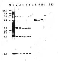
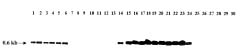
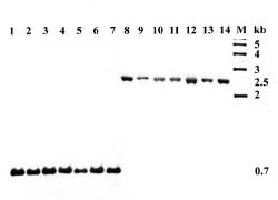
Abstract. A DNA probe was developed for identification of strains of Xanthomonas campestris pv. campestris from Taiwan. EcoRI restriction fragments of total DNA from X. campestris pv. campestris strain Xcc70 were cloned into pBluescript II KS and transformed into Escherichia coli DH10B. The recombinant plasmid DNAs from clones randomly selected were labeled with digoxigenin and screened for their specificity for X. campestris pv. campestris. In Southern hybridization, one of the clones (pXcc70-8) hybridized to several EcoRI-digested fragments of total DNA from only strains of X. campestris pv. campestris. Digestion of the insert DNA of the clone pXcc70-8 with EcoRI yielded fragments of 2.7, 1.6 and 0.6 kb. When a subclone containing the 0.6 kb fragment was used as a probe (Xcc70-8-l), it hybridized with all 51 strains of X. campestris pv. campestris and all seven strains of X. campestris pv.armoraciae, but not with the 60 strains of other bacteria tested. This probe, however, distinguishably detected a single fragment of 0.7 kb in strains of X. campestris pv. campestris and a single fragment of 2.5 kb in strains of X. campestris pv. armoraciae when their total DNAs were digested with KpnI. The detection limits of the probe Xcc70-8-l for the amount of DNA was 25 pg and for the number of cells was about 6 × 10⁴ CFU in dot blot assays. The colony and dot blot hybridizations with the probe were used to detect X. campestris pv.campestris in extracts of infected leaves of cabbage and seeds of several crucifers. The results indicate that the DNA probe can be used to detect X. campestris pv. campestris in plant tissues but probably not in seeds. The probe could be a useful tool for rapid identification of the pathogen in epidemiological studies in Taiwan.

Keywords: Crucifers (Brassica spp.); Detection; DNA probe; Identification; Xanthomonas campestris pv. campestris.

Introduction

Black rot of crucifers caused by Xanthomonas campestris pv. campestris (Pammel) Dowson is a destructive disease of worldwide importance (Williams, 1980). In Taiwan, black rot affects many cruciferous crops and can be observed at all growing seasons, particularly in most cabbage and cauliflower fields. The black rot pathogen is seed-borne (Walker and Tisdale, 1920; Williams, 1980; Schaad, 1982). Seed testing to ensure absence of the pathogen and seed treatments to eradicate the pathogen are important measures for preventing the occurrence of the disease (Williams, 1980). The success of these measures depends on an efficient and reliable method to detect the pathogen in seeds and to verify the efficacy of seed treatments. The pathogen also survives in infected crop debris, infested cruciferous weeds, and infected plant residues in soil (Alvarez and Cho, 1978; Schaad and White, 1974b; Schaad and Dianese, 1981). The importance of these sources of inoculum in the annual recurrence of

the disease in Taiwan remains to be investigated. This kind of ecological study also requires an effective method to detect the pathogen in fields.

Selective media and serology are the commonly used methods for the detection of X. campestris pv. campestris in seed, plant, or soil samples (Chun and Alvarez, 1983; Franken, 1992; Fukui et al., 1994; Schaad and White, 1974a; Schaad and Donaldson, 1980). The detection efficiency of selective media varies greatly with the source of samples. Most of the available selective media are not efficient for detection of the pathogen in seeds and soils under Taiwan conditions (Huang and Hsu, 1987). Furthermore, the identity of the suspect colonies on the media needs to be confirmed either by the pathogenicity test, which is time-consuming, or by serology. Serological assays are useful in the confirmation test for the isolated colonies (Schaad, 1979) and for direct detection of the pathogen in seeds (Franken, 1992; Schaad and Donaldson, 1980), infected leaves (Alvarez and Lou, 1985), and soil (Domen and Alvarez, 1978). Serology with monoclonal antibodies is particularly specific for the identification or differentiation of strains of X. campestris pv. campestris (Alvarez et al., 1985) and has been used suc

3Corresponding author. Fax: 886-4-2877585; E-mail: sthsu@mail.nchu.edu.tw


Botanical Bulletin of Academia Sinica, Vol. 41, 2000

cessfully in monitoring particular strains of the bacterium in fields in the epidemiological study (Yuen et al., 1987). Recently, DNA-based probes have offered another approach to the rapid and specific identification of plant pathogenic bacteria (Rasmussen and Reeves, 1992). A polymerase chain reaction (PCR) technique using primers designed from the sequence of a cloned DNA fragment specific to X. campestris pv. campestris has been developed for detection of the pathogen in crucifer seeds (Aline and Gabrielson, 1993). In this paper, we describe the development of a cloned DNA probe useful in Taiwan for identification and detection of X. campestris pv. campestris in plant tissues.

Materials and Methods

Bacterial Strains

The bacterial strains used in this study are listed in Table 1. All strains of X. campestris pv. campestris were isolated from various cruciferous plants in Taiwan except for strain NCPPB 528 (type strain of the pathovar), which was originated from the United Kingdom and was obtained from the Culture Collection and Research Center, Food Industry Research and Development Institute, Hsinchu, Taiwan. Xanthomonads were subcultured on potato dextrose agar (PDA, Difco), fluorescent pseudomonads were subcultured on King’s B medium (King et al., 1954), Ralstonia solanacearum (formerly Pseudomonas solanacearum) was subcultured on TZC medium (Kelman, 1954), Escherichia coli was subcultured on Luria-Bertani (LB) agar medium (Sambrook et al., 1989), and other bacteria were subcultured on nutrient agar (Difco Laboratories, Detroit, MI). All cultures were incubated at 28~30°C except for E. coli which was grown at 37°C. All strains were stored in LB broth containing 15% glycerol at -70°C.

Isolation of Total DNA from Bacteria

Total DNA was prepared by a modification of the method of Lazo et al. (1987). Each of the bacterial cultures was grown in 40 ml of LB broth at 28°C overnight and harvested by centrifugation at 8,000 g for 10 min. The cell pellet was washed in 4 ml of STE buffer (100 mM NaC1, 10 mM Tris-HC1, 1 mM EDTA, pH 8.0) and resuspended in 4 ml of STE buffer. The suspension was then added with a sodium dodecyl sulfate (SDS) solution at a final concentration of 1%, incubated at 65°C for 1.5 h, and cooled to room temperature. Subsequently, a proteinase K solution was added to a final concentration of 100 µg per ml, and the mixture was incubated at 37°C for more than 4 h. After addition of 10 ml of STE buffer, the mixture was extracted twice with an equal volume of phenol-chloroform-isoamyl alcohol (25:24:1, phenol equilibrated to pH 8 with 100 mM Tris) and twice with an equal volume of chloroform-isoamyl alcohol (24:1). All of the phenol or chloroform extractions were done gently and mixed well until emulsifying, and emulsified suspensions were separated by centrifugation at 12,000 g for 15 min. The supernatant containing DNA was transferred to a clean tube and precipitated by adding two volumes of ice- cold 95%

ethanol, mixing gently, and incubating at -20°C for 30 min. The DNA was spooled out of the precipitation mixture with a sterile glass bar, rinsed with ice- cold 70% ethanol, and then air-dried. The DNA was resuspended in TE buffer (10 mM Tris, 1 mM disodium EDTA) containing 10 µg/ml of DNase-free ribonuclease A.

Recombinant DNA Manipulations

Restriction enzyme digestion, preparation of plasmid DNA using alkaline extraction method, DNA ligation, agarose gel electrophoresis, bacterial transformation (CaCl2 procedure), and cloning of DNA fragments were performed as described by Sambrook et al. (1989). Strain Xcc70 of X. campestris pv. campestris was used for constructing a genomic library. Total DNA of Xcc70 was digested with EcoRI and ligated into the vector pBluescript II KS (Stratagene, USA). The ligation mixture was subsequently transformed into competent cells of E. coli DH10B. DNA fragments used in subcloning were separated by electrophoresis through a 0.8% agarose gel and then purified by electroelution from excised gel pieces in an Elutrap apparatus (Schleicher & Schuell, USA).

DNA Hybridizations

The recombinant plasmid DNAs and cloned DNAs for use as probes were labeled with digoxigenin-11-dUTP (DIG-dUTP) using the random primed method according to the manufacturer’s instructions (Boehringer Mannheim, Germany). Dot and Southern blots were made on Hybond N filters (Amersham International, Buckinghamshire, England) as recommended by the manufacturer. Generally, 10 µg of DNA was added to each dot in dot blots performed with Bio-dot apparatus (Bio-Rad, USA) and 4 µg of DNA was added to each lane in Southern blots. DNA hybridization procedures and immunodetection of hybridization signals were performed according to the user’s guide provided by the manufacturer (Boehringer Mannheim, Germany). Prehybridization and hybridization were conducted in hybridization buffer (5X SSC, 0.1% N-lauroyl sarcosine, 0.02% SDS, 1% blocking reagent) at 55°C for at least 5 h and at 68°C for at least 10 h, respectively. After hybridization, the filters were washed twice (10 min each) in 2X SSC containing 0.1% SDS at room temperature and twice (15 min each) in 0.1X SSC containing 0.1% SDS at 68°C. The hybridization signals were detected using the DIG luminescent detection kit (Boehringer Mannheim, Germany). The substrate for anti-DIG- conjugant alkaline phosphatase was Lumingen PPD (4-methoxy-4-[3-phosphate-phenyl]-spiro [l,2-dioxtane-3,2'-adamantane] disodium salt).

Specificity and Sensitivity of Cloned DNA Probes

The specificity of the probes was determined by Southern blot hybridization. Total DNA (4.5 µg) from each of the bacteria listed in Table 1 was digested with EcoRI. In addition, the total DNAs of X. campestris pv. campestris and X. campestris pv. armoraciae were also digested with KpnI. The DNA fragments separated by electrophoresis


Shih et al. — DNA probe for Xanthomonas campestris

Table 1. Strains of bacteria used in this study.

Bacterium Strain Host Location Sourcea

Burkholderia caryophylli NCPPB2151 Carnation U. S. A. 2

PCAR1, 6 Carnation Nantou, Yunlin 1

B. glumae PG1 Rice Taichung 1

Erwinia chrysanthemi HAS11, 12 Welsh onion Changhua 1

E. carotovora subsp. carotovora SP45 Potato Kaohsiung 1

Cel-2 Celery Taichung 1

Escherichia coli ATCC11775 2

Pseudomonas cichorii PC13-1, PC6 Celery Taichung 1

P. syringae pv. syringae Pss64 Wheat U. S. A. 7

P. putida YLFP1, 3 Cabbage Tainan 1

YLFP37 Mustard Taoyuan 1

P. fluorescens YLFP98 Radish Taichung 1

Ralstonia solanacearum PS63 Pepper Nantou 1

PS95, PSS4 Tomato Taipei, Tainan 1

Xanthomonas campestris

pv. campestris Xcc4, 70, 77,78, 79, 94 Cabbage Changhua 1

Xcc7, 45 Cabbage Hualien, Ilan 1

Xcc34, 90, 91, 92, 93 Cauliflower Changhua 1

Xcc40 Kohlrabi Taichung 1

Xcc61, 111 Cabbage Tainan, Taitung 1

Xcc104, 105, 112, 113, 114 Cabbage Tainan 3

Xcc62, 68, 99 Cabbage Taichung 1

Xcc107 Cabbage Taichung 3

Xcc71, 72, 73, 74 Cabbage Hsinchu 1

Xcc75, 76 Broccoli Kaohsiung 3

Xcc80, 81, 82, 83, 84, 85, 87 Cabbage Nantou 1

Xcc108, 109, 110 Cabbage Nantou 3

Xcc86 Mustard Yunlin 1

Xcc88 Cabbage Nantou 1

Xcc89 Broccoli Nantou 1

Xcc95, 102, 115 Cauliflower Tainan 3

NCPPB528 Brussels sprouts U. K. 2

Xcc101 Kohlrabi Tainan 3

Xcc103 Broccoli Tainan 3

Xcc106 Cabbage Kaohsiung 3

pv. armoraciae 417, 756 Cabbage seeds East Asia 4

A342 Broccoli U. S. A. 4

G3-27, XLS2, XLS6, XLS10 Cabbage U. S. A. 4

pv. begoniae ICMP194 Tuberous begonia New Zealand 2

pv. citri XW23, 32 Orange (Liucheng) Chiayi, Kaohsiung 5

XW27 Orange (Wentan) Chiayi 5

XW35 Orange (Jaffa) Hsinchu 5

XW47 Orange (Valencia) Hualien 5

pv. dieffenbachiae A044, 049, 058 Anthurium Tainan 8

pv. glycines XPS1, 3 Soybean Yunlin 1

pv. mangiferaeindicae 3, 4, 12, 30 Mango Tainan 7

pv. phaseoli XP1, 4 Bean Taichung 1

pv. pruni 3, 4, 5 Peach Taichung 7

pv. uppalii NCPPB586 Ipomoea muricata India 2

J-3, T-12-1 I. nil Chiayi, Taichung 1

pv. vesicatoria XV7, 12 Tomato Taitung, Yunlin 1

XV30, 48 Tomato Tainan, Changhua 1

XV42, 51 Pepper Pingtung, Hualien 1

X. albilineans ICMP196 Sugarcane Fiji 2

X. fragariae NCPPB1469 Strawberry U. S. A. 2

X. oryzae pv. oryzae 21, 84 Rice Taichung, Nantou 6

117 Rice Pingtung 6

Unidentified bacteria Sb1, 3, 4 Cabbage leaves Taichung 1

Sb2 Cabbage leaves Nantou 1

Sb5, 6, 7 Cabbage seeds Taichung 1

a1: Phytobacteriology laboratory, Department of Plant Pathology, National Chung Hsing University (NCHU), Taichung, Taiwan; 2: Culture Collection and Research Center, Food Industry Research and Development Institute, Hsinchu, Taiwan; 3: J. F. Wang, Asian Vegetable Research and Development Center, Tainan, Taiwan; 4: A. M. Alvarez, Department of Plant Pathology, University of Hawaii, Honolulu, U. S. A.; 5: W. C. Wu, NCHU; 6: S. P. Y. Hsieh, NCHU; 7: H. S. Huang, NCHU; and 8: S. H. Hseu, Taiwan Agricultural Research Institute, Taichung, Taiwan.


Botanical Bulletin of Academia Sinica, Vol. 41, 2000

through the agarose gel were denatured and transferred to Hybond N filters. The filters were then hybridized with the DIG-labeled DNA probe as described above.

The dot blot hybridization was used for probe sensitivity assays. Total DNA and cells of X. campestris pv. campestris Xcc70 cultured in LB broth were both two-fold serially diluted and spotted on Hybond N filters. Each spot contained the amount of total DNA ranging from 0.8 ng to 6.25 pg or the amount of cells ranging from 1.0 × 10⁶ to 3.1 × 10⁴ colony-forming units (CFU). The filters were then hybridized with the DIG-labeled DNA probe.

Detection of X. campestris pv. campestris in Leaves and Seeds

Direct detection of the pathogen in leaves and seeds was performed by dot blot hybridization. Leaves of cabbage with black rot-like lesions were collected from fields in Taichung. About 0.1 g of leaf lesions was placed in an Eppendorf tube and macerated in 100 µl sterile distilled water. Aliquots (5 µl) of the resulting suspension were spotted on the Hybond N filter. For seed detection, seven crucifer seed samples (three of cabbage and one each of Chinese kale, mustard, Chinese cabbage, and cauliflower) obtained from the Known-you Seed Company were assayed. Each seed sample (0.1 g) was ground in 0.25 ml of phosphate-buffered saline containing 0.1% Tween 80 in a mortar, and 5 µl of the resulting suspension was spotted on Hybond N filters. The blots for leaf and seed samples were then hybridized with the DIG-labeled DNA probe as previously described.

The same leaf and seed samples were also used in colony hybridization tests for rapid identification of the pathogen in the samples. After the samples were macerated, l0-fold serial dilutions were made, and 50 µl of appropriate dilutions were plated on PDA for leaf samples and on PDA containing 80 mg/ml of cycloheximide for seed samples. Colonies appearing on the plates after incubation at 30°C for 3 days were blotted on Hybond N filters, irradiated in a microwave oven, denatured with 0.5 M NaOH/I.5 M NaC1, and neutralized with 1 M Tris-HC1 (pH 7.2)/3 M NaC1 according to the procedure of Datta et al. (1988). After neutralization, the filters were air-dried, UV cross-linked, and hybridized with the DIG labeled DNA probe as previously described. Colonies randomly selected from the original plates were tested for their pathogenicity on leaves of cabbage plants grown in pots in a greenhouse at 25~35°C by the notched leaf inoculation method (Alvarez et al., 1994).

Results

Selection of the DNA Probe

Approximately 1000 transformant clones were obtained in the cloning of EcoRI-digested total DNA of X. campestris pv. campestris strain Xcc70. Recombinant plasmids of randomly selected 10 clones were isolated, labeled with digoxigenin and used as probes. These probes hybridized to total DNA of five strains of X. campestris pv.

campestris tested, but also to that of some other pathovars of X. campestris in dot blots (data not shown). However, in Southern hybridization analyses, one of the 10 probes, pXcc70-8 (containing a 4.9 kb insert DNA) hybridized to three DNA fragments that were only observed in EcoRI-digested total DNA from strains of X. campestris pv.campestris, but not in that from strains of other bacteria initially screened (Figure 1). This clone (pXcc70-8) was therefore selected for subcloning. Digestion of the insert DNA of the clone pXcc70-8 with EcoRI yielded fragments of 2.7, 1.6 and 0.6 kb. The 0.6 kb fragment was cloned into pBluescript II KS and then transformed into E. coli DH10B. The cloned 0.6 kb EcoRI fragment, designated probe Xcc70-8-1, was further evaluated for its specificity against X. campestris pv. campestris.

Specificity and Sensitivity of Probe Xcc 70-8-1

The specificity of the probe Xcc70-8-1 labeled with digoxigenin was tested by the Southern hybridization with EcoRI-digested total DNA from various bacteria. All of the 51 strains of X. campestris pv. campestris and the seven strains of X. campestris pv. armoraciae (Table 1) gave a positive hybridization signal, whereas the 60 strains of other bacteria including other pathovars of X. campestris, Xanthomonas spp., distantly related bacteria, and unidentified saprophytic bacteria from cabbage leaves and seeds (Table 1) were all negative (representative results are shown in Figure 2). The probe hybridized to a single DNA fragment of 0.6 kb in all strains of X.

Figure 1. Southern hybridization of EcoRI-digested bacterial total DNAs probed with the digoxigenin-labeled clone pXcc70-8 selected from a genomic library of Xanthomonas campestris pv. campestris. Lane M, HindIII-digested lambda marker (the sizes in kb shown in the margin); lane 1, plasmid pXcc70-8 DNA; lanes 2-6, X. campestris pv. campestris strains Xcc70, Xcc78, Xccl02, Xccl03 and Xccl04; lane 7, unidentified bacterium Sbl; lanes 8-9, X. campestris pv. vesicatoria strains XV48 and XV7; lane 10, X. campestris pv. citri XW47; lane 11, X. oryzae pv.oryzae 21; lane 12, Erwinia carotovora subsp. carotovora SP45; lane 13, Ralstonia solanacearum PSS4.


Shih et al. — DNA probe for Xanthomonas campestris

Figure 2. Southern blots of EcoRI-digested total DNAs from various bacteria hybridized with the digoxigenin-labeled probe Xcc70-8-1 containing a 0.6 kb insert DNA of Xanthomonas campestris pv. campestris. Lanes 1-6, X. campestris pv. campestris strains Xcc70, NCPPB528, Xccl0l, Xccl02, Xccl03, and Xccl04; lanes 7-8, X. campestris pv. vesicatoria strains XV7 and XV51; lane 9, X. oryzae pv. oryzae 21; lane 10, X. campestris pv.citri XW47; lane 11, X. campestris pv. mangiferaeindicae 30; lane 12, Erwinia carotovora subsp. carotovora SP45; lane 13,Ralstonia solanacearum PS95; lane 14, X. campestris pv.campestris Xcc88; lane 15, plasmid pXcc70-8-1 DNA; lanes 16-22, X. campestris pv. campestris strains Xccl07, Xccl08, Xccl09, Xccll0, Xcc71, Xcc72 and Xcc99; lanes 23-24, X. campestris pv.armoraciae strains 417 and XLS10; lane 25, X. campestris pv. begoniae ICMP194; lane 26, X. campestris pv. uppalii NCPPB586; lane 27, X. albilineans ICMP196; lane 28, Burkholderia caryophylli NCPPB2151; lane 29, Ralstonia solanacearum PSS4; lane 30, Pseudomonas putida YLFPI. The size (in kb) is shown in the margin.

campestris pv. campestris and X. campestris pv. armoraciae tested. However, when total DNAs of these two pathovars were digested with KpnI, the probe detected a single 0.7 kb fragment in all strains of X. campestris pv. campestris and a single 2.5 kb fragment in all strains of X. campestris pv. armoraciae tested (Figure 3). The sensitivity of the probe was determined by dot blot to a dilution series of total DNA and cells of strain Xcc70. The detection limits were 25 pg of DNA and 6.2 × 10⁴ CFU of cells.

Detection of X. campestris pv. campestris in Infected Leaves and Seeds of Crucifers

The hybridization signals were observed on the Xcc70-8-l-probed dot blots from extracts of infected cabbage leaves and also from extracts of two (Chinese kale and Chi

nese cabbage) of the seven seed samples tested. Yellow, mucoid colonies could be isolated on the medium from the extracts of the hybridization positive samples, and these colonies were identified as X. campestris pv. campestris by the pathogenicity test.

Several types of colonies were formed on the media plated with the suspensions of macerated tissues of leaves and seeds. In colony hybridization probed with Xcc70-8-1, the signals were observed on some colonies formed from the cabbage leaves (Figure 4) and also from the two (Chinese kale and Chinese cabbage) of seven seed samples. These hybridization positive colonies produced black rot symptoms when inoculated into cabbage leaves, confirming that they were X. campestris pv. campestris, whereas those hybridization negative colonies were not pathogenic to cabbage.

Figure 3. Southern blots of KpnI-digested total DNAs from Xanthomonas campestris pv. campestris and X. campestris pv. armoraciae hybridized with the digoxigenin-labeled probe Xcc70-8-1 containing a 0.6 kb insert DNA of X. campestris pv. campestris. Lanes 1-7, X. campestris pv. campestris strains Xcc34, Xcc70, Xcc88, Xcc92, Xccl04, Xccl05 and Xccl06; lanes 8-14, X. campestris pv. armoraciae strains 417, A342, G3-27, 756, XLS2, XLS6 and XLSI0; lane M, 1 kb marker (Stratagene, USA). The sizes (in kb) are shown in the margin.

Figure 4. Colony hybridization analysis of the bacterial colonies formed on a potato dextrose agar plate spread with the extract of the black rot-infected cabbage leaves. A, plate before the colonies were blotted; B, hybridization of the colonies blotted on a Hybond N filter with the digoxigenin-labeled probe Xcc70-8-1. Hybridization signals were observed only for colonies of Xanthomonas campestris pv. campestris. The Xcc-70-8-1 plasmid DNA was blotted in the arrow place as CK.


Botanical Bulletin of Academia Sinica, Vol. 41, 2000

Discussion

Development of specific DNA probes for identification and detection has been reported for a number of plant pathogenic bacteria (Rasmussen and Reeves, 1992; Hartung, 1992; Darrasse et al., 1994). Some of these DNA probes are developed by screening randomly cloned fragments of total genomic or plasmid DNA for their specificity (Gilbertson et al., 1989; Johansen et al., 1989; Thompson et al., 1989; Manulis et al., 1991; Hartung, 1992). In this study, the same approach was used to obtain a DNA probe specific for X. campestris pv. campestris. Unlike some pathovars of X. campestris, strains of X. campestris pv. campestris are variable in plasmid content (Lazo and Gabriel, 1987). Therefore, we used the total genomic DNA from a strain (Xcc70) of X. campestris pv. campestris to construct a genomic library from which a recombinant clone (pXcc70-8) containing partly specific insert DNA was identified. When the recombinant plasmid of this clone, pXcc70-8, was used to probe Southern blots of genomic digests from strains of X. campestris pv. campestris, restriction fragment length polymorphism was evident within these strains. This result agrees with the reports of Alvarez et al. (1994) and Chiu et al. (1996), who showed that strains of X. campestris pv. campestris are genetically heterogeneous based on RFLP analysis of total DNA. However, the hybridization pattern of EcoRI-digested total DNA probed with pXcc70-8 revealed a 0.6 kb fragment that was observed in strains of X. campestris pv. campestris but not in strains of other bacteria initially tested. A subclone (pXcc70-8-1) containing this 0.6 kb fragment was subsequently proved to be specific for X. campestris pv. campestris, except for X. campestris pv. armoraciae. Further tests showed that this probe distinguished between the X. campestris pv. campestris and pv. armoraciae by revealing a single DNA fragment of different size detected in their total DNAs digested with KpnI. Because the DNA probe was tested against strains of X. campestris pv. campestris only from Taiwan except one strain, the specificity of the probe to strains from other geographical regions remains to be determined. However, for use in Taiwan, the probe should be useful for rapid identification of X. campestris pv. campestris.

The probe Xcc70-8-1 developed in this study identified and distinguished X. campestris pv. campestris colonies from colonies of other bacteria on the agar media plated with extracts of infected leaves using the colony hybridization assay and detected the X. campestris pv. campestris directly in infected leaves using the dot blot hybridization method. In colony hybridization tests, colonies that showed hybridization signals were confirmed to be X. campestris pv. campestris as determined by the pathogenicity test. Likewise, in the dot blot assays, X. campestris pv. campestris was isolated from the hybridization positive samples. These results indicate that the hybridization procedures can be used for detection or confirmation of the presence of X. campestris pv. campestris in plant tissues. Although X. campestris pv. campestris in infected plant tissues usually have populations high enough to be

readily recovered on selective media, the DNA probe assay is more rapid and specific. Furthermore, the DNA probe is feasible for assaying large numbers of samples in a relatively short time.

Serological tests have been used for rapid identification and detection of X. campestris pv. campestris. However, polyclonal antibodies against strains of X. campestris pv. campestris often cross-react with one or a few other pathovars of X. campestris (Alvarez and Lou, 1985; Franken et a1., 1992; Schaad, 1978; Thaveechai and Schaad, 1984, 1986). For example, strains of X. campestris pv. vesicatoria often react with the polyclonal antibodies of X. campestris pv. campestris (Alvarez and Lou, 1985; Franken et al., 1992; Schaad, 1978; Thaveechai and Schaad, 1984, 1986). Xanthomonas campestris pv. vesicatoria is a widely distributed pathogen causing bacterial spot disease on tomato and pepper in Taiwan (Hartman et al., 1990). The chance of introducing this bacterial spot pathogen to the fields of cruciferous crops in Taiwan exists because of the planting multiple crops within a relatively small area and the spread of the pathogen by wind-driven rain. Therefore, differentiation between X. campestris pv. campestris and pv. vesicatoria would still be desired in studies involving, for example, the recovery of X. campestris pv. campestris from field samples that might be contaminated with X. campestris pv. vesicatoria. Our results showed that the probe Xcc70-8-l did not hybridize with any of the six strains of X. campestris pv. vesicatoria tested and can be used for their differentiation. Monoclonal antibodies produced against X. campestris pv. campestris are highly specific although strains of X. campestris pv. armoraciae may possess some of the same antigenic determinants (Alvarez et al., 1985, 1994). Our results also showed that DNAs from strains of X. campestris pv. armoraciae hybridized to the probe Xcc70-8-l. These two pathovars are considered to be very closely related (Alvarez et al., 1994). However, pv. campestris could be distinguished from pv. armoraciae by this probe when the total DNAs were digested with KpnI. The seven strains of X. campestris pv. armoraciae used in our DNA hybridization tests represented strains that also reacted with monoclonal antibodies generated to X. campestris pv. campestris (Alvarez et al., 1994). Thus, our DNA probe offers an advantage over serological methods for specific identification or differentiation of these two pathovars. The ability of the DNA probe to rapidly distinguish X. campestris pv. campestris from pv.armoraciae is valuable because these two pathovars infect crucifers, and the pathogens recovered from cruciferous plant and seed samples previously were reliably differentiated only by time-consuming pathogenicity tests (Alvarez et al., 1994).

A diagnostic tool’s effectiveness depends largely on its sensitivity in addition to its specificity. The sensitivity of the probe Xcc70-8-l for detecting X. campestris pv. campestris was about 104 CFU, similar to that of the enzyme-linked immunosorbent assay method (Alvarez and Lou, 1985), but not as sensitive as either the immunofluorescence method (Domen and Alvarez, 1978) or the miniplate ELISA enrichment method (Arias et al., 1996).


Shih et al. — DNA probe for Xanthomonas campestris

Darrasse A., A. Kotoujansky, and Y. Bertheau. 1994. Isolation by genomic substraction of DNA probes specific for Erwinia carotovora subsp. atroseptica. Appl. Environ. Microbiol. 60: 289-306.

Datta, A.R., B.A. Wentz, D. Shook, and M.W. Trucksess. 1988. Synthetic oligodeoxyribonucleotide probes for detection of Listeria monocytogenes. Appl. Environ. Microbiol. 54: 2933-2937.

Domen, H.Y. and A.M. Alvarez. 1978. Detection of Xanthomonas campestris in soil using a direct immunofluorescent technique. In Proc. 4th Int. Conf. Plant Pathog. Bact., Angers, France, pp. 301-305.

Franken, A.A.J.M. 1992. Comparison of immunofluorescence microscopy and dilution-plating for the detection of Xanthomonas campestris pv. campestris in crucifer seeds. Neth. J. Plant Pathol. 98: 169-178.

Franken, A.A.J.M., J.F. Zilverentant, P.M. Boonekamp, and A. Schots. 1992. Specificity of polyclonal and monoclonal antibodies for the identification of Xanthomonas campestris pv. campestris. Neth. J. Plant Pathol. 98: 81-94.

Fukui, R., R. Arias, and R. Alvarez. 1994. Efficacy of four semiselective media for recovery of Xanthomonas campestris pv. campestris from tropical soils. J. Appl. Bacteriol. 77: 534-540.

Gilbertson, R.L., D.P. Maxwell, D.J. Hagedom, and S.A. Leong. 1989. Development and application of a plasmid DNA probe for detection of bacteria causing common bacterial blight of bean. Phytopathology 79: 518-525.

Hartman, G.L., C.H. Yang, and J.F. Wang. 1990. Races of Xanthomonas campestris pv. vesicatoria in Taiwan and an inoculation technique to evaluate host resistance. Plant Prot. Bull. 32: 112-124.

Hartung, J.S. 1992. Plasmid-based hybridization probes for detection and identification of Xanthomonas campestris pv. citri. Plant Dis. 76: 889-893.

Huang, T.C. and S.T. Hsu. 1987. Efficiency of various selective media for detection of Xanthomonas campestris pv. campestris in Taiwan. Plant Prot. Bull. 29: 203-215.

Johansen, I.E., O.F. Rasmussen, and M. Heide. 1989. Specific identification of Clavibacter michiganense subsp. sepedonicum by DNA-hybridization probes. Phytopathology 79: 1019-1023.

Kelman, A. 1954. The relationship of pathogenicity in Pseudomonas solanacearum to colony appearance on a tetrazolium medium. Phytopathology 44: 693-695.

King, E.O., M.K. Ward, and D.E. Raney. 1954. Two simple media for the demonstration of pyocyanin and fluorescein. J. Lab. Clin. Med. 44: 301-307.

Lazo, G.R. and D.W. Gabriel. 1987. Conservation of plasmid DNA sequences and pathovar identification of strains of Xanthomonas campestris. Phytopathology 77: 448-453.

Lazo, G.R., R. Roffey, and D.W. Gabriel. 1987. Pathovars of Xanthomonas campestris are distinguishable by restriction fragment-length polymorphism. Int. J. Syst. Bacteriol. 37: 214-221.

Manulis, S., Y. Gafni, E. Clark, D. Zutra, Y. Ophir, and I. Barash. 1991. Identification of a plasmid DNA probe for detection of strains of Erwinia herbicola pathogenic on Gypsophila paniculata. Phytopathology 81: 54-57.

Rasmussen, O.F. and J.C. Reeves. 1992. DNA probes for the detection of plant pathogenic bacteria. J. Biotechnol. 25: 203-220.

The probe Xcc70-8-l may not be sensitive enough to detect X. campestris pv. campestris with populations normally present in alternative weed hosts, soils, and seeds. Although a small sample size (5 µl taken from the extract of 25 seeds ground in 250 µl buffer) was taken, X. campestris pv. campestris was detected in two of the seven seed samples tested by the DNA hybridization assay used in the present study. Therefore, these two seed samples were likely to have been naturally infested with unusually high numbers of the pathogen. In seed assays, a DNA probe should also detect strains of X. campestris pv. campestris from different geographical regions because the seed-borne pathogen could be imported from different regions. The specificity of our DNA probe to non-local strains has not been determined. Therefore, unless specificity and sensitivity can be satisfied, the DNA probe in its present form is not well-suited for seed assays. Its best use would be in providing a useful tool for rapid identification of X. campestris pv. campestris in epidemiological studies in Taiwan. An improvement in detection sensitivity may be achieved by the development of an amplification technique such as PCR using a primer set which can be designed from the sequence of the cloned DNA fragment (Saiki et al., 1988). Such developments are currently underway.

Acknowledgments. This study was supported by Grants NSC83-0409-B005-029 and NSC84-2321-B005-070 from the National Science Council, Republic of China.

Literature Cited

Aline Jr., R. and R.L. Gabrielson. 1993. PCR detection of Xanthomonas campestris pathovar campestris. Abstr. 6th Int. Cong. Plant Pathol., Montreal, Canada, pp. 46.

Alvarez, A.M. and J.J. Cho. 1978. Black rot of cabbage in Hawaii: Inoculum source and disease incidence. Phytopathology 68: 1456-1459.

Alvarez, A.M. and K. Lou. 1985. Rapid identification of Xanthomonas campestris pv. campestris by ELlSA. Plant Dis. 69: 1082-1086.

Alvarez, A.M., A.A. Benedict, and C.Y. Mizumoto. 1985. Identification of xanthomonads and grouping of strains of Xanthomonas campestris pv. campestris with monoclonal antibodies. Phytopathology 75: 722-728.

Alvarez, A.M., A.A. Benedict, C.Y. Mizumoto, J.E. Hunter, and D.W. Gabriel. 1994. Serological, pathological, and genetic diversity among strains of Xanthomonas campestris infecting crucifers. Phytopathology 84: 1449-1457.

Arias, R.S., G. Mochizuki, R. Fukui, and A.M. Alvarez. 1996. Most probable number method to enumerate a bioluminescent Xanthomonas campestris pv. campestris in soil. Soil Biol. Biochem. 28: 1725-1728.

Chiu, S.P., C.M. Chang, M.Y. Chen, and Y.A. Lee. 1996. Genetic diversity among strains of Xanthomonas campestris pv. campestris in Taiwan. (Abstr.) Plant Pathol. Bull. 5: 211-212.

Chun, W.W.C. and A.M. Alvarez. 1983. A starch-methionine medium for isolation of Xanthomonas campestris pv. campestris from plant debris in soil. Plant Dis. 67: 632-635.


Botanical Bulletin of Academia Sinica, Vol. 41, 2000

Saiki, R.K., D.H. Gelfand, S. Stoffel, S.J. Scharf, R. Higuchi, G.T. Horn, K.B. Mullis, and H.A. Erlich. 1988. Primer-directed enzymatic amplification of DNA with thermostable DNA polymerase. Science 239: 487-491.

Sambrook, J., E.F. Fritsch, and T. Maniatis. 1989. Molecular cloning: A laboratory manual, 2nd edn. Cold Spring Harbor Laboratory Press, Cold Spring Harbor, New York.

Schaad, N.W. 1978. Use of direct and indirect immunofluorescence tests for identification of Xanthomonas campestris. Phytopathology 68: 249-252.

Schaad, N.W. 1979. Serological identification of plant pathogenic bacteria. Ann. Rev. Phytopathol. 17: 123-147.

Schaad, N.W. 1982. Detection of seedborne bacterial plant pathogens. Plant Dis. 66: 885-890.

Schaad, N.W. and J.C. Dianese. 1981. Cruciferous weeds as sources of inoculum of Xanthomonas campestris in black rot of crucifers. Phytopathology 71: 1215-1220.

Schaad, N.W. and R.C. Donaldson. 1980. Comparison of two methods for detection of Xanthomonas campestris in infected crucifer seeds. Seed Sci. Technol. 8: 383-391.

Schaad, N.W. and W.C. White. 1974a. A selective medium for soil isolation and enumeration of Xanthomonas campestris. Phytopathology 64: 876-880.

Schaad, N.W. and W.C. White. 1974b. Survival of Xanthomonas campestris in soil. Phytopathology 64: 1518-1520.

Thaveechai, N. and N.W. Schaad. 1984. Comparison of different immunogen preparations for serological identification of Xanthomonas campestris pv. campestris. Phytopathology 74: 1065-1070.

Thaveechai, N. and N.W. Schaad. 1986. Serological and electrophoretic analysis of a membrane protein extract of Xanthomonas campestris pv. campestris from Thailand. Phytopathology 76: 139-147.

Thompson, E., J.V. Leary, and W.W.C. Chun. 1989. Specific detection of Clavibacter michiganense subsp. michiganense by a homologous DNA probe. Phytopathology 79: 311-314.

Walker, J.C. and W.B. Tisdale. 1920. Observations on seed transmission of the cabbage black rot organism. Phytopathology 10: 175-177.

Williams, P.H. 1980. Black rot: A continuing threat to world crucifer. Plant Dis. 64: 736-742.

Yuen, G.Y., A.M. Alvarez, A.A. Benedict, and K.J. Trotter. 1987. Use of monoclonal antibodies to monitor the dissemination of Xanthomonas campestris pv. campestris. Phytopathology 77: 366-370.

Ų©w¥xÆW¤Q¦rªá¬ì½­µæ¶Â»G¯fµß¤§®Ö»Ä±´°w

¥Û«H¼w1 ªL¤¸¬K1 ¶À¨q¬Ã2 ´¿°ê´Ü1 ®}¥@¨å1

1 °ê¥ß¤¤¿³¤j¾Ç´Óª«¯f²z¾Ç¨t

2 °ê¥ß¤¤¿³¤j¾Ç¹A·~¥Íª«¬ì§Þ¾Ç¬ã¨s©Ò
¤Q¦rªá¬ì½­µæ¶Â»G¯fµß (Xanthomonas campestris pv. campestris) Xcc70 µßªL¤§¥þ DNA ¸g EcoRI ¤Á³Î«á¡A¿ï´Þ©ó½èÅé pBluescript II KS ¤º¡A¦AÂà§Î¦Ü¤j¸z±ìµß DHl0B ¤¤¡C¥Ñ¦¹ÀH¾÷¿ï¨ú¤§¿ï´Þ®è¡A±N¨ä­«²Õ½èÅé¥H digoxigenin ¼ÐÃÑ«á§@¬°±´°w¡A¥Î©ó¿z¿ï¨ä¹ïŲ©w¶Â»G¯fµß¥xÆWµß®è¤§±M¤@©Ê¡C¦b«n¤èÂø°t¤ÀªR ¤W¡A¨ä¤¤¤@­Ó¿ï´Þ®è (pXcc70-8) ¶È¯à»P¶Â»G¯fµß¥þ DNA ¥H EcoRI ¤Á³Î«á¤§¼Æ­Ó¤ù¬q²£¥ÍÂø°t°T¸¹¡C¦¹¿ï´Þ®è¤º¤§¶Â»G¯fµß´O¤J DNA ¤ù¬q¥H EcoRI ¤Á³Î«á§t¦³ 2.7¡A1.6 ¤Î 0.6 kb ¤T­Ó¤ù¬q¡A±N¨ä¤¤¤§ 0.6 kb ¤ù¬qÂǥѦ¸¿ï´Þ«á©Ò»s³Æªº±´°w (Xcc70-8-1) »P¶Â»G¯fµß©Ò¦³ 51 ­Óµß®è¤Î½­µæ²Óµß©Ê¸­´³¯fµß (X. campestris pv. armoraciae) 7 ­Óµß®è§¡¦³Âø°t°T¸¹¡A¦Ó»P¨ä¥L²Óµß¤§ 60 ­Ó¨Ñ¸Õµß®è¡A«hµL¥ô¦óÂø°t¤ÏÀ³¡C·í¶Â»G¯fµß¤Î²Óµß©Ê¸­´³¯fµßªº¥þ DNA ¥H KpnI ¤Á³Î«á¡A¦¹±´°w¦b©Ò´ú¸Õªº 51 ­Ó¶Â»G¯fµß®è¡A§¡¥iÂø¦X¥X³æ¤@±ø 0.7 kb ¤ù¬q¡A¦Ó¦b©Ò´ú¸Õªº 7 ­Ó¸­´³¯fµß®è¡A«h¥iÂø¦X¥X³æ¤@±ø 2.5 kb ¤ù¬q¡C¦¹±´°w Xcc70-8-1 ¯à°»´ú¶Â»G¯fµß DNA ¶q¤Î²Ó­M¼Æ¤§³Ì§C­­«×¤À§O¬° 25 pg ¤Î¬ù 6 x l04 CFU¡C§Q¥Îµß¸¨¤ÎÂIº{Âø°tªk°»´ú¿©¯f¥ÌŸ­¤ù¤Î¼ÆºØ¤Q¦rªá¬ì½­µæºØ¤l¿i¸H²G¤¤ªº¶Â»G¯fµß¡Aµ²ªGÅã¥Ü¦¹ DNA ±´°w¥i¥Î©ó°»´ú´Óª«²Õ´¤ºªº¶Â»G¯fµß¡A¦ý¸û¤£¾A¥Î©óºØ¤l¤§°»´ú¡C¦b¥xÆW¶Â»G¯f¬y¦æ¯f¾Ç¬ã¨s¤W¡A¦¹±´°w¥i§@¬°§Ö³t ¤Î¥¿½TŲ©w¶Â»G¯fµß¤§¦³¥Î¤u¨ã¡C

ÃöÁäµü¡G¤Q¦rªá¬ì½­µæ¡F¶Â»G¯fµß¡F®Ö»Ä±´°w¡F°»´ú¡FŲ©w¡C