Bot. Bull. Acad. Sin. (2000) 41: 191-196

Hou et al. — Dioscorins with both carbonic anhydrase and trypsin inhibitor activities

Dioscorins from different Dioscorea species all exhibit both carbonic anhydrase and trypsin inhibitor activities

Wen-Chi Hou, Hsien-Jung Chen and Yaw-Huei Lin1

Institute of Botany, Academia Sinica, Nankang, Taipei 115, Taiwan, Republic of China

(Received January 31, 2000; Accepted February 24, 2000)

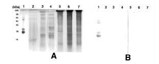
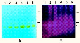
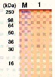
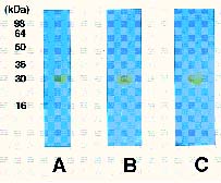
Abstract. Dioscorins, the storage proteins of yam tubers, from six cultivars of three Dioscorea species including D. batatas, D. alata, and D. pseudojaponica were detected in crude extracts of yam tubers but not leaves by immuno stainings on PVDF membrane with a polyclonal antibody which was raised against 28 kDa dioscorin from Dioscorea batatas Decne. It was demonstrated that the dioscorins from the above-mentioned yam species exhibited both carbonic anhydrase (CA) and trypsin inhibitor activities by activity stainings on 15% SDS-PAGE gels. Dioscorins were identified as glycoproteins by ConA-peroxidase activity staining. Dioscorins from D. batatas Decne contained no zinc atoms detectable by atomic absorption spectrophotometry. Neither 5 mM acetazolamide nor 2,6-pyridinedicarboxylic acid inhibited the CA activity of dioscorins.

Keywords: Acetazolamide; Carbonic anhydrase; Dioscorin; Pyridinedicarboxylic acid; Storage proteins; Trypsin inhibitor; Yam.

Abbreviations: CA, carbonic anhydrase; 2-ME, 2-mercaptoethanol; PVDF, polyvinylidene difluoride; SDS-PAGE, sodium dodecylsulfate-polyacrylamide gel electrophoresis; TI, trypsin inhibitor.

Introduction

Carbonic anhydrases (CAs) catalyze the simple interconversion of CO2 and HCO3-. They are found in almost all organisms and are notable for extremely high turnover numbers (Tashian, 1989; Badger and Price, 1994). In cyanobacteria and plants, CAs facilitate the interchange of CO2 and HCO3- and play a key role in CO2 fixation of photosynthesis (Hatch and Burnell, 1990; Badger and Price, 1992). In mammals, CAs also facilitate the interchange of CO2 and HCO3- and play a key role in respiration (Tashian and Hewett-Emmett, 1984; Forster, 1988). According to Hewett-Emmett and Tashian (1996), CA is encoded by three independent CA gene families (a-CA, b-CA, and g-CA). Most CAs of mammals and green algae belonged to the a-CA family, those of plants and eubacteria to the b-CA family, and those of archaebacteria to the g-CA family. Surprisingly, based on the cDNA deduced amino acid sequence alignment, the storage protein of yam (Dioscorea cayenensis L.) tuber, dioscorin, was found to be a a-CA related protein (Hewett-Emmett and Tashian, 1996). However, Hewett-Emmett and Tashian (1996) also mentioned that dioscorin was unlikely to have classical CA activity due to the active site alternations.

Patatin, the potato tuber storage protein, has been reported to contain both lipid acyl hydrolase and acyltransferase activities, both of which are involved in tuber tissue response to wounding (Andrews et al., 1988). The soybean vegetative storage proteins VSP a and VSP b both have acid phosphatase activity (Dewald et al., 1992). We have demonstrated that the root storage proteins of sweet potato not only have trypsin inhibitor (TI) activity (Hou and Lin, 1997a), but also dehydroascorbate reductase and monodehydroascorbate reductase activities, which might be involved in response to environmental stresses (Hou and Lin, 1997b). Tarin, the storage protein of taro (Colocasia esculenta L. Schott), was homologous to mannose-binding lectin and curculin in deduced amino acid sequences (Bezerra et al., 1995). Recently, it was reported that yam bean (Pachyrhizus erosus L. Urban) storage proteins YGB 1 and YGB 2 exhibited cysteine protease activities and YBP 2 showed sequence homology to protease inhibitors (Gomes et al., 1997). We reported recently that the purified dioscorins from tubers of Dioscorea batatas Decne (imported from Japan) showed both CA and TI activities (Hou et al., 1999a) and both dehydroascorbate reductase and monodehydroascorbate reductase activities (Hou et al., 1999b). In this report, we extend the work to show that both CA and TI activities could be found in dioscorin samples from six cultivars of three Dioscorea species including D. batatas, D. alata and D. pseudojaponica grown in Taiwan by activity staining on 15% SDS-PAGE gels. Some properties of dioscorins were also included.

1Corresponding author. Tel: 886-2-2789-9590 ext. 320; Fax: 886-2-2782-7954; E-mail: boyhlin@ccvax.sinica.edu.tw


Botanical Bulletin of Academia Sinica, Vol. 41, 2000

Materials and Methods

Plant Materials and Chemicals

Fresh tubers and leaves of yam grown in Hualien, Taiwan—including D. batatas Decne (imported from Japan); D. batatas Decne var. Shoufeng; D. alata L. var. Tainong 1; D. alata L. var. Tainong 2; D. alata L. var. Zhongguochang and D. pseudojaponica var. Keelung—were kindly provided by Dr. Peng Hwang, Research Fellow of Hualien District Agricultural Improvement Station. Electrophoresis grade acrylamide, N,N'-methylene-bis-acrylamide, N,N,N'N'-tetramethyl-ethylenediamine, ammonium persulfate, 2,6-pyridinedicarboxylic acid and trypsin (TPCK-treated, 40 U/mg) were from E. Merck Inc. (Darmstadt, Germany); Immobilon PVDF membrane was from Millipore (Bedford, MA); Seeblue prestained markers for SDS-PAGE were from Novex (San Diego, CA); other chemicals of reagent grade were purchased from Sigma Chemical Co. (St. Louis, MO).

Soluble Protein Extractions from Tubers and Leaves of Different Yam Species

Extraction processes were performed at 4°C according to Harvey and Boulter (1983) with minor modifications. After cleaning with water, tubers were peeled and cut into strips immediately before extraction. Cleaned samples of tuber or leaf were homogenized with four volumes (W/V) of 50 mM Tris-HCl buffer (pH 8.3) instead of the same buffer containing 10 mM 2-ME (Harvey and Boulter, 1983). After centrifugation at 12,500 g for 30 min, the supernatants were saved as crude extracts.

Electroblotting and Immuno Staining of Dioscorins

The crude extracts from tuber or leaf of different yam species were subjected to 15% SDS-PAGE according to Laemmli (1970). After electrophoresis, gels were equilibrated in the buffer of 25 mM Tris and 192 mM glycine (pH 8.3) for at least 10 min (Towbin et al., 1979), and then transferred onto Immobilon PVDF membranes using a semi-dry electroblotting cell for immuno staining with peroxidase-hydrogen peroxide-aminoethyl carbazole system. Polyclonal antibody against the 28 kDa dioscorin (Hou et al., 1999a) of Dioscorea batatas Decne was raised from rabbit.

CA Activity Staining of Dioscorins

The CA activity staining of dioscorins from different yam species on 15% SDS-PAGE gels was achieved by color change of bromothymol blue (Edwards and Patton, 1966) having CA activity with yellow bands against a blue background of bromothymol blue. Four volumes of sample were mixed with one volume of sample buffer (60 mM Tris-HCl buffer pH 6.8 containing 2% SDS, 25% glycerol and 0.1% bromophenol blue) at room temperature overnight. After electrophoresis, the gel was cut into two parts. One was fixed with 12.5% trichloroacetic acid for protein stain

ing with Coomassie Brilliant Blue G-250 (Neuhoff et al., 1985); the other was washed with 25% isopropanol in 10 mM Tris-HCl buffer (pH 7.9) to remove SDS (Hou and Lin, 1998) before activity staining. For CA activity inhibition experiments, the gel was placed in 100 mM Tris buffer (pH 8.5) containing 5 mM of acetazolamide (requiring protection against light) or 2,6-pyridinedicarboxylic acid with gentle shaking for 2 h or overnight. After washing with 100 mM Tris buffer (pH 8.5) several times, the gels were stained for CA activity.

TI Activity Staining of Dioscorins

Activity staining of TI for dioscorins from different Dioscorea spp. on 15% SDS-PAGE gels was according to the method of Hou et al. (1999a) and Hou and Lin (1998).

Detection of Dioscorins as Glycoproteins on Gels

The detection of glycoprotein was performed on 15% SDS-PAGE gels according to the method of Spatz et al. (1985) using the ConA-peroxidase activity staining method.

Measurement of Dioscorin Zinc Contents

The zinc contents of purified dioscorin (six determinations) from D. batatas Decne were measured by an atomic absorption spectrophotometer (Model 2380, Perkin-Elmer Co., Connecticut) after dry ashing according to the method of Osborne and Voogt (1978).

Results and Discussion

We were the first to report that dioscorins purified from tubers of D. batatas Decne (imported from Japan) exhibited both CA and TI activities (Hou et al., 1999a). In this report, we extend the work to show that both CA and TI activities could be detected in dioscorin samples from six cultivars of three Dioscorea species, including D. batatas, D. alata, and D. pseudojaponica, grown in Taiwan. The soluble proteins extracted from fresh tubers of different yam cultivars grown in Hualien, Taiwan—including D. batatas Decne; D. batatas Decne var. Shoufeng; D. alata L. var. Tainong 1; D. alata L. var. Tainong 2; D. alata L. var. Zhongguochang, and D. pseudojaponica var. Keelung—were analyzed with polyclonal antibody raised against the 28 kDa dioscorin from Dioscorea batatas Decne (5000-fold dilution). In Figure 1A, the purified dioscorins, a positive control, from tuber of D. batatas Decne are shown in lane 1. Lanes 2 to 6 are crude tuber extracts of D. batatas Decne var. Shoufeng; D. pseudojaponica var. Keelung; D. alata L. var. Tainong 1; D. alata L. var. Tainong 2, and D. alata L. var. Zhongguochang, respectively. A comparison of Figure 1A (protein staining) with Figure 1B (immuno staining) clearly shows the dioscorins as the major soluble proteins (over 90%) in six cultivars from three species of yam tubers. Harvey and Boulter (1983) estimated that about 85% of the total soluble proteins in crude extracts were dioscorins.


Hou et al. — Dioscorins with both carbonic anhydrase and trypsin inhibitor activities

Figure 1. Protein staining (A) and immuno staining (B) of yam tuber crude extracts. Lane 1 was purified dioscorins from tubers of D. batatas Decne (Hou et al., 1999a) as positive control; Lanes 2 to 6 were tuber crude extracts of D. batatas Decne var. Shoufeng; D. pseudojaponica var. Keelung; D. alata L. var. Tainong 1; D. alata L. var. Tainong 2 and D. alata L. var. Zhongguochang, respectively. "M" indicate the prestained molecular markers. 5 µg protein was loaded in each well. Arrows indicate the dioscorin bands.

As Figure 1B shows, at least three major protein bands were detected in all species that cross-reacted with polyclonal antibody raised against the 28 kDa dioscorin. The intra- and inter-molecular disulfide bridges to form oligomers have been reported in dioscorin at different pH values, ionic strength, and protein concentrations (Harvey and Boulter, 1983). Conlan et al. (1998) used two-dimensional non-reduced/reduced electrophoresis and isoelectric focusing to show the existence of different dioscorin molecules in yam tuber. Based on these results, the multiple dioscorin bands detected in Figure 1 appear due to (a) the existence of different dioscorin molecules, (b) the formation of oligomers due to the intra- and inter- molecular disulfide bridges, or (c) both.

In Figure 2, the purified dioscorins, a positive control, from tubers of D. batatas Dence are shown in Lane 1. Lanes 2 to 7 are crude leaf extracts of D. batatas Decne; D. batatas Decne var. Shoufeng; D. pseudojaponica var. Keelung; D. alata L. var. Tainong 1; D. alata L. var.

Tainong 2 and D. alata L. var. Zhongguochang, respectively. Comparing Figure 2A (protein staining) with Figure 2B (immuno staining), we conclude that dioscorins are undetectable in leaf samples. These results suggest that dioscorins are tuber-specific proteins in yam. This is in agreement with Conlan et al. (1995) who claimed that dioscorins were not expressed in leaf tissues according to the results of Northern blot hybridization. The tuber-specific expressions of yam dioscorin storage proteins are possibly used as vital nitrogen sources for tuber sprouting.

Figure 3 shows both CA and TI activity stainings of crude tuber extracts from various yam species. The purified dioscorins (Hou et al., 1999a) in Lane 1 were used as positive controls. Lanes 2 to 6 were crude tuber extracts of D. batatas Decne var. Shoufeng; D. pseudojaponica var. Keelung; D. alata L. var. Tainong 1; D. alata L. var. Tainong 2 and D. alata L. var. Zhongguochang, respectively. In Figure 3A, three CA activity bands (yellow

Figure 2. Protein staining (A) and immuno staining (B) of yam leaf crude extracts. Lane 1 was purified dioscorins from tubers of D. batatas Decne (Hou et al., 1999a) as positive control; Lanes 2 to 7 were leaf crude extracts of D. batatas Decne (imported from Japan); D. batatas Decne var. Shoufeng; D. pseudojaponica var. Keelung; D. alata L. var. Tainong 1; D. alata L. var. Tainong 2 and D. alata L. var. Zhongguochang, respectively. 10 µg protein was loaded in each well.


Botanical Bulletin of Academia Sinica, Vol. 41, 2000

Figure 3. Carbonic anhydrase activity staining (A) and trypsin inhibitor activity staining (B) of yam tuber crude extracts. Lane 1 was purified dioscorins from tubers of D. batatas Decne (Hou et al.,1999a) as positive control; Lanes 2 to 6 were the same as Figure 1. 5 µg protein was loaded in each well. Arrows indicate the dioscorin bands.

bands on a blue background) detected in all samples matched the immuno stained bands of dioscorins in Figure 1B, and all exhibited TI activity (Figure 3B). According to our previous report (Hou et al., 1999a), ca. 1.9 µg trypsin could be inhibited by 100 µg dioscorins added. Based on these results, we conclude that the dioscorins exhibiting both CA and TI activities are likely a general property of yam tubers.

Use of the Con A-peroxidase analysis system suggests yam dioscorins could be glycoproteins (Spatz et al., 1985) (Figure 4). Harvey and Boulter (1983), however, reported that dioscorins from another yam species Dioscorea rotundata were not glycoproteins. The reasons for the disagreement were not clear but are possibly due to (a) different types of dioscorins, (b) different cultivars or species of yam, or (c) both.

The CAs in general require zinc as the cofactor to achieve higher enzyme activity (Tashian and Hewett-Emmett, 1984; Tashian, 1989; Badger and Price, 1994). Several factors that could affect CA activity have been reported. Lower zinc content in Zn-deficient rice leaves, for example, reduced CA activity (Sasaki et al., 1998). Hewett-Emmett and Tashian (1996) pointed out that one of the three Zn-liganding sites (all three in a conserved active-site region containing 15 amino acid residues) was mismatched (histidine119 ® glutamine119) when the amino acid sequence of dioscorin was compared with those of other a-CAs (some deduced from cDNA). In addition, a different amino acid residue (glutamine92 ® arginine92) was found in the conserved active-site region of dioscorin; 18 different amino acid residues were found in a total of 36 in the whole active-site region. However, we did detect CA activity in dioscorin molecules from tubers of three Dioscorea species with no appreciable amount of zinc atom as determined by atomic absorption spectrophotometry (data not shown). The CA of bovine erythrocytes used as positive control (C-4831, lot 126H9401, Sigma Co., Louis, MO) was found to contain zinc atoms (data not shown). Matsumoto et al. (1984) reported that ubiquitin from bovine erythrocytes also exhibited CA

Figure 4. Glycoprotein detection of dioscorins purified from Dioscorea batatas Decne. 10 µg protein was loaded in each well.

Figure 5. Inhibition experiments of carbonic anhydrase activity of dioscorins from Dioscorea batatas Decne without inhibitors (A, control) or with 5 mM acetazolamide (requiring protection against light) (B); or with 5 mM pyridinedicarboxyl acid (C) in 100 mM Tris buffer (pH 8.5) with gentle shaking for 2 h followed by carbonic anhydrase activity staining. 10 µg protein was loaded in each well.


Hou et al. — Dioscorins with both carbonic anhydrase and trypsin inhibitor activities

Baird, T.T.Jr., A. Waheed, T. Okuyama, W.S. Sly, and C.A. Fierke. 1997. Catalysis and inhibition of human carbonic anhydrase IV. Biochemistry 36: 2669-2678.

Bezerra, I.C., L.A.B. Castro, G. Neshich, and E.R.P. de Almeida. 1995. A corm-specific gene encodes tarin, a major globulin of taro (Colocasia esculenta L. Scott). Plant Mol. Biol. 28:137-144.

Chegwidden, W.R. and I.M. Spencer. 1996. Carbonic anhydrase provides bicarbonate for de novo lipogensis in the locust. Comp. Biochem. Physiol. 15B: 247-254.

Chen, D., L.C. Waite, and W.M.Jr. Pierce. 1998. In vitro bone resorption is dependent on physiological concentration of zinc. Biol. Trace. Elem. Res. 61: 8-18.

Conlan, R.S., L.A. Griffiths, J.A. Napier, P.R. Shewry, S. Mantel, and C. Ainsworth. 1995. Isolation and characterisation of cDNA clones representing the genes encoding the major tuber storage protein (dioscorin) of yam (Dioscorea cayenensis Lam.). Plant Mol. Biol. 28: 369-380.

Conlan, R.S., L.A. Griffiths, M. Turner, R. Fido, A. Tatham, C. Ainsworth, and P. Shewry. 1998. Characterisation of the yam tuber storage protein dioscorin. J. Plant Physiol. 153: 25-31.

Dewald, D.B., H.S. Mason, and J.E. Mullet. 1992. The soybean vegetative storage proteins VSP a and VSP b are acid phosphatases active on polyphosphates. J. Biol. Chem. 267: 15958-15964.

Edwards, L.J. and R.L. Patton. 1966. Visualization of carbonic anhydrase activity in polyacrylamide gels. Stain Technol. 41: 333-334.

Forster, R.E. 1988. Velocity of CO2 exchange. Annu. Rev. Physiol. 50: 623-717.

Gomes, A.V., G. Sirju-Charran, and J.A. Barnes. 1997. Major proteins of yam bean tubers. Phytochemistry 46: 185-193.

Harvey, P.J. and D. Boulter. 1983. Isolation and characterization of the storage protein of yam tubers (Dioscorea rotundata). Phytochemistry 22: 1687-1693.

Hatch, M.D. and J.N. Burnell. 1990. Carbonic anhydrase activity in leaves and its role in the first step of C4 photosynthesis. Plant Physiol. 93: 380-383.

Hewett-Emmett, D. and R. E. Tashian. 1996. Functional diversity, conervation, and convergence in the evolution of the a-, b-, and g-carbonic anhydrase gene families. Mol. Phylo. Evol. 5: 50-77.

Hou, W.C. and Y.H. Lin. 1997a. Polyamine-bound trypsin inhibitors in sweet potato (Ipomoea batatas [L.] Lam cv. Tainong 57) storage roots, sprouted roots and sprouts. Plant Sci. 126: 11-19.

Hou, W.C. and Y.H. Lin. 1997b. Dehydroascorbate reductase and monodehydroascorbate reductase activities of trypsin inhibitors, the major sweet potato (Ipomoea batatas [L.] Lam ) root storage protein. Plant Sci. 128: 151-158.

Hou, W.C. and Y.H. Lin. 1998. Activity staining on polyacrylamide gels of trypsin inhibitors from leaves of sweet potato (Ipomoea batatas [L.] Lam) varieties. Electrophoresis 19: 212-214.

Hou, W.C., J.S. Liu, H.J. Chen, T.E. Chen, C.F. Chang, and Y.H. Lin. 1999a. Dioscorins, the major tuber storage proteins of yam (Dioscorea batatas Decne) with carbonic anhydrase and trypsin inhibitor activities. J. Agric. Food Chem. 47: 2168-2172.

Hou, W.C., H.J. Chen, and Y.H. Lin. 1999b. Dioscorins, the

activity. Ubiquitin is a zinc-free protein. We do not know the real CA activity mechanisms of dioscorins or ubiquitin in the absence of zinc. Since we used different Dioscorea species from those used by Conlan et al. (1995, 1998), the amino acid residues in the whole active-site region may not be the same among all Dioscorea species. We are now cloning the dioscorin cDNAs of our Dioscorea species. Once the cDNA sequences are obtained, some questions regarding the real mechanisms for dioscorin with CA activity could be answered.

We analyzed the influence of inhibitors on the CA activity of dioscorins (Figure 5). These included sulfonamides (such as acetazolamide, ethoxzolamide and methazolamide), specific potent inhibitors of CAs (Baird et al., 1997; Chegwidden and Spencer, 1996; Maren and Sanyal, 1983), and pyridinedicarboxyl acid (or dipicolinic acid) (Chen et al., 1998; Scozzafava et al., 1999), a Zn-chelating agent. Figure 5A is a control. In Figure 5B and 5C, samples were put in 100 mM Tris buffer (pH 8.5) containing either 5 mM of acetazolamide (requiring protection against light) or 2,6-pyridinedicarboxylic acid with gentle shaking for 2 h before CA activity staining. The results showed that neither 5 mM acetazolamide nor 2,6-pyridinedicarboxylic acid could inhibit the CA activity of dioscorins. The time of incubation was extended overnight, and the result was the same (data not shown). It was therefore concluded that both acetazolamide and 2,6-pyridinedicarboxylic acid have no inhibitory effects on the CA activity of dioscorins. This is compatible with the fact that no zinc atoms were detected in our dioscorin samples. Maren and Sanyal (1983) found that the CAs from plant sources were 103-104 fold less sensitive than those from animals. Dioscorins purified from yam tubers were a-CA related proteins (Hewett-Emmett and Tashian, 1996), meaning that dioscorins were closed to CAs of animal sources and not those of plants. Hence, the insensitivity toward acetazolamide (one kind of sulfonamide) of the CA activity of our dioscorin samples is novel, interesting, and deserves further investigation.

Acknowledgements. The authors thank National Science Council, Republic of China (R.O.C.) for the financial support (NSC88-2311-001-043) and Dr. Peng Hwang, Research Fellow of Hualien District Agricultural Improvement Station for providing yam samples.

Literature Cited

Andrews, D.L., B. Beames, M.D. Summers, and W.D. Park. 1988. Characterization of the lipid acyl hydrolase activity of the major potato (Solanum tuberosum) tuber protein, patatin, by cloning and abundant expression in a baculovirus vector. Biochem. J. 252: 199-206.

Badger, M.R. and G.D. Price. 1992. The CO2 concentrating mechanism in cyanobacteria and green algae. Physiol. Plant. 84: 606-615.

Badger, M.R. and G.D. Price. 1994. The role of carbonic anhydrase in photosynthesis. Annu. Rev. Plant Physiol. Plant Mol. Biol. 45: 369-392.


Botanical Bulletin of Academia Sinica, Vol. 41, 2000

major tuber storage proteins of yam (Dioscorea batatas Decne), with dehydroascorbate reductase and monodehydroascorbate reductase activities. Plant Sci. 149: 151-156.

Laemmli, U.K. 1970. Cleavage of structural proteins during the assembly of the head of bacteriophage T4. Nature 227: 680-885.

Maren, T.H. and G. Sanyal. 1983. The activity of sulfonamides and anions against the carbonic anhydrases of animals, plants, and bacteria. Ann. Rev. Pharmacol. Toxicol. 23: 439-459.

Matsumoto, H., N. Taniguchi, and H.F. Deutsch. 1984. Isolation, characterization, and esterase and CO2 hydration activities of ubiquitin from bovine erythrocytes. Arch. Biochem. Biophys. 234: 426-433.

Neuhoff, V., R. Stamm, and H. Eibl. 1985. Clear background and highly sensitive protein staining with Coomassie blue dyes in polyacrylamide gels: a systematic analysis. Electrophoresis 6: 427-448.

Osborne, D.R. and P. Voogt. 1978. Ash, Elements, and Inorganic Constituents. In The Analysis of Nutrients in Food. Academic Press, New York. pp. 165-170.

Sasaki, H., T. Hirose, Y. Watanabe, and R. Ohsugi. 1998. Car

bonic anhydrase activity and CO2-transfer resistance in Zn-deficient rice leaves. Plant Physio. 118: 929-934.

Scozzafava, A., L. Menabuoni, F. Mincione, F. Briganti, G. Mincione, and C.T. Supuran. 1999. Carbonic anhydrase inhibitors. Synthesis of water-soluble, topically effective, intraocular pressure-lowering aromatic/heterocyclic sulfonamides containing cationic or anionic moieties: is the tail more important than the ring? J. Med. Chem. 42: 2641-2650.

Spatz, H.C., T.V. Waehneldt, and V. Neuhoff. 1985. A combination of Coomassie Blue and concanavalin A-based glycoprotein stains avoids ambiguities in the assignment of bands. Electrophoresis 6: 353-356.

Tashian, R.E. and D. Hewett-Emmett. 1984. Biology and chemistry of the carbonic anhydrase. Ann. New York Acad. Sci. 429: 1-840.

Tashian, R.E. 1989. The carbonic anhydrases: widening perspectives on their evolution, expression, and function. BioEssays 10: 186-192.

Towbin, H., T. Staehelin, and J. Gordon. 1979. Electrophoretic transfer of proteins from polyacrylamide gels to nitrocellulose sheets: procedure and some applications. Proc. Natl. Acad. Sci. USA 76: 4350-4354.

¤£¦P¤sÃÄ«~ºØ¤¤ªºÀxÂéʳJ¥Õ½è³£¨ã¦³

ºÒ»ÄÓþ ú@¤Î¯Ø³J¥Õ ú@§í¨î¦]¤lªº¬¡©Ê

«J¤åµX ³¯Åãºa ªLÄ£½÷

¤¤¥¡¬ã¨s°|´Óª«¬ã¨s©Ò

¥H¤TºØ¤sÃÄ«~ºØ (¥]¬A D. batatas, D. alata ¤Î D. pseudojaponica) ¤¤ªº¤»ºØ®â°öºØªº¶ô²ô»P¸­¤l¬°§÷ ®Æ¶i¦æ¥i·»©Ê³J¥Õ½èªº©â¨ú¡A§Q¥Î§K¬Ì¬V¦âªkµo²{ÀxÂéʳJ¥Õ½è (dioscorins) ¬é¦s¦b©ó¶ô²ô¤¤¦Ó¤£¦s¦b©ó ¸­¤l¤¤¡C¦b SDS-PAGE ½¦Åé¤W¡A§Q¥Î¬¡©Ê¬V¦âªkÃÒ¹ê©Ò¦³¤»ºØ®â°öºØªºÀxÂéʳJ¥Õ½è³£¨ã¦³ºÒ»ÄÓþ ú@ (carbonic anhydrase) ¤Î¯Ø³J¥Õú@ §í¨î¦]¤l (trypsin inhibitor) Âù­«¬¡©Ê¡CÀxÂéʳJ¥Õ½è§Q¥Î ConA-peroxidase ¬V¦âªkÃҹꬰÁÞ³J¥Õ½è¡C§Q¥Î­ì¤l§l¥ú¥ú«×ªk¤ÀªRµo²{±q D. batatas Decne ©Ò¯Â¤ÆªºÀxÂéʳJ¥Õ½è¨Ã¤£§t ¦³¾N­ì¤l¡C¦ÓÀxÂéʳJ¥Õ½è©Ò¨ã¦³ªººÒ»ÄÓþ ú@¬¡©Ê¤£¨ü 5 mM acetazolamide ¤Î 2,6-pyridinedicarboxylic acid §í¨î¡C

ÃöÁäµü¡G Acetazolamide¡FºÒ»ÄÓþ ú@¡FÀxÂéʳJ¥Õ½è ¡FPyridinedicarboxylic acid¡F¯Ø³J¥Õú@ §í¨î¦]¤l¡F¤sÃÄ¡C