Bot. Bull. Acad. Sin. (2000) 41: 197-202

Lee and Chou — Microcystins from cultured Microcystis aeruginosa

Isolation and identification of seven microcystins from a

cultured M.TN-2 strain of Microcystis aeruginosa

Tzong-Huei Lee1 and Hong-Nong Chou2,3

1Institute of Botany, Academia Sinica, Nankang, Taipei, Taiwan 11529

2Institute of Fisheries Science, National Taiwan University, Taipei, Taiwan 10617

(Received October 30, 1999; Accepted March 6, 2000)

Abstract. Seven microcystins (MCYSTs), three major and four minor ones, were isolated from a cultured Microcystis aeruginosa strain M.TN-2 isolated from an eel pond in southern Taiwan in the summer of 1993. Microcystins were separated by a column separation of LH-20 gel filtration chromatography, a subsequent silica gel flash column chromatography, and then purified by a reversed phase high performance liquid chromatography. MCYST-LR, -RR, -RA, -FR, -WR, [D-Asp3] MCYST-FR, and [D-Asp3] MCYST-WR were characterized and determined by amino acid analysis, MALDI-TOF mass spectrometry, 1H and DQF-COSY nuclear magnetic resonance spectroscopy and UV spectroscopy. Among them, [D-Asp3] MCYST-FR and [D-Asp3] MCYST-WR, two new compounds, were the desmethyl variants of MCYST-FR and MCYST-WR, respectively.

Keywords: Cyanobacteria; Hepatotoxin; Microcystin; Microcystis aeruginosa.

Introduction

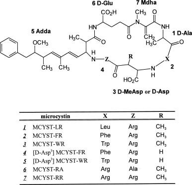
Toxic cyanobacteria have caused fatal poisoning of mammals, birds, and fish (Codd and Poon, 1988; Carmichael, 1992; Kotak et al., 1996; Tencalla and Dietrich, 1997), and may also have exerted adverse effects on human health (Gorham and Carmichael, 1988; Carmichael, 1994, 1997). Microcystins, a vast group of cyanobacterial toxins, are known to be hepatotoxic cyclic heptapeptides produced by some species within the genera of Microcystis (Shirai et al., 1991; Namikoshi et al., 1992; Luukkainen et al., 1994; Lee et al., 1998), Oscillotoria (Meriluoto et al., 1989; Luukkainen et al., 1993), Nostoc (Namikoshi et al., 1990; Sivonen et al., 1990; Beattie et al., 1998), and Anabaena (Harada et al., 1991a; Sivonen et al., 1992; Namikoshi et al., 1998). Toxic Microcystis species are widely distributed, mainly in the fresh water world, and form blooms frequently. The general structure of microcystins is composed of (2S, 3S, 8S, 9S)-3-amino-9-methoxy-2,6,8-trimethyl-10-phenyldeca-4,6-dienoic acid (Adda), g-linked D-glutamic acid (D-Glu), N-methyldehydroalanine (Mdha), and D-alanine (D-Ala). Four special amino acids are linked in sequential order followed by two variable L-amino acids with D-erythro-b-methylaspartic acid (D-MeAsp) inserted between them, forming a cyclic structure (Figure 1). There have been more than 60 different components isolated and determined. These compounds differ primarily in their L-amino acids at the number 2 and number 4 position of the

peptide ring skeleton, and in the presence or absence of the methyl group on D-MeAsp and/or Mdha of the 3rd and 7th amino acid, respectively (Namikoshi et al., 1992). L-amino acids in microcystins at the 2nd and 4th position are known to be arginine, leucine, tyrosine, phenylalanine, tryptophan and others. Among the identified microcystins, more than 70% of the structures contained L-Arg at the 4th position. Among these 7 cyclic-linked amino acids in microcystins, Adda has been thought of

3Corresponding author. Tel: +886-2-23639291-125; Fax: +886- 2-23629919; E-mail: unijohn@ccms.ntu.edu.tw

Figure 1. Structures of microcystins identified in this research.


Botanical Bulletin of Academia Sinica, Vol. 41, 2000

separation were combined and chromatographed on a 3 cm i.d. flash column, packed with 45 g silica gel for flash column (Baker, USA) and eluted with the same solution as the developer used in TLC, flow rate 36 ml/min. Toxic fractions were pooled into two major partitions, P-I (70 mg) and P-II (10 mg), according to their composition as determined by TLC analysis. Both partitions were subjected for further separation by reversed phase HPLC. P-I was first separated by a semi-preparative C18 column (10 × 250 mm, Vydac, USA) using 0.01 M ammonium acetate: acetonitrile (76:24, v/v) or 0.01 M ammonium acetate: methanol (50:50, v/v) as eluent to give semi-purified fractions. All these fractions were further purified using 0.05% aqueous trifluoroacetic acid: acetonitrile (62:38, v/v) as eluent on the same column to yield pure components: 1 (0.5 mg), 2 (2.5 mg), 3 (2.2 mg), 4 (0.3 mg), 5 (0.3 mg), and 6 (0.8 mg). From P-II, compound 7 (2.0 mg) was purified by elution of 0.01 M ammonium acetate: acetonitrile (78:22, v/v) on an Alltima C18 column (4.6 × 250 mm, Alltech, USA). All purified compounds were vacuum-dried and stored at -20°C before structural analysis.

Structural Elucidation

Structures of the compounds were elucidated by comparing their DQF-COSY nuclear magnetic resonance (NMR) spectra with that of MCYST-LR. The identification of MCYST-LR 1, isolated in this study was also confirmed by comparing its chromatographic characteristics with those of authentic MCYST-LR. NMR spectra of compound 1, 6, and 7 were measured on an FT-NMR spectrometer (AM-400, Bruker, Germany) using methanol-d4 as solvent and internal standard (d3.3), while the spectra of compounds 2, 3, 4, and 5 were acquired on a Bruker DMX-500 SB FT-NMR spectrometer. Molecular weight of each toxin was determined by G2025A matrix-assisted desorption and ionization-time of flight mass spectrometry (MALDI-TOF/MS, Hewlett-Packard, USA) using a-cyano-4-hydroxycinnamic acid (a-CHC) as the matrix. UV spectra of compounds 1 to 7 were measured on a U-2000 Spectrophotometer (Hitachi, Japan).

Amino Acid Analysis

Each toxin component was hydrolyzed by means of gas-phase hydrolysis in 500 µl of 7 M HCl with 10% trifluoroacetic acid containing 0.1% phenol at 158°C for 30 min (Chang and Liu, 1988). The released amino acids were derivatized with 4-dimethylaminoazobenzene-4'-sulfonyl chloride (DABS-Cl) to form colored DABS-amino acids, prior to HPLC analysis. HPLC separation of amino acids was performed on an Alltima C18 column (4.6 × 250 mm, Alltech, USA), eluted initially with a gradient of 15 to 40% of acetonitrile in 35 mM sodium acetate solution containing 4% N', N-dimethylformamide for 20 min, then eluted with a gradient of 40% to 70% for 12 min, and maintained at 70% for 2 min at a 1 ml/min flow rate. The DABS-derivatives of amino acids were detected at 436 nm for absorption.

as a key structural component for their biological activities (Dahlem, 1989; Harada et al., 1990a, b; Carmichael, 1992; Saito et al., 1994). Inhibitory activity on the protein phosphatase 1 and 2A of microcystins was generally recognized as the mechanism by which microcystins exerted their hepatotoxicity (Nishiwaki-Matsushima et al., 1992). As a result of these enzyme inhibitions in liver, hepatocyte shrinks and liver tissue dissociates, followed by a deadly internal hemorrhage (Runnegar and Falconer, 1986). Bury et al. (1996) and Zambrano and Canelo (1996) reported the fish mass mortality in the Microcystis blooming was due to the toxic effect of microcystins on the disruption of ion homeostasis of the internal medium in fish by blockage of gill function.

In this paper, we report the isolation and identification of seven different microcystins from cultured M. aeruginosa strain M.TN-2 isolated from an eel pond in southern Taiwan.

Materials and Methods

Culture of Microcystis aeruginosa M.TN-2

Microcystis aeruginosa strain M.TN-2 was isolated from an eel pond at Duujia in Tainan County in Taiwan, July 1993. This strain was identified according to description of the species in the Plankton Algae of Reservoirs in Taiwan (Moriwaka and Chyi, 1996) and in Mizuno (1980). Its toxicity was determined by mouse assay (Lee et al., 1998). The cyanobacteria culture was maintained in modified Fitzgerald media (Hughes et al., 1958) and kept at 23±1°C with an illumination of 26.4 µEin•m-2s-1, 12 h by fluorescent light bulbs. Cells of M.TN-2 were harvested in their late exponential phase and concentrated by continuous centrifugation, followed by lyophilization before extraction and isolation of microcystins, and high performance liquid chromatography (HPLC) analysis.

Microcystin Extraction and Purification

Dried cells (50 g) were extracted three times with 400 ml methanol for 30 min with sufficient mixing by a magnetic stirrer. The methanol extract was adjusted to 85% of the aqueous solution for hexane partition. The lower layer of the polar solution was then vacuum-evaporated to dryness (3.0 g). For each batch of chromatographic separations, 200 mg dried extract in 10 ml of methanol was applied.

The first separation was carried out by a Sephadex LH-20 column (3 × 55 cm, Pharmacia Biotech., Sweden) of gel filtration chromatography and eluted by MeOH with a flow rate of 13 ml/min. Fractions collected were checked for their compositions by thin layer chromatography (TLC) using Silica gel 60 F254 (Merck, Germany) plate (200 µm thickness) and ethyl acetate: isopropanol: H2O (8:5:3, v/v/v) for development. Contents of the toxic fractions were compared with an authentic sample of MCYST-LR (Sigma, USA). Vanillin-sulfuric acid charring and dark-box observation of UV absorption were applied in the detection of microcystins. Subsequently, toxin fractions from above


Lee and Chou — Microcystins from cultured Microcystis aeruginosa

Chiral Analysis of the Constituent Amino Acids

Configuration analysis of the amino acids of the acid hydrolysate was performed using a PFP-IPA amino acid derivatization kit (Alltech, USA), and the derivatives were analyzed using a gas chromatography/mass detector (GC/MS) system (5890 series II, Hewlett-Packard, USA). The amino acids derivatives were injected into a Heliflex Chirasil-Val glass capillary column (0.25 mm × 25 m, Alltech, USA) and eluted with helium (0.6 ml/min) for 4 min at 90°C, followed by a linear gradient of 90°C to 210°C for 30 min.

Results

Structure Elucidation of Compound 1~7

The structures of compounds 1 to 7 isolated from cultured M.TN-2 strain are shown in Figure 1. Compounds 1, 6, and 7 were identified as MCYST-LR, -RA, and -RR, respectively according to their DQF-COSY NMR and FAB/MS data assignments, which were repforted previously (Lee et al., 1998) and will not be described in detail here. However, before the compounds 2 to 5 could be elucidated, proton chemical shifts of MCYST-LR had to be fully assigned according to the homonuclear and heteronuclear correlation of proton and carbon-13 nuclei on this compound through extensive TOCSY, HMQC, and HMBC experiments of modern NMR spectroscopy. Structure of the newly isolated microcystins were then deduced from such an established database of NMR signals, including chemical shifts and splitting patterns of the authentics. In addition, the composition and stereochemistry of the resolved amino acids in these microcystins, based on high resolution liquid chromatographic and gas chromtographic separation of the acid hydrolysate of purified toxins, were further evidence in support of their structure determination.

Compounds 2 to 5 were collected from several batches of separation until they were sufficient in quantity for NMR experiments. NMR data of each compound from DQF-COSY experiments confirmed the presence of amino acid residues revealed in their amino acid analysis. It was noticed that the resonance levels of each H-2 in the MeAsp, Adda, and Glu moieties of compounds 2~5 were

slightly shifted to the levels of the corresponding protons in MCYST-LR measured in an earlier 1H NMR experiment (with the residual ammonium effect). A similar result was obtained in MCYST-LR when the compound was free from the contamination of ammonium acetate (Table 1).

Amino acid analysis of compound 2 revealed a composition of MeAsp, Glu, Ala, Arg, and Phe (Table 2), of which MeAsp, Glu, and Ala were determined as the D-amino acids according to the chiral separation of compound 2 acid hydrolysate by GC/MS. Adda and Mdha, the existence of which cannot be resolved from the amino acid analysis, were confirmed by comparing the NMR data with the previous peak assignment of authentic MCYST-LR. We concluded that compound 2 is MCYST-FR. The molecule ion peak of compound 2 was recorded at 1029 by MALDI/TOF mass spectrometer, which matched that of MCYST-FR as reported by Namikoshi et al. (1992).

Amino acid analysis of compound 3 gave only four amino acids, a phenylalanine less than compound 2, even with a molecular weight of 1068, higher than MCYST-FR from the MALDI-TOF/MS analysis (Table 2). 1H NMR data also showed the presence of Adda and Mdha in compound 3, just as in compound 2. In addition, 1H NMR peaks of d4.45 (H-2), d3.29 (H2-3), d7.30 (H-5), d7.63 (H-7), d6.99 (H-8), d7.06 (H-9), and d7.31 (H-10) of the compound suggested the presence of tryptophan molecule, which was supported by the absorption maximum (lmax) at 223 nm and the molecular weight of the compound. Moollan et al.

Table 1. Chemical shift differences (Dd > 0.1) of some specific protons in D-MeAsp3, Adda5, and D-Glu6 moieties of MCYST-LR caused by the existence of residual ammonium acetate in methanol-d4 while taking a NMR measurement at 500 Hz.

Proton location Solvent

MeOH-d4a

[D-MeAsp3] -H-2 -0.26

Dd [Adda5] -H-2 +0.35

[D-Glu6] -H-2 -0.25

[D-Glu6] -H-4a -0.12

aWith residual CH3COONH4.

Table 2. Stereochemistry of the amino acids in different microcystins (denoted as compounds 1~7 accordingly) listed with the protonated molecular ions and lmax from MALDI-TOF mass and UV spectroscopic measurements.

Microcystins Amino acid analysisa M+H+ lmax (m/z) (nm)

1 MCYST-LR D-MeAsp D-Glu D-Ala Arg L-Leu 995 238

2 MCYST-FR D-MeAsp D-Glu D-Ala Arg L-Phe 1029 238

3 MCYST-WR D-MeAsp D-Glu D-Ala Arg 1068 223

4 [D-Asp3] MCYST-FR D-Asp D-Glu D-Ala Arg L-Phe 1015 238

5 [D-Asp3] MCYST-WR D-Asp D-Glu D-Ala Arg 1054 223

6 MCYST-RA D-MeAsp D-Glu D-Ala Arg L-Ala 953 238

7 MCYST-RR D-MeAsp D-Glu D-Ala Arg Arg 1038 238

aAmino acid composition and stereochemistry were determined by the high performance liquid and gas chromatographic separation on the acid hydrolysate of microcystins. Stereochemistry of arginine moiety in microcystins could not be assigned by the method in this research.


Botanical Bulletin of Academia Sinica, Vol. 41, 2000

(1996) have reported the tryptophan chromophore in microcystin causes a shift from 238 nm, a typical MCYST lmax, to 222 nm. Compound 3 was thus assigned as MCYST-WR.

Compound 4 had a molecular weight of 1015, 14 daltons (CH2) less than that of MCYST-FR. Amino acid analysis of compound 4 showed the same composition as MCYST-FR from both LC and GC studies except its D-MeAsp was replaced by D-Asp (Table 2). Due to the small quantity of sample available and the embedded signals, peaks of H-2 and H-3 of D-Asp could not be clearly identified in its 1H NMR spectrum. However, all other proton signals from Ala, Phe, Arg, Adda, Glu, and Mdha amino acid moieties of compound 4 were able to be assigned completely and be in consistency with those of MCYST-FR. In addition, missing of the significant H3-5 signal in the 1H NMR spectrum and the crossed signal of H-3 and H3-5 coupling peaks of MeAsp in the DQF-COSY NMR spectrum revealed compound 4 [D-Asp3] MCYST-FR, a desmethyl variant of compound 2.

Compound 5 showed a UV maximal absorption at 223 nm, the same as MCYST-WR, suggesting a similar presence of tryptophan in the amino acid composition as MCYST-WR of compound 5. Molecular weight (1054, 12 daltons less than 1068 of MCYST-WR) from MALDI-TOF/MS and the 1H NMR signals of tryptophan moiety similar to those of MCYST-WR showed in the experiments of compound 5, confirming it as a desmethyl variant of MCYST-WR, [D-Asp3] MCYST-WR.

Discussion

Structures of compounds 1~7 were mainly determined by their 1H NMR and DQF-COSY NMR data. The elucidated structures were further confirmed by their MALDI-TOF/MS and amino acid analysis. Two-dimensional DQF-COSY NMR spectra gave strong evidence in determining the correlated protons within the molecule of these cyclic heptapeptides. Thus the proton signals within and the nature of each amino acid moiety of the toxin were assigned. The amino acid sequence of the cyclic peptides was speculated to be identical in all the toxins due to the similar proton signals among the respective amino acids in different toxins. Identification of the isolated microcystins was confirmed by comparing the chemical shifts and splitting patterns of peaks with those of the published ones (Namikoshi et al., 1990; Harada et al., 1991b; Namikoshi et al., 1992).

Table 1 showed the difference in chemical shifts of each H-2 proton in the MeAsp, Adda and Glu moieties of MCYST-LR, with and without the residual ammonium effect during its NMR measurement. This is an experiment designed to explain the shifted energy levels of the respective protons in compounds 2~5, while they were measured for the chemical shifts right after the isolated using ammonium acetate as eluent (Lee et al., 1998). The difference in chemical shifts of certain protons was due to the deprotonation reaction of the residual ammonium ions on

the free carboxylic acid of MeAsp and Glu. It was reported that deprotonation of carboxylic acid in Glu and MeAsp moieties occurred when ND3/D2O solution was added to the deuteriated solvent in the NMR measurment, and led to the signal shifts of certain protons near the affected functional group (Rudolph-Böhner et al., 1993). In this study we observed an up-field shift of the H-2 proton signals in Glu and MeAsp and a down-field shift of the H-2 proton signal in Adda of the microcystins deprotonated. We speculate that a conformational change may have happened to the molecule due to the coincident shift of the H-2 of Adda while deprotonations were occurring on the free acid groups. The spatial distance of the H-2 of Adda to the nearby carboxylic acid group seems to rule out bond transmission. What kind of conformational change induced the signal shift of the H-2 in Adda due to deprotonation of the carboxylic acid groups needs further study.

Both MCYST-FR and its desmethyl variant [D-Asp3] MCYST-FR were found in the same culture in this study, as were MCYST-WR and its desmethyl variant [D-Asp3] MCYST-FR. Although MCYST-WR has been described by Namikoshi et al. (1992), and MCYST-FR by Luukkainen et al. (1994), the [D-Asp3] MCYST-FR and [D-Asp3] MCYST-WR found in this study are new, emerging compounds to this vast group of heptapeptides. Ratio of the content of microcystin to its desmethylaspartic acid variant seems to be regulated by the same factors in living cells since they are identical in both MCYST-FR and MCYST-WR pairs. However, this hypothesis needs further confirmation. The question has been raised as to whether the desmethylaspatic acid variant of MCYST-LR or MCYST-RR is produced in the same culture. A careful review of the toxin profile confirmed that desmethylaspartic acid derivatives of MCYST-LR and MCYST-RR were not produced by the cell culture. It is not necessary for the peptide synthetase responsible for the formation of [D-Asp3] MCYST-FR and [D-Asp3] MCYST-WR to have the same function on MCYST-LR and MCYST-RR in the same organism, although [D-Asp3] MCYST-LR and [D-Asp3] MCYST-RR are quite common in certain strains of M. aeruginosa (Rinehart et al., 1994). From the list summarized by Rinehart et al. (1994), we can also find that most of the reported desmethyl replacement in MeAsp and Mdha residue of microcystins are found in cyanobacteria from Finland and Norway, while a small portion of the desmethyl microcystins were in Microcystis from Japan. Our report enriched the toxin list studied in Asia.

It was reported that MCYST-LR is the most common component among the known microcystins in the studied cyanobacterial strains all over the world (Carmichael et al., 1988; Watanabe et al., 1991; Namikoshi et al., 1992). We also found that MCYST-RR was also a major component, in addition to MCYST-LR, in most of the Microcystis strains isolated in Taiwan (Lee et al., 1999). However, in M.TN-2 strain MCYST-LR was found to be a minor component. This was the reason we selected this strain in our search for new toxin molecules.


Lee and Chou — Microcystins from cultured Microcystis aeruginosa

Recent studies showed a non-ribosomal thio-template mechanism, namely the peptide synthetase, a multifunctional enzyme, was responsible for the production of different microcystins (Arment and Carmichael, 1996), and its genes were identified (Dittmann et al., 1997; Nishizawa et al., 1999). With the help of various Microcystis strains that produce diferent microcystins, the biosynthetic regulation of the synthesis of various toxin derivatives will be clarified.

Acknowledgement. Grant support from the National Science Council (NSC88-2312-B-002-039) of Taiwan was appreciated. We are grateful to Miss Huang Shoou-Ling for the NMR data acquisition in the Instrumentation Center of College of Science, NTU.

Literature Cited

Arment, A.R. and W.W. Carmichael. 1996. Evidence that microcystin is a thio-template product. J. Phycol. 32: 591-597.

Beattie, K.A., K. Kaya, T. Sano, and G.A. Codd. 1998. Three dehydrobutyrine-containing microcystins from Nostoc. Phytochemistry 47: 1289-1292.

Bury, N.R., G. Flik, F.B. Eddy, and G.A. Codd. 1996. The effects of cyanobacteria and the cyanobacterial toxin microcystin-LR on Ca2+ transport and Na+/K+-ATPase in the Tilapia gills. J. Exp. Biol. 199: 1319-1326.

Carmichael, W.W., J.-W. He, J. Eschedor, Z.-R. He, and Y.-M. Juan. 1988. Partial structural determination of hepatotoxic peptides from Microcystis aeruginosa (cyanobacterium) collected in ponds of central China. Toxicon 26: 1213-1217.

Carmichael, W.W. 1992. Cyanobacteria secondary metabolites - the cyanotoxins. J. Appl. Bacteriol. 72: 445-459.

Carmichael, W.W. 1994. The toxins of cyanobacteria. Sci. Amer. 270: 64-72.

Carmichael, W.W. 1997. The cyanotoxins. Adv. Bot. Res. 27: 211-256.

Chang, C.-S. and C.-S. Liu. 1988. A picomole-level amino acid analysis by means of gas-phase hydrolysis and DABS-Cl/HPLC method. J. Chinese Biochem. Soc. 17: 12-19.

Codd, G.A. and G.K. Poon. 1988. Cyanobacterial toxins. Proc. Phytochem. Soc. Eur. 28: 283-296.

Dahlem, A.M. 1989. Ph.D. thesis. University of Illinois, Urbana-Champaign.

Dittmann, E., B.A. Neilan, M. Erhard, H.V. Döhren, and T. Börner. 1997. Insertional mutagenesis of a peptide synthetase gene that is responsible for hepatotoxin production in the cyanobacterium Microcystis aeruginosa PCC 7806. Mol. Microbiol. 26: 779-787.

Gorham, P.R. and W.W. Carmichael. 1988. Hazards of freshwater blue green algae (Cyanobacteria). In C. A. Lembi and J. R. Waaland (eds.), Algae and Human Affairs, Cambridge, pp. 403-431.

Harada, K.-I., K. Matsuura, M. Suzuki, M.F. Watanabe, S. Oishi, A.M. Dahlem, V.R. Beasley, and W.W. Carmichael. 1990a. Isolation and characterization of the minor components associated with microcystins LR and RR in the cyanobacterium (blue-green algae). Toxicon 28: 55-64.

Harada, K.-I., K. Ogawa, K. Matsuura, H. Murata, M. Suzuki,

M.F. Watanabe, Y. Itezono, and N. Nakayama. 1990b. Structural determination of geometrical-isomers of microcystin-LR and -RR from cyanobacteria by two-dimensional NMR spectroscopic techniques. Chem. Res. Toxicol. 3: 473-481.

Harada, K.-I., K. Ogawa, Y. Kimura, H. Murata, M. Suzuki, P.M. Thorn, W.R. Evans, and W.W. Carmichael. 1991a. Microcystins from Anabaena flos-aquae NRC 525-17. Chem. Res. Toxicol. 4: 534-540.

Harada, K.-I., K. Ogawa, K. Masuura, H. Nagai, H. Murata, M. Suzuki, Y. Itezono, N. Nakayama, M. Shirai, and M. Nakano. 1991b. Isolation of two toxic heptapeptide microcystins from an axenic strain of Microcystis aeruginosa, K-139. Toxicon 29: 479-489.

Hughes, E.O., P.R. Gorham, and A. Zehnder. 1958. Toxicity of a unialgal culture of Microcystis aeruginosa. Can. J. Microbiol. 4: 225-236.

Kotak, B.G., S. Semalulu, D. L. Fritz, E.E. Prepas, S.E. Hrudey, and R.W. Coppock. 1996. Hepatic and renal pathology of intraperitoneally administered microcystin-LR in rainbow trout (Oncorhynchus mykiss). Toxicon 34: 517-525.

Lee, T.-H., Y.-M. Chen, and H.-N. Chou. 1998. First report of microcystins in Taiwan. Toxicon 36: 247-255.

Lee, T.-H., Y.-M. Chen, and H.-N. Chou. 1999. Toxicity assay of cyanobacterial strains using Artemia salina in comparison with the mouse bioassay. Acta Zool. Taiwan. 10: 1-9.

Luukkainen, R., K. Sivonen, M. Namikoshi, M. Färdig, K.L. Rinehart, and S.I. Niemelä. 1993. Isolation and identification of eight microcystins from thirteen Oscillatoria agardhii strains and structure of a new microcystin. Appl. Environ. Microbiol. 59: 2204-2209.

Luukkainen, R., M. Namikoshi, K. Sivonen, K.L. Rinehart, and S.I. Niemelä. 1994. Isolation and identification of 12 microcystins from four strains and two bloom samples of Microcystis spp.: structure of a new hepatotoxin. Toxicon 32: 133-139.

Meriluoto, J.A.O., A. Sandström, J.E. Eriksson, G. Remaud, A.G. Craig, and J. Chattopadhyaya. 1989. Structure and toxicity of a peptide hepatotoxin from the cyanobacterium Oscillatoria agardhii. Toxicon 27: 1021-1034.

Mizuno, T. 1980. Illustrations of the freshwater plankton of Japan. Hoikusha Publishing, Osaka.

Moollan, R.W., B. Rae, and A. Verbeek. 1996. Some comments on the determination of microcystin toxins in waters by high-performance liquid chromatography. Analyst 121: 233-238.

Moriwaka, M. and J. Chyi. 1996. Plankton Algae of Reservoirs in Taiwan. National Institute Environmental Analysis, EPA, Taipei, R.O.C.

Namikoshi, M., K.L. Rinehart, R. Sakai, K. Sivonen, and W.W. Carmichael. 1990. Structures of three new cyclic heptapeptide hepatotoxins produced by the cyanobacterium (blue-green alga) Nostoc sp. strain 152. J. Org. Chem. 55: 6135-6139.

Namikoshi, M., K.L. Rinehart, R. Sakai, R.R. Stotts, A.M. Dahlem, V.R. Beasley, W.W. Carmichael, and E.R. Evans. 1992. Identification of 12 hepatotoxins from a Homer bloom of the cyanobacteria Microcystis aeruginosa, Microcystis viridis, and Microcystis wesenbergii: nine new microcystins. J. Org. Chem. 57: 866-872.

Namikoshi, M., M. Yuan, K. Sivonen, W.W. Carmichael, K.L. Rinehart, L. Rouhiainen, F. Sun, S. Brittain, and A. Otsuki.


Botanical Bulletin of Academia Sinica, Vol. 41, 2000

Shirai, M., A. Ohtake, T. Sano, S. Matsumoto, T. Sakamoto, A. Sato, A. Aida, K.-I. Harada, T. Shimada, M. Suzuki, and M. Nakano. 1991. Toxicity and toxins of natural blooms and isolated strains of Microcystis spp. (cyanobacteria) and improved procedure for purification of cultures. Appl. Environ. Microbiol. 57: 1241-1245.

Sivonen, K., W.W. Carmichael, M. Namikoshi, K.L. Rinehart, A.M. Dahlem, and S.I. Niemelä. 1990. Isolation and characterization of hepatotoxic microcystin homologs from the filamentous freshwater cyanobacterium Nostoc sp. strain 152. Appl. Environ. Microbiol. 56: 2650-2657.

Sivonen, K., M. Namikoshi, W.R. Evans, W.W. Carmichael, F. Sun, L. Rouhiainen, R. Luukkainen, and K.L. Rinehart. 1992. Isolation and characterization of a variety of microcystins from seven strains of the cyanobacterial genus Anabaena. Appl. Environ. Microbiol. 58: 2495-2500.

Tencalla, F. and D. Dietrich. 1997. Biochemical characterization of microcystin toxicity in rainbow trout (Oncorhychus mykiss). Toxicon 35: 583-595.

Watanabe, M.F., M. Watanabe, T. Kato, K.-I. Harada, and M. Suzuki. 1991. Composition of cyclic toxins among strains of Microcystis aeruginosa (blue-green algae, cyanobacteria). Bot. Mag. Tokyo 104: 49-57.

Zambrano, F. and E. Canelo. 1996. Effects of microcystin-LR on the partial reactions of the Na+-K+ pump of the gill of carp (Cyprinus carpio Linneo). Toxicon 34: 451-458.

1998. Seven new microcystins possessing two L-glutamic acid units, isolated from Anabaena sp. strain 186. Chem. Res. Toxicol. 11: 143-149.

Nishiwaki-Matsushima, R., T. Ohta, S. Nishiwaki, M. Suganuma, K. Kohyama, T. Ishikawa, W.W. Carmichael, and H. Fuji. 1992. Liver tumor promotion by the cyanobacterial cyclic peptide toxin microcystin-LR. J. Cancer Res. Clin. Oncol. 118: 420-424.

Nishizawa, T., M. Asayama, K. Fujii, K.-I. Harada, and M. Shirai. 1999. Genetic analysis of the peptide synthetase genes for a cyclic heptapeptide microcystin in Microcystis spp. J. Biochem. 126: 520-529.

Rinehart, K.L., M. Namikoshi, and B.W. Choi. 1994. Structure and biosynthesis of toxins from blue-green algae (cyanobacteria). J. Appl. Phycol. 6: 159-176.

Rudolph-Böhner, S., J.-T. Wu, and L. Moroder. 1993. Identification and characterization of microcystin-LY from Microcystis aeruginosa (strain 298). Biol. Chem. Hoppe-Seyer 374: 635-640.

Runnegar, M.T.C. and I.R. Falconer. 1986. Effect of toxin from the cyanobacterium Microcystis aeruginosa on ultrastructural morphology and actin polymerization in isolated hepatocytes. Toxicon 24: 109-115.

Saito, S., Y. Nakano, K. Kushida, M. Shirai, K.-I. Harada, and M. Nakano. 1994. Cross-reactivity and neutralizing ability of monoclonal antibodies against microcystins. Microbiol. Immunol. 38: 389-392.

»Éºñ·LÅnĦ®è M.TN-2 ©Ò§t¤CºØ·LÅnĦ¬rªº¤ÀÂ÷

»P¤Æ¾Çµ²ºcŲ©w

§õ©vÀ²1 ©P§»¹A2

1 ¤¤¥¡¬ã¨s°|´Óª«¬ã¨s©Ò

2 °ê¥ß»OÆW¤j¾Çº®·~¬ì¾Ç¬ã¨s©Ò

1993 ¦~®L©u³æÂ÷¦Û»OÆW«n³¡ÅÁ³½¦À»Éºñ·LÅnĦµØ (Microcystis aeruginosa) ¤§ M.TN-2 Ħ®è¡A¸g¤j¶q °ö¾i«á¡AĦÅ餺©Ò§t¤§·LÅnĦ¬r (microcystin, MCYST) ¸g¤@¨t¦Cªº LH-20 ºÞ¬W½¦Âo¼hªR¡Bª¿½¦§lªþ©Ê³t ¤ÀºÞ¬W¼hªR¤Î°f¬Û°ª®Ä²G¬Û¼hªRµ¥ªº¤ÀÂ÷¡B¯Â¤Æ¡A¦@¤ÀÂ÷¥X¤CºØ¤£¦Pªº·LÅnĦ¬r¡A¨äµ²ºcÄò¸gµµ¥~¥úÃСB ¤¶½è»²§U¿E¥ú¥h§lªþ¤ÎÂ÷¤l¤Æ­¸¦æ®É¶¡½èÃÐ (MALDI-TO/FMS)¡B1H ©M DQF-COSY ®ÖºÏ¦@®¶ªiÃЪº¸Ñ ªR¡A¦õ¥H®ò°ò»Ä²Õ¦¨ªº°ª®Ä²G¬Û¼hªR»P¤â©Ê (chirality) ¤ÀÂ÷ªº®ð¬Û¼hªR¡A©l½T»{¨äµ²ºc¤À§O¬° MCYST-LR¡B-RR¡B-RA¡B-FR¡B-WR ¤Î [D-Asp3] MCYST-FR¡B-WR¡A¨ä¤¤ [D-Asp3] MCYST-FR ¤Î -WR ¬°¹L¥h¤åÄm¤¤©Ò¥¼´¿µoªíªº·s¤Æ¦Xª« ¡C

ÃöÁäµü¡G »Éºñ·LÅnĦ¡F·LÅnĦ¬r¡F¨xŦ¬r¯À¡FÂźñ²Óµß ¡C