Bot. Bull. Acad. Sin. (2000) 41: 315-322

Chen and Tzean — Conioscypha taiwaniana sp. nov. and several new records of the genus from Taiwan

Conioscypha taiwaniana sp. nov. and several new records of the genus from Taiwan

Jin-Liang Chen1 and Shean-Shong Tzean2

1Department of Hospital and Health Care Administration, Chia-Nan University of Pharmacy and Science, Tainan, Taiwan 717, ROC

2Department of Plant Pathology, National Taiwan University, Taipei, Taiwan 106, ROC

(Received October 18, 1999; Accepted February 14, 2000)

Abstract. Five dematiaceous hyphomycetes of Conioscypha, which were collected in Taiwan, are presented in this study. Conioscypha taiwaniana sp. nov. is described and illustrated. Conioscypha hoehnelii, C. japonica, and C. lignicola are recorded for the first time in Taiwan, and C. bambusicola is proposed as an additional record.

Keywords: Conioscypha taiwaniana sp. nov.; Conioscypha bambusicola; Conioscypha hoehnelii; Conioscypha japonica; Conioscypha lignicola; Hyphomycetes; Taxonomy; Taiwan.

Introduction

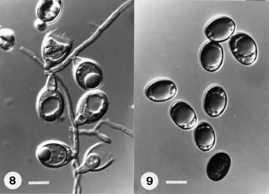
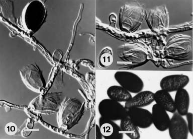
Höhnel (1904) established a new genus Conioscypha with Conioscypha lignicola Höhnel, as the type species. Shearer (1973) reviewed previous studies and provided a revised description of C. lignicola, then published a second species C. varia Shearer (Shearer, 1973). Notable characteristics of Conioscypha include enteroblastic conidiogenesis, compact, erumpent colonies; immersed mycelium; hyaline, lateral or terminal, short-stalked sessile or intercalary, percurrent conidiogenous cells with a conspicuous multilayered cup-like collarette and dark brown, 1-celled conidia (Shearer, 1973). Later, five species, C. bambusicola Matsushima, C. dimorpha Matsushima, C. fabiformis Matsushima, C. hoehnelii P.M. Kirk and C. japonica S.I. Udagawa & N. Toyazaki, were added to this genus. This brought the total number of species in Conioscypha to seven (Matsushima, 1975, 1993, 1996; Udagawa and Toyazaki, 1983; Kirk, 1984). Conioscypha bambusicola, is the only species initially described from Taiwan (Matsushima, 1980).

During studies of hyphomycetes from rotten vegetation in Taiwan, four species of Conioscypha were collected from different sources. Conioscypha bambusicola and C. lignicola were from rotten twigs or leaves of Phyllostachys pubescens. Conioscypha hoehnelii and C. japonica were from herbaceous rotten stems, and a previously undescribed fungus was isolated from decaying stems in Jenai, Nantou Hsien. This new fungus fitted the generic description of Conioscypha and was easily distingnished from other known species of this genus. Therefore, Conioscypha taiwaniana sp. nov. is proposed. Conioscypha hoehnelii, C. japonica and C. lignicola are recorded for the first time in Taiwan, and an additional record of C. bambusicola, is provided with detailed description.

Materials and Methods

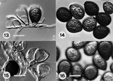
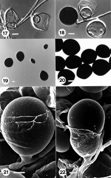
Samples collected from various rotten vegetation in Taiwan were incubated in moist chambers (plastic boxes, 30 × 20 × 12 cm, with three layers of moistened papers) for fungal sporulation. Pure culture was established by isolating a single spore or spores on 3% water agar with a sterile glass microneedle. A piece of agar containing isolated spores was transferred to oat meal agar (OMA) slants or plates under a stereomicroscope. Details of fungal characteristics and conidiogenesis were recorded and photographed with an Olympus light microscope (BH-2). Material preparation for scanning electron microscopy was as described previously by Tzean and Estey (1978). All specimens are deposited in Department of Plant Pathology, National Taiwan University, Taipei, Taiwan, ROC (TNTU).
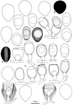
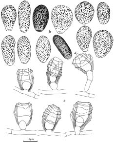
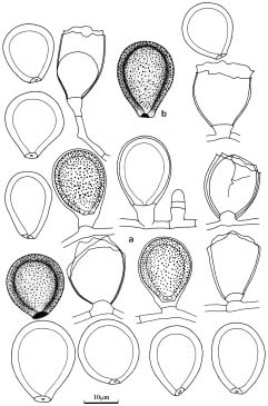
Taxonomy

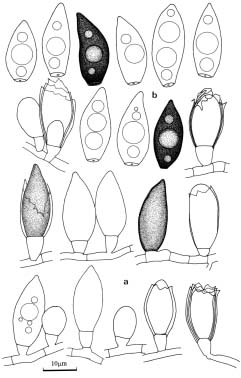
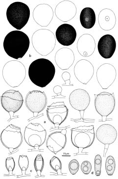
Conioscypha bambusicola Matsushima, 1975. Icones Microfungorum a Matsushima Lectorum (I). p. 38.
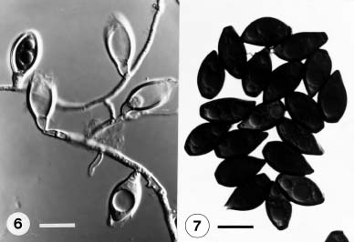
(Figures 1, 6-7)

Colony diameter on oat meal agar, 27 mm in 33 days at 25°C, velvety, olive brown to brownish grey, white at the margin; reverse brownish grey to olive brown or greyish brown. Mycelium immersed, composed of branched, septate, smooth, hyaline to subhyaline, 0.8-2.6 µm wide hyphae. Conidiophores semimacronematous, micronematous, mononematous, smooth, hyaline. Conidiogenous cells percurrent, cuneiform, smooth, hyaline, 1.6-8.0 × 2.3-4.8 µm, often with conspicuously multilayered collarette remaining at the apex; multicollarette cup-shaped, 6.8-8.8 µm wide. Conidia ovoid or broadly obclavate, truncate at the base, often ta


Botanical Bulletin of Academia Sinica, Vol. 41, 2000

Figure 1. a-b, Conioscypha bambusicola. (a) young conidia and conidiogenous cells initiated from the hyphae; (b) mature conidia with 2-4 oil droplets. Scale bar = 10 µm.

Figure 2. a-b, Conioscypha hoehnelii. (a) young conidia initiated from the hyphae and conidiogenous cells; (b) mature conidia with 1 oil droplets. Scale bar = 10 µm.

pering towards a point at the apex, smooth, olive brown to yellowish brown or dark brown, 14.1-20.0 × 6.4-8.0 µm.

Specimens examined. On a twig of Phyllostachys pubescens, Huisun, Nantow Pref., Feb. 10 1993. leg. J.L. Chen. TNTU 1040 (dried culture); on rotten stem, Hsiaoyehliu, Jul. 13 1997. Leg. J.L. Chen. CTN-68 (dried culture).

Note. Most of Conioscypha bambusicola isolates were collected from Phyllostachys spp. in Taiwan. Our species is similar to the type species of C. bambusicola (conidia up to 16 µm long; Matsushima, 1975), but has longer conidia (TNTU 1040: up to 20 µm long; CTN-68: up to 25.6 µm long).

Conioscypha hoehnelii Kirk, 1984. Trans. Br. Mycol. Soc. 82(1): 177-178. (Figures 2, 8-9)

Colony diameter on oat meal agar, 55 mm in 128 days at 25°C, plane, granulate, brown to dark brown or black; reverse dark brown to black. Mycelium immersed, composed of branched, septate, smooth, hyaline to subhyaline, 0.8-4.0 µm wide hyphae. Conidiophores semimacronematous to micronematous, mononematous. Conidiogenous cells cuneiform, cylindrical, often with a conspicuous cup-shaped multicollarette at the apex,

Figure 3. a-b, Conioscypha japonica. (a) conidiogenous cells; (b) mature conidia with pigments deposited irregulatly at the periphery of the wall to give the apearance of roughness. Scale bar = 10 µm.


Chen and Tzean — Conioscypha taiwaniana sp. nov. and several new records of the genus from Taiwan

ellipsoid, smooth but with pigments deposited, pale brown to yellowish brown or dark brown, truncated with a central pore at the base, 11.2-20.8 × 5.6-13.6 µm.

Specimens examined. On a rotten herbaceous stem, Wulai, Taipei Pref., May 22, 1993, leg. J.L. Chen. TNTU 1103 (dried culture).

Note. The conidia of our isolate (11.2-20.8 × 5.6-13.6 µm) are larger than the type species of C. japonica (7-14 × 4.5-10 µm; Udagawa & Toyazaki, 1983).

Conioscypha lignicola Höhnel, 1904. Ann. Mycol. 2: 58-59. (Figures 4, 13-16)

Colony diameter on oat meal agar, 33 mm 39 days at 25°C, velvety, olive brown, margin yellowish white to yellowish grey; reverse yellowish white to brownish grey or dark olive brown. Mycelium immersed, composed of branched, septate, smooth, hyaline to subhyaline, 0.8-2.6 µm wide hyphae. Conidiophores micronematous, semimacronematous, mononematous. Conidiogenous

Figure 4. a-b, Conioscypha lignicola. (a) young conidia initiated from the hyphae and conidiogenous cells; (b) mature conidia thick-walled, smooth with pigmented roughness. Scale bar = 10 µm.

percurrent, smooth, hyaline, 2.8-15.2 × 3.2-4.0 µm. Conidia endogenous, smooth, obovoid, napiform, subglobose, ellipsoidal, brown to dark brown, often truncated at the base, 10.8-17.2 × 10.4-14.4 µm.

Specimens examined. On a herbaceous stem, Huisun, Nantow Pref., Feb. 10 1993. leg. J.L. Chen. TNTU 1039 (dried culture).

Note. Although characteristics of the Taiwanese isolate are close to the type species of C. hoehnelii (conidia 10-20 × 8-15 µm; Kirk, 1984), the Taiwanese isolate has smaller conidia (10.8-17.2 × 10.4-14.4 µm).

Conioscypha japonica Udagawa & Toyazaki, 1983. Mycotaxon 18: 131-137. (Figures 3, 10-12)

Colony diameter on oat meal agar, 47-51 mm in 58 days at 25°C, effuse, plane, zonate, orange white to brownish grey brown; reverse pale orange to greyish brown. Mycelium immersed, composed of branched, septate, smooth, hyaline to subhyaline, 0.8-3.2 µm wide hyphae. Conidiophores micronematous, semimacronematous, mononematous. Conidiogenous cells terminal to lateral or lateral arising directly from the hyphae, percurrent, smooth, hyaline, 4.0-17.6 × 3.2-3.8 µm, with a multilayered cup-like collarette at the apex. Conidia endogenous, obpyriform, obovoid to broad ellipsoidal or elongate-

Figure 5. a-d, Conioscypha taiwaniana. (a) young conidia initiated from the hyphae and conidiogenous cells; (b) mature conidia flattened in one plane, napiform, globose to subglobose, black brown to black with truncate the base; (c-d) smaller conidiogenous cells and conidia with 1-2 oil droplets. Scale bar = 10 µm.


Botanical Bulletin of Academia Sinica, Vol. 41, 2000

cells mostly cuneiform, doliiform, smooth, hyaline, 1.6-4.8 × 2.8-6.8 µm, percurrent, often with a cup-shaped multicollarette up to 16.0 µm wide at the apex. Conidia ovoid to obpyriform or subglobose, thick-walled, smooth, but with conspicuous pigmented roughness or scattered warts in maturity, pale olivaceous brown to dark brown, 11.0-21.6 × 10.6-16.8 µm, often truncated and with a dark scar at the base.

Specimens examined. On a rotten leaf of Phyllostachys pubescens, Huisun, Nantow Pref., Feb. 10 1993. leg. J.L. Chen. TNTU 1042 (dried culture).

Note. The Taiwanese isolate resembles the type species of C. lignicola (conidia 12-22.6 × 11-22 µm; Shearer, 1973), but has smaller conidia (11.0-21.6 × 10.6-16.8 µm).

Figures 6-7. Conioscypha bambusicola. (6) conidiogenous cells; (7) mature conidia with oil droplets. Scale bar = 10 µm.

Figures 8-9. Conioscypha hoehnelii. (8) conidiogenous cells; (9) conidia with oil droplets. Scale bar = 10 µm.


Chen and Tzean — Conioscypha taiwaniana sp. nov. and several new records of the genus from Taiwan

Conioscypha taiwaniana J.L. Chen and S.S. Tzean sp. nov. (Figures 5, 17-22)

Coloniae diametro in OMA, 30-32 mm in 7 diebus ad 25°C, effusae, velutinuae, albusae ad aterae; reversae pallidulae flavidae albusae ad atrogriseae vel olivaceae griseae. Mycelium immersum, ex hyphis ramosis, septatis, lenibus, hyalinis, 1.1-2.8 µm latis compositum. Conidiophora micronematoidea, semimacronematoidea,

mononematoidea. Cellulae conidiogenae cuneiformes, percurrentes, laeves, hyalinae, 2.8-6.4 × 4.0-7.2 µm, cum collis capularibus multiplicibus usque ad 25.0 µm latis ad apicem. Conidia enteroblastica, 1-cellularia, laevia, basilaria truncata, applanata in uno planis, in aspectu frontali: napiformia, globosa ad subglobosa vel ovoidea, atrobrunnea vel atra, 17.0-29.0 × 15.0-24.0 µm; in aspectu laterali: late ellipsoidea, usque ad 12.0-18.0 µm crassa;

Figures 10-12. Conioscypha japonica. (10-11) conidiogenous cells; (12) mature conidia smooth with pigmented roughness. Scale bar = 10 µm.

Figures 13-16. Conioscypha lignicola. (13,15) conidiogenous cells; (14,16) mature conidia thick-walled, smooth with pigmented roughness . Scale bar = 10 µm.


Botanical Bulletin of Academia Sinica, Vol. 41, 2000

Figures 17-22. Conioscypha taiwaniana. (17-18) conidiogenous cells; (19-20) mature conidia napiform, globose to subglobose, black brown to black; (21-22) conidiogenous cells (SEM). Scale bars: 17-20 = 10 µm; 21-22 = 2 µm.


Chen and Tzean — Conioscypha taiwaniana sp. nov. and several new records of the genus from Taiwan

conidia secunda ellipsoidea, subglobosa ad globosa vel obovoidea, laevia, subflava brunea ad brunea, 10.8-19.2 × 5.6-13.4 µm.

Holotypus in caudicibus putrescentibus, Jenai, Nantou, Taiwan, 10 Feb. 1993. TNTU 1053.

Colony diameter on oat meal agar, 30-32 mm in 48 days at 25°C, effuse, velvety, black at the submargin, white at the center and margin; reverse pale yellowish white at the center and margin, dark grey or olive grey at the submargin. Mycelium immersed, composed of branched, septate, smooth, hyaline 1.1-2.8 µm wide hyphae. Conidiophores micronematous, semi-macronematous, mononematous. Conidiogenous cells cuneiform, percurrent, smooth, hyaline, 2.8-6.4 × 4.0-7.2 µm, with a multilayered cup-like collarette up to 25.0 µm wide at the apex. Conidia enteroblastic, 1-celled, smooth, truncate at the base with a central pore, moderately flattened in one plane, in front view: napiform, globose to subglobose or ovoid, black brown or black, 17.0-29.0 × 15.0-24.0 µm, in lateral view: broadly ellipsoidal, up to 12.0-18.0 µm wide, pale brown to brown at the center, dark or black brown at both sides; often with smaller, ellipsoidal, subglobose to globose or obovoid, smooth, yellowish brown to brown secondary conidia, 10.8-19.2 × 5.6-13.4 µm.

Specimens examined. On a decaying stem, Jenai, Nantou Pref., Taiwan, ROC Feb. 10 1993, holotype TNTU 1053 (dried culture), deposited in the Department of Plant Pathology, National Taiwan University, Taipei, Taiwan, ROC.

Conioscypha taiwaniana is similar to C. hohnelii, but can be distinglished by the larger, black conidia, the wider multilayered cup-like collarette, and secondary conidia. In 1996, Matsushima published a new species of

Conioscypha, C. dimorpha Matsushima, which has two kinds of conidia. Conioscypha taiwaniana is closely related to C. dimorpha, but C. taiwaniana can be distinguished by the larger conidia and lack of microconidia. Differences between C. taiwaniana and other species of Conioscypha include the shape, size, color, and walled-ornamentation of conidia. Pigments which are roughly deposited and present in the walls of the conidia of C. lignicola and C. japonica cannot be observed in the conidial walls of C. taiwaniana. Conioscypha bambusicola is the only species having more or less ovate conidia, which are apically pointed. The conidia of C. varia are smaller, ovoid, flammiform, naviculiform, or subellipsoid. A comparative summary of the conidial morphology of Conioscypha taiwaniana and the seven closely related Conioscypha species is presented in Table 1.

Acknowledgements. We are grateful for the support of the National Science Council, ROC, by grant (NSC-82-0409-B002-099) to S.S. Tzean.

Literature Cited

Höhnel, F.X.R. von. 1904. Mykologische Fragmente. Annales Mycologici 2: 38-60.

Kirk, P.M. 1984. New or interesting microfungi XII. A new species of Conioscypha (Hyphomycetes). Trans. Br. Mycol. So. 82: 177-178.

Matsushima, T. 1975. Icones Microfungorum a Matsushima Lectorum. Matsushima, Kobe, Japan.

Matsushima, T. 1980. Matsushima Mycological Memoirs No. 1. Matsushima, Kobe, Japan.

Matsushima, T. 1993. Matsushima Mycological Memoirs No. 7. Matsushima, Kobe, Japan.


Botanical Bulletin of Academia Sinica, Vol. 41, 2000

Matsushima, T. 1996. Matsushima Mycological Memoirs No. 9. Matsushima, Kobe, Japan.

Shearer, C.A. and J.J. Motta. 1973. Ultranstructure and conidiogenesis in Conioscypha (Hyphomycetes). Can. J. Bot. 51: 1747-1751.

Shearer, C.A. 1973. Fungi of the Chesapeake Bay and its tribu

taries II. The geus Conioscypha. Mycologia 65: 128-136.

Tzean, S.S. and R.H. Estey. 1978. Schizophyllum commune Fr. As a destructive mycoparasite. Can. J. Microbiol. 24: 780-784.

Udagawa, S. I. and N. Toyazaki. 1983. A new species of Conioscypha. Mycotaxon 18: 131-137.

¥xÆW²£ Conioscypha taiwaniana ·sºØ»P¨äÄݤº¤§·s°O¿ýºØ

³¯«°ãS1¡@´¿Å㶯2

1 ¹Å«nÃIJz¬ì§Þ¤j¾ÇÂå°ÈºÞ²z¨t

2°ê¥ß¥xÆW¤j¾Ç´Óª«¯f²z¾Ç¨t

¥»¤å¤¤´y­z¤@ºØ¥xÆW²£·sºØ Conioscypha taiwaniana J.L. Chen & S.S. Tzean ¤§µ·ÌUºõ¤£§¹¥þµß¡A ¦P®É¥ç°O­z¥|ºØ¸ÓÄݤº¤§¯uµß¡A¨ä¤¤°£¤F Conioscypha hoehnelii¡AConioscypha japonica »P Conioscypha lignicola ³Q¦C¬°¥xÆW·s°O¿ýºØ¥~¡A¨Ã´£¨Ñ¥xÆW²£µßºØ Conioscypha bambusicola §¹¾ã¤§§Î ºA¤Î¤ÀÃþ¯S¼x´y­z¡C

ÃöÁäµü¡G ¤£§¹¥þµß¡Fµ·ÌUºõ¡F·sºØ (Conioscypha taiwaniana sp. nov.)¡F·s°O¿ýºØ (Conioscypha hoehnelii¡BConioscypha japonica¡BConioscypha lignicola)¡F¥xÆW¡C