Bot. Bull. Acad. Sin. (2001) 42: 39-44

Chiang et al. — Fungal rDNA sequences from Miscanthus

Detection of leaf-associated fungi based on PCR and nucleotide sequence of the ribosomal internal transcribed spacer (ITS) in Miscanthus

Yu-Chung Chiang1,2, Chang-Hung Chou1, Pei-Ray Lee1, and Tzen-Yuh Chiang3,*

1Institute of Botany, Academia Sinica, Nankang, Taipei 115, Taiwan

2Department of Biology, National Taiwan Normal University, Taipei 100, Taiwan

3Department of Biology, Cheng-Kung University, Tainan 701, Taiwan

(Received November 23, 1999; Accepted July 10, 2000)

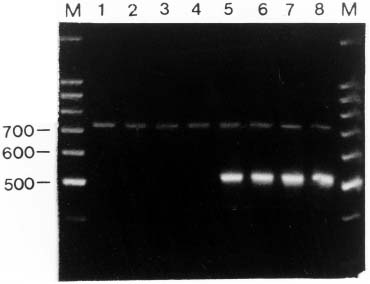
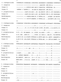
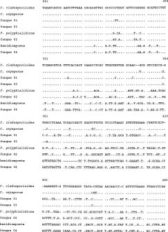
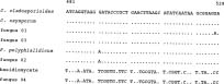
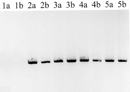
Abstract. PCR amplification of the internal transcribed spacer region (ITS) of the nuclear ribosomal DNA from the genomic DNA isolated from the leaf tissue of Miscanthus sinensis varieties and M. floridulus resulted in two amplified fragments. Via FASTA search, the shorter length of about 500 base pairs was identified as the ITS region of fungi symbiotic in the grass foliage. Two Cladosporium species, one Fusarium and one basidiomycete were added to the mycoflora of Miscanthus grassland. Using a Southern blotting and hybridization, fungal ITS was also detected from the genomic DNAs isolated from sterilized seedling, a suggestive seed transmission of endophytic fungi. The "contaminated" genomic DNA may be a hindrance to the phylogenetic analysis of Miscanthus itself, but may provide other insights into the biodiversity of grassland ecosystems. The endophytic foliage fungi were not detected in M. sinensis var. condensatus, probably a result of its apomictic reproduction.

Keywords: Contamination; Foliage fungi; Miscanthus sinensis; Mycoflora; Phylogeny; rDNA ITS.

Introduction

The association between fungi (both endophytic and epiphytic) and higher plants is thought to be ubiquitous (Clay, 1988; Legault et al., 1989; Petrini et al., 1992). Nevertheless, the prevalence of symptomless fungi on healthy plants has become clear only lately (cf. Camacho et al., 1997). Bills (1996) even suggested that endophytic fungi are basic components of microbiota and are distinct from pathogens ecologically. In grasslands, where Poaceae are dominant, symbiotic fungi, both endophytic and epiphytic, may play critical roles in plant-microbe interactions. Diverse taxa of fungi have been isolated and identified from the leaf tissue of many grasses (Harada, 1987; Nakase et al., 1987; Watson and Dallwitz, 1992).

Molecular techniques have been applied widely in ecological studies (e.g., Wirgin et al., 1997) and wildlife conservation (Avise and Hamrick, 1996; O'Corry-Crowe et al., 1997) in the past several years. In many studies the polymerase chain reaction (PCR) has proved powerful in detecting cryptic genetic variation, e.g., the DNA of symbionts that can not be cultured and separated from their cosymbionts (Haddad et al., 1995), and the microbial floristic composition (cf. Hansen and Hanson, 1996). For systematic and ecological studies, many universal prim

ers have been designed, which may be used across a large range of taxa (Dumolin-Lapegue et al., 1997; Chiang et al., 1998; Chow and Hazama, 1998). The primer sequences of the internal transcribed spacer (ITS) region of the nuclear ribosomal DNA have been widely used for resolving phylogenetic relationships at the species or generic levels (White et al., 1990; Baldwin, 1992). Two recent reports documented the amplification of fungal sequences from plant foliage (Liston et al., 1996; Klein and Smith, 1996). Employing phylogenetic analysis, Camacho et al. (1997) was able to identify the endophytic fungi in Picea based on the ITS nucleotide sequences. In addition, sequences of the nuclear ribosomal internal transcribed spacer region are known providing species-specific genotypes in many fungi (Egger, 1996; Norman and Egger, 1996; Holst-Jensen et al., 1997; Zhang et al., 1997; Vralstad et al., 1998).

A species complex of Miscanthus sinensis Anders. consists of several intraspecific taxa that occur in different elevations on Taiwan (Chou et al., 1999a). The history of adaptive radiation and secondary contacts in the species complex has been reconstructed based on genetic variation of both nrITS and cpDNA atpB-rbcL spacer sequences (Chou et al., 1999b). Most taxa play a dominant role in the floristic composition of grasslands at different elevations. Examples include M. sinensis var. transmorrisonensis (Hayata) Lee in high mountains, M. sinensis var. formosanus Hack. at middle elevations, and M. sinensis var. glaber (Nakai) Lee in lowlands. In contrast to these taxa with a wide range in distribution, M.

*Corresponding author. Tel: +886-6-2757575 ext. 65525; Fax: +886-6-2742583. E-mail: tychiang@mail.ncku.edu.tw


Botanical Bulletin of Academia Sinica, Vol. 42, 2001

sinensis var. condensatus (Hack.) Makino is restricted to the coasts of southeast Taiwan and two offshore islands (Hsu, 1978; Koyama, 1987). In addition, apomixis with sterile pollen has been documented in M. sinensis var. condensatus (Adati, 1958), while most other taxa of the complex were found to have outcrossing (Chou et al., 1999a).

In this study we detected the presence of four symbiotic fungal species in leaf tissue of the taxa of Miscanthus sinensis complex and another related species, M. floridulus. We also found evidence, when the template DNA was extracted from seedlings after seed sterilization, for systemic infection in most Miscanthus taxa.

Materials and Methods

Plant Samples and DNA Extraction

Young and healthy shoots of taxa of the Miscanthus sinensis complex, i.e., M. sinensis var. transmorrisonensis, M. sinensis var. formosanus, M. sinensis var. glaber, and M. sinensis var. condensatus (cf. Lee, 1995), as well as a closely related species, M. floridulus, were collected in the field (Table 1). Seedlings were also cultivated using the seeds of the same individuals used in leaf collections and were harvested for DNA isolation. One seed from each individual was planted in the experimental greenhouse of the Academia Sinica, Taipei. Both leaves and seeds were surface-sterilized with 0.5% sodium hypochlorite for 2 min and rinsed three times with sterile water (Holme and Petersen, 1996). Leaf tissue was powdered in liquid nitrogen and stored at -70°C until use. Genomic DNA was isolated following the CTAB protocol of Murray and Thompson (1980) and gel-quantified.

PCR, Sequencing and FASTA Search, and Hybridization

PCR for amplifying the internal transcribed spacer region (ITS) of nuclear ribosomal DNA was performed in a volume of 100 µl using 10 ng of template DNA, 10 µl of 10X reaction buffer (Promega, Madison, USA), 10 µl MgCl2 (25 mM), 10 µl dNTP mix (8 mM), 10 pmole of each primer,

10 µl of 10% NP-40, and 2 U of Taq polymerase (Promega, Madison, USA). The reaction was programmed on an MJ Thermal Cycler (PTC 100) as one cycle of denaturation at 95°C for 4 min, 30 cycles of 45s denaturation at 92°C, 1 min 15s annealing at 52°C, and 1 min 30s extension at 72°C, followed by 10 min 72°C extension. Template DNA was denatured with the reaction buffer, MgCl2, NP-40 and ddH2O for 4 mins (first cycle), and cooled on ice immediately. A pair of universal primers, ITS1 and ITS4, for amplifying the nrDNA ITS (White et al., 1990), dNTP and Taq polymerase were added to the above ice-cold mixture. Reaction was restarted at the first annealing at 52°C.

PCR fragments were eluted using a High Pure PCR Product Purification Kit (Roche Molecular Biochemicals, Basel, Switz.), ligated in a p-GEM-T easy Vector (Promega, Madison, USA), and cloned in competent Escherichia coli DH5a. Plasmid DNA was purified using Wizard Plus SV Minipreps DNA Purification Systems (Promega, Madison,

Figure 1. Agarose electrophoresis of the PCR products of nrDNA ITS amplified from genomic DNA isolated from foliage (1, 2-7) or seedlings (2, 8, representative sample) of Miscanthus. 1-4, M. sinensis var. condensatus; 5, M. sinensis var. glaber; 6, M. sinensis var. formosanus; 7-8, M. sinensis var. transmorrisonensis; M, 100 bp ladder marker.

Table 1. Materials collected for amplification of the nrDNA ITS region.

Taxon Locality Source Isolate Fungal contamimation

M. sinensis var. glaber Taipei (121°40'E, 25°09'N) Leaf tissue Chiang 1588 +

Taipei (121°40'E, 25°09'N) Seedling Chiang 1588 +

M. sinensis var. formosanus Alishan (120°40'E, 23°29'N) Leaf tissue Chiang 1673 +

Alishan (120°40'E, 23°29'N) Seedling Chiang 1673 +

M. sinensis var. transmorrisonensis Mt. Yushan (120°48'E, 23°28'N) Leaf tissue Chiang 2499 +

Mt. Yushan (120°48'E, 23°28'N) Seedling Chiang 2499 +

M. sinensis var. condensatus Taitung (121°00'E, 22°42'N) Leaf tissue Chiang 2534 _

Taitung (121°00'E, 22°42'N) Seedling Chiang 2534 _

Islet Orchid (121°30'E, 22°05'N) Leaf tissue Chiang 2544 _

Islet Green (121°30'E, 22°40'N) Leaf tissue Chiang 2545 _

M. floridulus Wushe (121°10'E, 24°00'N) Leaf tissue Chiang 2573 +

Wushe (121°10'E, 24°00'N) Seedling Chiang 2573 +


Chiang et al. — Fungal rDNA sequences from Miscanthus

USA). Purified plasmid DNAs were sequenced in both directions by standard methods of the Taq dye deoxy terminator cycle sequencing kit (Perkin Elmer) on an Applied Biosystems Model 377A automated sequencer (Applied Biosystems). Primers for sequence determination were the SP6 and T7 promoters located on the p-GEM-T easy Vector termination site. In this study, we sequenced the nrDNA ITS amplified from the leaf-tissue DNAs. DNA sequences were compared to other homologous sequences registered in GenBank using the FASTA search, a function implemented in the GCG software package (Devereux et al., 1991).

In order to identify the PCR amplification of the fungal ITS from the seedling DNAs, a conserved region between sites 120 and 317 was amplified from the leaf-tissue DNAs of the isolate Chiang 1673 with two additional primers (5'-CTTTGCAGTCTGAGTAAACT-3' and 5'-GGTGAAATGACGCTCGAACAG-3') and was labeled using the PCR DIG Probe Synthesis Kit (Roche Molecular Biochemicals, Basel, Switz). The synthesized probe was used to hybridize with the PCR products of the nrDNA ITS, which were agarose-gel electrophoresed and amplified from both leaf-tissue and seedling DNAs of Miscanthus. Southern blotting and hybridization were carried out and modified as described in Tregear et al. (1996).

Results

Two DNA fragments, 725 bp and 500 bp, of the internal transcribed spacer (ITS) region, consisting of ITS1, ITS2, and 5.8S rRNA gene, were PCR amplified from most Miscanthus except for a single PCR band in M. sinensis var. condensatus (Table 1; Figure 1). In the taxa with two amplified DNA bands, genomic DNAs obtained from both foliage and seedlings all yielded two ITS bands (Table 1).

DNA fragments of both sizes were eluted, ligated in a T-vector, and sequenced. Via FASTA searches the larger fragments were identified as the nrDNA ITS sequences of angiosperms, i.e. Miscanthus in this study (cf. Chou et al., 1999b); and the sequences of the smaller fragments appeared to be closely related to fungal ITS sequences. Clone fungus01 (AJ289867), isolated from the foliage of an individual of M. sinensis var. glaber (isolate Chiang 1588), and clone fungus03 (AJ289869), isolated from the foliage of an individual of M. sinensis var. formosanus


Figure 2. Consensus alignment of nucleotide sequences of nrDNA ITS of fungal clones (Fungus01-04) and the related taxa.


Botanical Bulletin of Academia Sinica, Vol. 42, 2001

specific genes of the nuclear DNA provides markers independent from the contaminant fungal genome for systematic and ecological studies. Otherwise, precautions ought to be taken when contaminated DNA is manipulated. According to this and many other studies, PCR amplification of the internal transcribed spacer (ITS) using universal primers could possibly identify the hidden contamination, even in healthy-looking leaf tissue.

In addition to the documented mycoflora in foliage of Miscanthus, including Puccinia, Sphacelotheca, Ustilago (Watson and Dallwitz, 1992) and Sporobolomyces (Nakase et al., 1987), two Cladosporium species, one Fusarium, and one basidiomycete were recorded for the first time. Among them, basidiomycetes seem widespread in the Poaceae (cf. Zhang et al., 1997). Interestingly, according to the ITS amplification carried out with the sterilized seedlings and Southern blotting, the infection of fungi in Miscanthus seems to be systemic and may indicate seed-borne transmission. Accumulated evidence has suggested that microbial interactions are critical determinants of plant biodiversity in successional fields (cf. Clay, 1992; Clay et al., 1993; Tsai et al., 1994). Clay and Holah's (1999) experiments indicated that fungal endophytes altered plant community structure and reduced the species richness significantly.

Fungal infection was not detected in M. sinensis var. condensatus, a taxon distributed in saline habitats. According to previous cytological (Adati, 1958) and RAPD fingerprinting (Chou et al., 1999a) investigations, the reproductive mode in M. sinensis var. condensatus is obligate apomixis. That is, the formation of seeds does not accompany the regular gametic fusion, but rather proceeds from a diploid ovule. Some recent investigations have revealed that apomixis may provide an opportunity for avoiding the transmission of systematic pathogens in many crops (Nassar et al., 1998). On the other hand, the absence of genomic contamination in M. sinensis var. condensatus might be associated with environmental selection as well.

Although contaminated DNA causes difficulties in detecting the genetic variability of host plants, it may provide another opportunity for detecting the presence of cryptic parasitic or symbiotic organisms (Camacho et al., 1997). The merit of sensitivity of polymerase chain reaction and the universal presence and high identity of the ribosomal RNA genes (both 18S and 28S) across phyla can be used for the detection of mycoflora. Via this screening, more understanding of the ecological association between fungi and host plants could be obtained, as a supplement to standard-culturing and morphological identification of fungal species.

Acknowledgements. This research was supported by grants to CH Chou (NSC 84-2311-B001-016, 85-2311-B001-014, 86-2311-B001-013, NSC-87-2311-B001-009-B27, and NSC-88-2311-B001-026-B27) and TY Chiang (NSC-89-2311-B006- 002-B27 and NSC-88-2311-B006-001-B27) from the National Science Council, Taiwan. We are grateful to two anonymous reviewers for providing valuable comments on our manuscript.

Figure 3. Southern blotting of the PCR products of the ITS amplified with both foliage (a) and seedling DNAs (b), using the probe of the shorter fragment of fungal ITS. 1, Miscanthus sinensis var. condensatus; 2, M. sinensis var. glaber; 3, M. sinensis var. formosanus; 4, M. sinensis var. transmorisonensis; 5, M. floridulus.

(isolate Chiang 1673), shared 86.6% and 98.1% similarity with Cladosporium oxysporum (L25432) and Cladosporium cladosporioides (L25429) (Loculoascomycetes), respectively. Likewise, clone fungus02 (AJ289870), isolated from the foliage of an individual of M. floridulus (isolate Chiang 2573), shared 79.3% similarity with Fusarium polyphialidicum (X94172) (Pyrenomycetes); and the clone fungus04 (AJ289868), isolated from M. sinensis var. transmorrisonensis (isolate Chiang 2499), shared 84.6% identity with a basidiomycete (U65599), which, however, was not indicated with species name.

The probe based on the sequence (sites 120-317, Figure 2) of the clone fungus03 from isolate Chiang 1673) hybridized with the shorter ITS fragments only. Both leaf-tissue and seedling DNAs of all Miscanthus, except for M. sinensis var. condensatus, yielded fungal nrDNA ITS (Figure 3).

Discussion

Contaminant fungal DNA has been reported previously from gymnosperms (Klein and Smith, 1996; Camacho et al., 1997) and grasses (Zhang et al., 1997). Outside of this study, the first to focus on Miscanthus, the problem of DNA contamination has been frequently encountered when doing PCR of nrITS in various plants, including mosses (Chiang and Schaal, 1999), Begonia (Peng and Chiang, 1999), and ferns (cf. Hsu et al., 2000).

Genomic contamination will cause noise in PCR-based fingerprinting, such as RAPD amplification. Genomic loci, annealing to the oligo primers, of both green plants and parasitic fungi will be amplified with no discrimination. Under such circumstances, an overestimate of the genetic variation based on PCR-based fingerprints becomes inevitable (cf. Hudson, 1995; Camacho et al., 1997). Using cpDNA, which is absent in the fungal genome, or more


Chiang et al. — Fungal rDNA sequences from Miscanthus

pompejana. Appl. Envir. Microbiol. 61: 1679-1787.

Hansen, R.S. and T.E. Hanson. 1996. Methanotropic bacteria. Microbiol. Rev. 60: 439-471.

Harada, Y. 1987. Aecial hosts for three graminicolous Puccinia species (Uredinales) in Japan, with a designation of biological forms in Puccinia phragmitis. Trans. Mycol. Soc. Jap. 28: 197-208.

Holme, I.B. and K.K. Petersen. 1996. Callus induction and plant regeneration from different explant types of Miscanthus × ogiformis Honda `Giganteus'. Pl. Cell Tiss. Org. Cult. 45: 43-52.

Holst-Jensen, A., L.M. Kohn, K.S. Jakobsen, and T. Schumacher. 1997. Molecular phylogeny and evolution of Monilinia (Sclerotiniaceae) based on coding and noncoding rDNA sequences. Amer. J. Bot. 84: 686-701.

Hsu, C.C. 1978. Gramineae (Poaceae). In H.L. Li, T.S. Liu, T. Koyama, and C.E. DeVol (eds.), Flora of Taiwan. 1st Ed. Vol. V, Epoch, Taipei, pp. 372-783.

Hsu, J.W., S.J. Moore, and T.Y. Chiang. 2000. Genetic variability of Archangiopteris itoi based on RAPD fingerprints. Bot. Bull. Acad. Sin. 41: 15-18.

Hudson, A.J. 1995. Fungal cytochrome c gene from plants. J. Mol. Evol. 41: 1170-1171.

Klein, A.S. and D.E. Smith. 1996. Phylogenetic inference on the relationship of North American and European Picea species based on nuclear ribosomal 18S sequences and the internal transcribed spacer 1 region. Mol. Phylogen. Evol. 5: 286-287.

Koyama, T. 1987. Grasses of Japan and Its Neighboring Regions. An Identification Manual. The New York Botanical Garden, New York.

Lee, S. J. 1995. Taxonomy of Miscanthus of Taiwan. Ph. D. Dissertation. Department of Botany, National Taiwan University, Taipei.

Legault, D., M. Dessureault, and G. Laflamme. 1989. Mycoflore des aiguiles de Pinus banksiana et Pinus resinosa. I. Champignons endophytes. Can. J. Bot. 72: 1108-1113.

Liston, A., W.A. Robinson, J.M. Oliphant, and E.R. Alvarez-Buylla. 1996. Length variation in the nuclear ribosomal DNA internal transcribed spacer region of non- flowering seed plants. Syst. Bot. 21: 109-120.

Murray, M.G. and W.F. Thompson. 1980. Rapid isolation of high molecular weight DNA. Nucl. Acids Res. 8: 4321-4325.

Nakase, T., M. Itoh, and M. Suzuki. 1987. Sporobolomyces falcatus sp. nov., isolated from a dead leaf of Miscanthus sinensis in Japan. Trans. Mycol. Soc. Jap. 28: 295-301.

Nassar. N.M.A., M.A. Vieira, C. Vieira, and D. Grattapaglia. 1998. A molecular and embryonic study of apomixis in cassava (Manihot esculenta Crantz). Euphytica 102: 9-13.

Norman, J.E. and K.N. Egger. 1996. Phylogeny of the genus Plicaria and its relationship to Peziza inferred from ribosomal DNA sequence analysis. Mycologia 88: 986-995.

O'Corry-Crowe, G.M., R.S. Suydam, A. Rosenberg, K.J. Frost, and A.E. Dizon. 1997. Phylogeography, population structure and dispersal patterns of the beluga whale Delphinapterus lucas in the western Nearctic revealed by mitochondrial DNA. Mol. Ecol. 6: 955-970.

Peng, C.-I and T.Y. Chiang. 1999. Hybridity and parentage of Begonia ×taipeiensis. Ann. Missouri Bot. Gard. (in press)

Literature Cited

Adati, S. 1958. Studies on the genus Miscanthus with special reference to the Japanese species suitable for breeding purpose as fodder crops. Bull. Fac. Agron. Mie Univ. 17: 1-112.

Avise, J.C. and J.L. Hamrick. 1996. Conservation Genetics. Case Histories from Nature. Chapman & Hall, New York.

Baldwin, B.G. 1992. Phylogenetic utility of the internal transcribed spacers of nuclear ribosomal DNA in plants: an example from the Compositae. Mol. Phylogen. Evol. 1: 3-16.

Bills, G.F. 1996. Isolation and analysis of endophytic fungal communities from woody plants. In S. C. Redlin and L. M. Carris (eds.), Fungi in Grasses and Woody Plants, Systematics, Ecology, and Evolution. APS Press, St. Paul., pp. 31-66.

Camacho, F.J., D.S. Gernandt, A. Liston, , J.K. Stone, and A.S. Klein. 1997. Endophytic fungal DNA, the source of contamination in spruce needle DNA. Mol. Ecol. 6: 983-988.

Chiang, T.Y. and B.A. Schaal. 1999. The internal transcribed spacer 2 region of the nuclear ribosomal DNA and the phylogeny of the moss family Hylocomiaceae. Pl. Syst. Evol. (in press)

Chiang, T.Y., B.A. Schaal, and C.I. Peng. 1998. Universal primers for amplification and sequencing a noncoding spacer between the atpB and rbcL genes of chloroplast DNA. Bot. Bull. Acad. Sin. 39: 245-250.

Chou, C.H., S. Huang, S.H. Chen, C.S. Kuoh, T.Y. Chiang, and Y.C. Chiang. 1999a. Ecology and evolution of Miscanthus of Taiwan. Natl. Sci. Coun. Month. 27: 1158-1169.

Chou, C.H., T.Y. Chiang, and Y.C. Chiang. 1999b. The genetic resource of subtropical grasses and their physiological adaptation to stresses. In J. C. Mounolou (ed.), Evolutionary Genetics and Plant Genetic Resources Assessment. (in press)

Chow, S. and K. Hazama. 1998. Universal PCR primers for S7 ribosomal protein gene introns in fish. Mol. Ecol. 7: 1255-1256.

Clay, K. 1988. Fungal endophytes of grasses: a defensive mutualism between plants and fungi. Ecology 69: 10-16.

Clay, K. 1992. Fungal endophytes of plants: biological and chemical diversity. Nat Toxins 1: 147-149.

Clay, K., S. Marks, and G.P. Cheplick. 1993. Effects of insect herbivory and fungal endophyte infection on competitive interactions among grasses. Ecology 74: 1767-1777.

Clay, K. and J. Holah. 1999. Fungal endophyte symbiosis and plant diversity in successional fields. Science 285: 1742-1744.

Devereux, J.P., P. Haeberli, and P. Marquess. 1991. Genetic Computer Group Manual. Version 7.0. University of Wisconsin, Madison.

Dumolin-Lapegue, S., M.H. Pemonge, and R.J. Petit. 1997. An enlarged set of consensus primers for the study of organelle DNA in plants. Mol. Ecol. 6: 393-397.

Egger, K.N. 1996. Molecular systematics of E-strain mycorrhizal fungi: Wilcoxina and its relationship to Tricharina (Pezizales). Can. J. Bot. 74: 773-779.

Haddad, A., F.J. Camacho, P. Durand, and S.C. Cary. 1995. Phylogenetic characterization of the epibiotic bacteria associated with the hydrothermal vent polychaete Alvinella


Botanical Bulletin of Academia Sinica, Vol. 42, 2001

Petrini, O., T.N. Sieber, L. Toti, and O. Viret. 1992. Ecology, metabolite production, and substrate utilization in endophytic fungi. Nat. Tox. 1: 185-196.

Tregear, J.W., S. Jouannic, N. Schwebel-Dugué, and M. Kreis. 1996. An unusual protein kinase displaying characteristics of both the serine/threonine and tyrosine families is encoded by the Arabidopsis thali-ana gene ATN1. Plant Sci. 117: 107-119.

Tsai, H.F., J.S. Liu, C. Staben, M.J. Christensen, G.C.M. Latch, M.R. Siegel, and C.L. Schardl. 1994. Evolutionary diversification of fungal endophytes of tall fescue grass by hybridization with Epichloë species. Proc. Natl. Acad. Sci. USA 91: 2542-2546.

Vralstad, T., A. Holst-Jensen, and T. Schumacher. 1998. The postfire discomycete Geopyxis carbonaria (Ascomycota) is a bitrophic root associate with Norway spruce (Picea abies) in nature. Mol. Ecol. 7: 609-616.

Watson, L. and M.F. Dallwitz. 1992. The Grass Genera of the World. CABI Press, New York.

White, T.J., T. Bruns, S. Lee, and J. Taylor. 1990. Amplification and direct sequencing of fungal ribosomal RNA genes for phylogenetics. In M.A. Innis, D.H. Gelfand, J.J. Sninsky, and T.J. White (eds.), PCR Protocols. A Guide to Methods and Applications. Academic Press, San Diego, pp. 315-324.

Wirgin, I., L. Maceda, J. Stabile, and K. Mesing. 1997. An evaluation of introgression of Atlantic coast striped bass mitochondrial DNA in a Gulf Mexican population using formalin-preserved museum collections. Mol. Ecol. 6: 907-916.

Zhang, C., J.F. Wendel, and L.G. Clark. 1997. Bamboozled again! Inadvertent isolation of fungal rDNA sequences from bamboos (Poaceae: Bambusoideae). Mol. Phylogen. Evol. 8: 205-217.

§Q¥Î PCR ÂX¥R¤Î¤À¤l§Ç¦C®Ö®Ö¿}Åé DNA ITS ¤ù¬q°»´ú¨~ÄÝ ´Óª«¸­¦@¥ÍªºµßÃþ

¦¿¤Í¤¤1,2 ©P©÷¥°1 §õ¨Øèû1 ½±Âí¦t3

1 ¤¤¥¡¬ã¨s°|´Óª«¬ã¨s©Ò
2 °ê¥ß¥x¥_®v½d¤j¾Ç¥Íª«¾Ç¨t
3 °ê¥ß¦¨¥\¤j¾Ç ¥Íª«¾Ç¨t

¥Ñ¤¤°ê¨~¦UÅܺؤΤ­¸`¨~¸­²Õ´µÑ¨úªº°ò¦]Åé DNA¡A§Q¥Î PCR ÂX¥R®Ö®Ö¿}Åé DNA ITS µo²{¨â­Ó¤j¤p¤£¦Pªº¤ù¬q¡A¥H FASTA ·j´M¡A¨ä¤¤¸û¤p¬ù 500 bp ªº¤ù¬q¡AŲ©w¨ä¸û¥i¯à¨Ó¦Û¦¹¥Ý¯ó¸­¤ù¤¤¦@¥Íªº µßÃþ¡A¨ä¤¤¥]§t¤GºØ Cladosporium¡A¤@ºØ Fusarium ¤Î¤@­Ó basidiomycete¡C§Q¥Î«n¤è¾¥ÂIÂø¦X¤ÏÀ³¡A¦¹ µßÃþ ITS ¤ù¬q¥ç¦s¦b©ó¸g®ø¬r³B²zªº¤p­]¡A«ü¥Ü¦¹µßÃþ¥i¸g¥ÑºØ¤l¶Ç»¼¡C¦¹¤@¡©¦Ã¬Vªº¡ª°ò¦]Åé DNA ¹ï©ó¨~ÄÝ´Óª«ªº¿Ë½t­««Ø¥i¯à³y¦¨»Ùê¡A¥t¤@¤è­±«o´£¨Ñ§Ú­Ì¤F¸Ñ¯ó­ì¥ÍºA¨t¦h¼Ë©Êªº°T®§¡C¤º¥Íµß¨Ã¥¼¦b ¤K¤V¨~¤¤³Q°»´ú¨ì¡A¥i¯à»P¨äµL°t¥Í´Þ¦³·¥¤j¬ÛÃö¡C

ÃöÁäµü¡G ¦Ã¬V¡F¸­²Õ´¦@¥Íµß¡F¤¤°ê¨~¡FµßÃþ»x¡F¿Ë½t¡F®Ö¿}Åé DNA ITS¡C