Bot. Bull. Acad. Sin. (2001) 42: 109-114

Wei et al. — Maize lines analysed by GISH

Comparative analyses of disease resistant and nonresistant lines from maize × Zea diploperennis by GISH

Wen-Hui Wei1, Rui Qin1, Yun-Chun Song1,*, Le-Qun Guo2, and Ming-Guang Gu2

1The Key Laboratory of MOE for Plant Developmental Biology, College of Life Sciences, Wuhan University, Wuhan Hubei 430072, The People's Republic of China

2Institute of Genetics, Chinese Academy of Sciences, Beijing 100101, The People's Republic of China

(Received April 27, 2000; Accepted October 9, 2000)

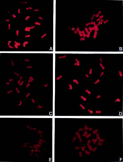
Abstract. After a cross between maize inbred line Lu9 and Zea diploperennis (DP) and a backcross between their F1 and Lu9, the BC1 was reproduced with parthenogenesis induced by chemicals. In this study a2-(1) and a2-(6), the two tested, stable alloplasmic sister lines, were obtained through selecting and selfing of the parthenogenetical progenies for over ten generations. The line a2-(1) showed DP characters, such as resistance to Helminthosporium turcium Pass, H. maydis Nisik, and H. carbonum ULLstrup while a2-(6) exhibited no such resistance. The introgressed DP DNA segments were successfully detected, and their physical location on chromosomes were compared in these two lines by genomic in situ hybridization (GISH). In line a2-(1), the hybridization signals were located on the long arms of chromosomes 1, 2 and 8, and on the short arm of chromosome 6; in line a2-(6), they were located only on the long arms of chromosomes 1, 2 and 8, with no signal on chromosome 6 at all. The distribution of introgressed DP segments on chromosomes, reasons for differences of disease resistance between these two lines, and the existence of resistance genes in the introgressed segments are discussed.

Keywords: Alien introgressed segments; Disease resistance genes; Genomic in situ hybridization (GISH); Maize; Zea diploperennis.

Introduction

Many fine crop varieties showing high yield, quality, and disease resistance have been obtained by distant hybridization, including maize (Guo et al., 1997; 1998), rice (Mohan et al., 1994), barley (Pickering et al., 1997), wheat (Schwarzacher et al., 1992), onion (Keller et al., 1996), and their wild relatives. Zea diploperennis (DP) found by Iltis et al. (1979) is the immune source for several maize viruses, and it is also resistant to maize Helminthosporium turcium Pass, H. maydis Nisik, H. carbonum ULLstrup, foliar and root pathogens as well as insect pests such as corn earworms, stalk borers, and rootworms. In addition, DP has stress tolerance to drought, excess water, and low temperature (Nault et al., 1981). Guo et al. (1997; 1998) crossed maize inbred lines Lu9 and 330 with Zea diploperennis (DP) and obtained several maize lines showing DP characters, such as disease resistance, stress resistance, high yield, and quality. Whether the DP DNA segments were steadily introgressed into the genome of these lines or not—and if so, where such segments are located—is still unknown.

Plant breeders are interested in detecting the alien DNA segments in distant hybrids. The technique of genomic in situ hybridization (GISH) has been widely used in the

analyses of genomic construction and the detection of alien chromatin (Durnam et al., 1985; Pinkel et al., 1986; Schwarzacher et al., 1989; Barre et al., 1998; Humphreys et al., 1998; Khrustaleva and Kik, 1998; Kamstra et al., 1999; Liu et al., 2000; Zhang et al., 2000). In this paper, we describe the detection and location of the introgressed alien segments in two alloplasmic pure lines from inbred Lu9 × DP, one disease resistant and the other susceptible.

Materials and Methods

Plant Materials

The materials were obtained by Guo et al. (1997; 1998). Maize inbred line Lu9 was crossed with Zea diploperennis (DP) in 1985. Subsequently the F1 was backcrossed with its maize parent and produced BC1 in 1986. BC1 was reproduced parthenogenetically by treatment with chemicals in 1987. Guo et al. further selected and bred maize lines of high yield and quality with some DP characters, such as disease and stress resistance, by selfing for over ten generations, eventually obtaining several stable alloplasmic lines. Two stable, pure lines a2-(1) and a2-(6) and their parents, maize inbred lines Lu9 and DP, were chosen as the tested materials. According to inoculated tests in field and greenhouse (Guo et al., 1997), line a2-(1) was resistant to maize Helminthosporium turcium Pass, H.

*Corresponding author. E-mail: ycsong@whu.edu.cn


Botanical Bulletin of Academia Sinica, Vol. 42, 2001

maydis Nisik and H. carbonum ULLstrup, while a2-(6) was sensitive. They were sister lines from the same ear in 1992 and showed no prominent phenotypic differences except for the disease resistance. In the present study, the tested seeds were produced in 1998.

Chromosome Preparation

Chromosome preparation methods were developed by using the protoplast technique described by Song and Gustafson (1995) with some modifications. Root tips were treated in a-bromonaphthalene for 2 h at room temperature. The fixed root tips were immediately digested in 1% cellulase (Shanghai Institute of Biochemistry, Chinese Academy of Sciences) and 1% pectinase (SERVA) at 28°C for 2.5-3 h. The root tips were subjected to a hypotonic treatment in distilled water before being squashed.

DNA Extraction

The extraction of DP and inbred line Lu9 genomic DNA was performed using the procedure described by Ding et al. (1999). Lu9 genomic DNA, used as blocking DNA, was autoclaved for 5 min to produce 300-500 bp sequences.

Biotin Labeling of DNA and In Situ Hybridization

The DP genomic DNA was biotin-labeled with standardized procedures described by Sino-American Biotechnology Company, China. 10 µl dNTP(equal dATP, dGTP and dCTP), 5 µl 10×Buffer, 5 µl DP genomic DNA (0.2 µg/µl ), 4 µl Bio-11-dUTP(0.45 µg/µl), 21 µl ddH2O, 5 µl enzyme (equal polymerase I and DNase I) were components in a 50 µl labeling cocktail. After 1.5-2.5 h at 14-15°C, the labeling was stopped by adding 5 µl 0.2 mol/L EDTA (pH 8.0). The labeled probe was then separated through a Sepharose column and the labeling result was evaluated by means of dot blots.

In situ hybridization was performed with the procedure described by Gustafson and Dillé (1992), with some modifications. Chromosome preparations were pretreated with RNase A (100 µg/ml) in 2×SSC (0.3 mol/L sodium chloride plus 0.03 mol/L sodium citrate) at 37°C for 1 h, rinsed briefly in 2×SSC, and post-fixed in freshly depolymerised 4% paraformaldehyde for 10 min. Chromosomal DNA was then denatured by immersing the slides in 70% deionized formamide in 2×SSC at 70°C for 3.5 min, dehydrated in an ice-cold ethanol series (70%, 95% and 100%), and air-dried. The hybridization mixture contained 50% deionized formamide, 10% (w/v) sodium dextran sulphate, 2×SSC (1×SSC : 0.15 mol/L NaCl plus 0.015 mol/L sodium citrate), 0.25% (w/v) SDS, 4 µg/µl sonicated herring sperm DNA, 5 ng/µl labeled probe DP genomic DNA, 75 ng/µl blocking Lu9 genomic DNA. The blocking ratio was 1 probe DNA:15 Lu9 DNA. The mixture was denatured in boiling water for 10 min and then placed on ice for at least 5 min; 40 µl of the hybridization mixture was applied per slide. The slides were denatured at 90°C for 10 min and hybridization was performed overnight at 37°C in a humid chamber.

Detection

The detection of fluorescent signals followed the procedure published by Dong and Quick (1995), with minor modifications. Slides were washed respectively for 10 min in 20% formamide, 2×SSC and 0.1×SSC at 42°C, in 0.1% TritonX-100 (diluted in 1×PBS) for 4 min and then in 1×PBS (0.13 mol/L NaCl, 0.007 mol/L Na2HPO4·12H2O plus 0.003 mol/L NaH2PO4·2H2O) at room temperature for 5 min before detection.

The signals were detected with 10 µg/ml sheep avidin-fluorescein isothiocyanate (Beijing Medical Academy), 5 µg/ml biotinylated-rabbit anti-sheep, and again 10 µg/ml sheep avidin-fluorescein isothiocyanate for 1 h at 37°C. The slides were washed in 1×PBS for 15 min between each of the above two steps at room temperature. The preparations were counterstained with 3 µg/ml PI (propidium iodide) and mounted in 10 µg/ml antifade (p-phenylenediamine dihydrochloride). Slides were examined with an Olympus BX-60 fluorescence microscope. Photographs were taken on Kodak 100 colour film.

A mean of the hybridization site measurements was taken by calculating the distance from the centromere to the detection site and using that as a percentage of the arm on which the site was located. The arm ratio of the chromosome showing a detection site was also measured in order to determine on which chromosome the site was located.

Results

The results of GISH with 1:15 blocking showed that all chromosomes were red and signal spots were yellow (Figure 1).

Line a2-(1)

Sites of hybridization were located on the long arms of chromosomes 1, 2 and 8, and the short arm of chromosome 6. The mean percent distance from the centromere to the detection site was 84.87 ± 0.08, 52.95 ± 3.12, 67.56 ± 2.43 and 73.57 ± 0.38 on chromosomes 1, 2, 8, and 6 (Figure 1A-D, Table 1), respectively. The signals were detected on two members of each among chromosomes 1, 2, 8 and 6.

Line a2-(6)

Hybridization signals were detected on the long arms of two members of each among chromosomes 1, 2 and 8. The mean percent distance of the hybridization signals on chromosomes 1, 2, and 8 was 90.63 ± 1.27, 59.26 ± 2.07, and 73.40 ± 1.87, respectively (Figure 1E and F, Table 1).

The physical locations of alien introgressed segments on chromosomes were similar between these two lines, except those on chromosome 6, where there was no hybridization signal at all in line a2-(6).

As a control, the chromosome preparations of inbred line Lu9 were used for GISH with the same procedure mentioned above. No signal of hybridization was detected.


Wei et al. — Maize lines analysed by GISH

Table 1. The locations of hybridization signals in lines a2-(1) and a2-(6).

Chromosome and Average percent distance Total number Number of Lines arm detected Arm ratios from the hybridized site of the cells the cells Detection rate signals to the centromere observed detected signals

a2-(1) 1L* 1.24±0.11*** 84.87±0.08*** 223 77 34.53

2L 1.25±0.07 52.95±3.12 287 105 36.59

8L 2.81±0.11 67.56±2.43 198 83 41.92

6S** 2.27±0.15 73.57±0.38 210 54 25.71

a2-(6) 1L 1.23±0.12 90.63±1.27 200 64 32.00

2L 1.27±0.06 59.26±2.07 258 96 37.29

8L 2.90±0.07 73.40±1.87 220 96 43.64

*Long arm; **Short arm; ***Standard deviation.

Figure 1. A-D were the detected results of alien introgressed segments in line a2-(1); E-F were the detected results of alien introgressed segments in line a2-(6). Double signals were detected on the homologous chromosomes. A, Hybridization signals were on the subterminal region of the long arm of chromosome 1; B, Hybridization signals were on the median part of the long arm of chromosome 2; C, Hybridization signals were on the median part of the short arm of chromosome 6; D, Hybridization signals were on the median part of the long arm of chromosome 8; E, Hybridization signals were on the subterminal region of the long arm of chromosome 1 and on the median part of the long arm of chromosome 2; F, Hybridization signals were on the median part of the long arm of chromosome 8.


Botanical Bulletin of Academia Sinica, Vol. 42, 2001

Discussion

In our laboratory, several important disease resistance genes have been mapped by in situ hybridization with linked RFLP markers in maize. They include Helminthosporium turcicum Pass, H. maydis Nisik, and H. carbonum ULLstrup resistance genes ht, htn, rhm and hm. The gene ht1 was located on the long arm of chromosome 2 (Li et al., 1998a), htn1 on the long arm of chromosome 8 (Li et al., 1998b), hm1 on the long arm of chromosome 1 (Li et al., 1998c), and rhm on the short arm of chromosome 6 (unpublished data). The physical locations on the chromosome arms corresponded to their positions in the genetic map (Coe, 1995). In this study, the results showed that the hybridization sites on the detected chromosome arms completely corresponded to those where H. turcicum Pass, H. maydis Nisik, and H. carbonum ULLstrup resistance genes ht, htn, rhm and hm were located. All the alien introgressions were located between the median and terminal regions, as were the disease resistant genes. DP was interfertile with maize (Iltis et al., 1979), meaning that during meiosis the pairing of homologous chromosomes could be performed in their hybrids. Therefore, the DP chromatin might be integrated into the maize genome by homologous or homoeologous recombination during meiosis. It can be deducted that the introgressed segments integrated in chromosomes 1, 2, 8 and 6 probably contain the genes hm, ht, htn and rhm, respectively. Chromosome microdissection developed recently is an efficient technique for the construction of specific library and the isolation of genes (Albani et al., 1993; Tian et al., 1999). We think that the introgressed segments can be isolated by microdissection based on their physical location and applied to the construction of a specific library. Then the related genes may be screened and characterized with the library if they are really positioned in these segments.

It has been demonstrated that active genes are mainly located on the distal chromosome regions (Pedersen et al., 1997). It is evident that the introgressed segments detected in this study all are located on these active gene regions, indicating that the introgressed segments may be functional genetically. We never found any signals on the centromere regions, and this can be explained by the following two reasons: (i) The synapses near the centromeres are hindered by some unknown factors (Khrustaleva and Kik, 1998). (ii) Most recombination events occur in gene-rich regions (Künzel et al., 2000).

Like a2-(1), the line a2-(6) also displayed the hybridization sites on chromosomes 1, 2 and 8. It did not display on chromosome 6, but it was sensitive to the diseases mentioned above. Probably resistance or sensitivity to disease depends on more than the existence or inexistence of the introgressed segments on chromosome 6 because, as we discuss above, the DP chromosome segments integrated in chromosomes 1, 2 and 8 should contain DP resistance genes. We think the line a2-(6) might lose some regular sequences activating the resistance gene expression and that these sequences were probably contained

in the introgressed segments on chromosome 6 or somewhere else. Whether this is true or not remains to be proved in further study.

The signals on each detected chromosome are all small spots instead of large blocks, a phenomenon that has also been reported by other researchers (Kamstra et al., 1997; Karlov et al., 1999; Poggio et al., 1999). We think it is to be expected that when tested stable alloplasmic maize lines are obtained by selection over many generations, the wild characters of DP, except for those associated with disease resistance, must be gradually thrown away during that process. The materials that carried large DP segments might be those which showed more wild characters and were even unstable.

As a rule, the DP DNA sequences homologous with maize should be blocked by maize genomic DNA, and most of the DP genes should be homologous with maize genes. How could the DP segments on chromosomes 1, 2, 8 and 6 not be blocked by maize genomic DNA and still show the DP DNA signals? The reason is that the unexpressed species-specific repetitive sequences probably are nonhomologous between DP and maize, and the introgressed Zea diploperennis-specific repetitive sequences might not be blocked by maize genomic DNA. Therefore, the signals observed should represent the unexpressed DP-specific repetitive sequences rather than the expressed ones. Studies on identifying alien chromatins and genomic construction with FISH (fluorescence in situ hybridization) of species-specific repetitive DNA sequences have been reported by some researchers (Mukai and Nakahara, 1993; Pedersen and Langridge, 1997; Kamstra et al., 1999), and they also detected the hybridization signals just like those we observed in this study. Because differences in the repetitive sequences between DP and maize were not absolute, the integrated DP chromatins were still blocked to a certain degree by maize genomic DNA with a high blocking ratio.

Acknowledgements. This work was supported by the National Natural Science Foundation of China (39870423) and the Doctorate Vesting Point Foundation of the Educational Ministry of China.

Literature Cited

Albani, D., M.J. Cote, and K.C. Armstrong. 1993. PCR amplification of microdissected wheat chromosome arms in a single "single tube" reaction. Plant J. 4: 899-903.

Barre, P., M. Layssac, A.D. Hont, J. Louarn, A. Charrier, S. Hamon, and M. Noirot. 1998. Relationship between parental chromosomic contribution and nuclear DNA content in the coffee interspecific hybrid C. pseudozanguebariae × C. liberica var. "dewevrei". Theor. Appl. Genet. 96: 301-305.

Coe, E.H. 1995. Gene list and working maps. Maize Genet. Coop. Newsl. 69: 247-258.

Ding, Y., X.P. Chen, and L. Yan. 1999. Botanical status of wild relatives to barley from the Qing-Zang plateau of China. Hereditas 130: 111-116.


Wei et al. — Maize lines analysed by GISH

Li, L.J., Y.C. Song, H.M. Yan, L. Wang, and L.H. Liu. 1998c. Physical location of Helminthosporium carbonum susceptibility gene hm1 by FISH of a RFLP marker umc119 in maize. Wuhan Univ. J. Natural Sci. 3(4): 495-498.

Liu, W.X., P.D. Chen, and D.J. Liu. 2000. Studies of the development of Triticum aestivum-Leymus racemosus translocation lines by pollen irradiation. Chinese Acta Genet. Sin. 27(1): 44-49.

Mohan, M., S. Nair, and J.S. Bentur. 1994. RFLP and RAPD mapping of the rice Gm2 gene that confers resistance to biotype 1 of gall midge (Orseolia oryzae). Theor. Appl. Genet. 87: 782-788.

Mukai, Y. and Y. Nakahara. 1993. Simultaneous discrimination of the three genomes in hexaploid wheat by multicolor fluorescence in situ hybridization using total genomic and highly repeated DNA probes. Genome 36: 489-494.

Nault, L.R. and W.R. Findley. 1981. Primitive relative offers new traits for corn improvement. Ohio-Rep. Res. Dev. 66(6): 90-92.

Pedersen, C., J. Zimny, and D. Becker. 1997. Location of introduced genes on the chromosomes of transgenic barley, wheat and triticale by fluorescence in situ hybridization. Theor. Appl. Genet. 94: 749-757.

Pedersen, C. and P. Langridge. 1997. Identification of the entire chromosome complement of bread wheat by two-colour FISH. Genome 40: 589-593.

Pickering, R.A., A.M. Hill, and R.G. Kynast. 1997. Characterization by RFLP analysis and genomic in situ hybridization of a recombinant and a monosomic substitution plant derived from Hordeum vulgare L. × Hordeum bulbosum L. Crosses. Genome 40: 195-200.

Pinkel, D., T. Straume, and J.W. Gray. 1986. Cytogenetic analysis using quantitative, high-sensitivity, fluorescence hybridization. Natl. Acad. Sci. USA 83: 2934-2938.

Poggio, L., V. Confalonieri, C. Comas, A. Cuadrado, N. Jouve, and C.A. Naranjo. 1999. Genomic in situ hybridization (GISH) of Tripsacum dactyloides and Zea mays ssp. Mays with B chromosomes. Genome 42(4): 687-691.

Schwarzacher, T., K. Anamthawat-Jonsson, G.E. Harrison, and A.K.M.R. Islam. 1992. Genomic in situ hybridization to identify alien chromosomes and chromosome segments in wheat. Theor. Appl. Genet. 84: 778-786.

Schwarzacher, T., A.R. Leitch, and M.D. Bennett. 1989. In-situ localization of parental genomes in a wide hybrid. Ann. Bot. London 64: 315-324.

Song, Y.C. and J. P. Gustafson. 1995. The physical location of fourteen RFLP markers in rice (Oryza stiva L.). Theor. Appl. Genet. 90: 113-119.

Tian, J., Y.F. Lu, J.X. Deng, B. Li, X.Y. Zhang, and G.T. Liu. 1999. The microdissection of addition chromosomes in Triticum aestivum-Th. intermedium addition line TAI-27 and the selection of specific probes. Sci. in China (Series C) 29(2): 174-179.

Zhang G.F., J.W. Liu, Y.Z. Huang, M. Ding, S.X. Tang, and X. Jia. 2000. Isolation and verification of Triticum aestivum-Eremopyrum orientale addition lines and substitution lines by GISH. Chinese Acta Genet. Sin. 27(1): 50-55.

Dong, H. and J.S. Quick. 1995. Detection of 2.6 kb single/low copy DNA sequence on chromosomes of wheat (Triticum aestivum) and rye (Secale cereale) by fluorescence in situ hybridization. Genome 38: 240-249.

Durnam, D.M., R. Gelinas, and D. Myerson. 1985. Detection of species specific chromosomes in somatic cell hybrids. Somatic Cell Mol. Genet. 11: 571-577.

Guo, L.Q., M.G. Gu, and T.X. Yang. 1997. Studies on alloplasmic pure line derived from chemically-induced parthenogenic maize distant hybrid and its breeding. Chinese Acta Genet. Sin. 24(6): 537-543.

Guo, L.Q., M.G. Gu, T.X. Yang, S.J. He, and Z. Zhang. 1998. Production of alloplasmic pure line through chemically-induced parthenogenesis of female gametophyte of maize distant hybrid. Develop. Reprod. Biol. 7(1): 55-62.

Gustafson, J.P. and J.E. Dillé. 1992. Chromosome location of Oryza sativa recombination linkage groups. Proc. Natl. Acad. Sci. USA 89: 8646-8650.

Humphreys, M.W., A.G. Zare, I. Pasakinskiene, and H. Thomas. 1998. Interspecific genomic rearrangements in androgenic plants derived from a Lolium multiflorum × festuca arundinacea (2n=5x=35) hybrid. Heredity 80: 78-82 .

Iltis, H.H., J.F. Doebley, R.M. Guzman, and R. Pazy. 1979. Zea diploperennis (Cramineae): a new teosinte from Mexico. Science 203: 186-188.

Kamstra, S.A., A.G. Kuipers, M.J. De Jeu, M.S. Ramanna, and E. Jacobsen. 1997. Physical localisation of repetitive DNA sequences in Alstroemeria: karyotyping of two species with species-specific and ribosomal DNA. Genome 40: 652-658.

Kamstra, S.A., A.G. Kuipers, M.J. De Jeu, and M.S. Ramanna. 1999. The extent and position of homoeologous recombination in assessment of first generation backcross progenies. Chromosoma 108(1): 52-63.

Karlov, G.I., L.I. Khrustaleva, K.B. Lim, and J.M. van Tuyl. 1999. Homoeologous recombination in 2n-gamete producing Interspecific hybrids of Lilium (Liliaceae) studies by genomic in situ hybridization (GISH). Genome 42(4): 681-686.

Keller, E.R.J., I. Schubert, J. Fuchs, and A. Meister. 1996. Interspecific crosses of onion with distant Allium species and characterization of the presumed hybrids by means of flow cytometry, karyotype analysis and genomic in situ hybridization. Theor. Appl. Genet. 92: 417-424.

Khrustaleva, L.I. and C. Kik. 1998. Cytogenetical studies in the bridge cross Allium cepa × (A. fistulosum × A. rylei). Theor. Appl. Genet. 96: 8-14.

Künzel, G., L. Korzun, and A. Meister. 2000. Cytologically integrated physical restriction fragment length polymorphism maps for the barley genome based on translocation breakpoints. Genetics 154: 397-412.

Li, L.J., Y.C. Song, H.M. Yan, L. Wang, and L.H. Liu. 1998a. The physical location of the gene ht1 (Helminthosporium turcium resistance 1) in maize (Zea mays L.). Hereditas 129: 101-106.

Li, L.J., Y.C. Song, and H.M. Yan. 1998b. Chromosome location of two RFLP markers umc84 and umc30 tightly linked to the htn1 gene in maize. Chinese Acta Phytopathol. Sin. 28(2): 117-121.


Botanical Bulletin of Academia Sinica, Vol. 42, 2001

¥É¦Ì¡Ñ¤G­¿Åé¦h¦~¥ÍÃþ¥É¦Ì§Ü¯f»P«D§Ü®è¨tªº GISH ¤ÀªR

ÃQ¤å½÷1 µ÷ ·ç1 §º¹B²E1 ³¢¼Ö¸s2 ½\©ú¥ú2

1 ªZº~¤j¾Ç´Óª«µo¨|¥Íª«¾Ç±Ð¨|³¡­«ÂI¹êÅç«Ç

2 ¤¤°ê¬ì¾Ç°|¿ò¶Ç¬ã¨s©Ò
¥É¦Ì¦Û¥æ¨t®È 9 »P¤G­¿Åé¦h¦~¥ÍÃþ¥É¦Ì¡]Zea diploperennis, DP¡^Âø¥æ¡AF1 »P®È 9 ¦^¥æ¡A¦^¥æ¤@¥N (BC1) ³q¹L¤Æ¾ÇÃĪ«»¤¾É¶i¦æ©t»Û¥Í´Þ¡A¥»¬ã¨s¥Î©ó¹êÅ窺¨â­Óí©w²§½è©j©f¨t a2-(1) ©M a2-(6) ¬O¹ï©t»Û¤@¥N¶i¦æ¦Û¥æ¨Ã¿ï¾Ü¤F¤Q¦h¥N¥H«á±o¨ìªº¡C®è¨t a2-(1) Åã¥Ü¥X DP ¤@¨Ç©Êª¬¡A¦p§Ü¥É¦Ì¤j´³¯f¡B¤p´³ ¯f©M¶é´³¯f¡A a2-(6) «h¤£§Ü¯f¡C§Q¥Î°ò¦]²Õ­ì¦ìÂø¥æ (GISH) ¦¨¥\¦aÀË´ú¤F³o¨â­Ó®è¨t¤¤º¯¤Jªº DP DNA ¤ù¬q¡A¨Ã¤ñ¸û¤F¥¦­Ì¦b¬V¦âÅé¤Wªºª«²z¦ì¸m¡C¦b a2-(1) ¤¤¡AÂø¥æ«H¸¹©w¦ì¦b 1¡A2¡A8 ¸¹¬V¦âÅéªøÁu¤Î 6 ¸¹¬V¦âÅéµuÁu¡A¦b a2-(6) ¤¤¡AÂø¥æ«H¸¹¥u©w¦ì¦b 1¡A2¡A8 ¸¹¬V¦âÅéªøÁu¡A 6 ¸¹¬V¦âÅéµuÁu¨S¦³ÀË¥XÂø¥æ «H¸¹¡C¹ï¬V¦âÅé¤W DP º¯¤J¤ù¬qªº¤À§G¡A¨â®è¨t¶¡§Ü¯f©Ê¤£¦Pªº­ì¦]¡A¥H¤Îº¯¤J¤ù¬q¤¤¦s¦bªº§Ü¯f°ò¦] ¶i¦æ¤F°Q½×¡C

ÃöÁäµü¡G ¥É¦Ì¡F¤G­¿Åé¦h¦~¥ÍÃþ¥É¦Ì¡F¥~·½º¯¤J¤ù¬q¡F§Ü¯f°ò¦]¡F°ò¦]²Õ­ì¦ìÂø¥æ¡C