Bot. Bull. Acad. Sin. (2001) 42: 123-129

Chen et al. — Sucrose phosphate synthase purification

Purification and characterization of sucrose phosphate synthase from sweet potato tuberous roots

Wei-Liang Chen1,3, Dong-Jiann Huang1,3, Pang-His Liu1, Heng-Long Wang2, Jong-Ching Su1,2, and Ping-Du Lee1,*

1Biochemistry Laboratory, Department of Agricultural Chemistry, National Taiwan University, 1 Roosevelt Road Section 4, Taipei, Taiwan 107, Republic of China

2Institute of Biological Chemistry, Academia Sinica, P.O. Box 23-106, Taiwan 107, Republic of China

(Received March 24, 2000; Accepted August 30, 2000)

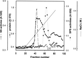
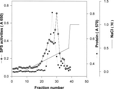
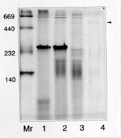
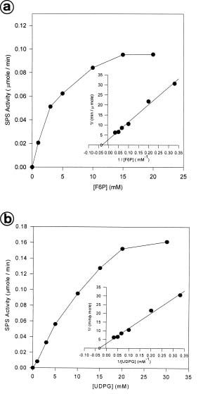
Abstract. Sucrose phosphate synthase (SPS) is one of the key enzymes in the sucrose biosynthesis pathway. SPS was purified 40 fold from crude extract of sweet potato tuberous roots by the methods of batch elution from DEAE-Sephacel, PEG precipitation, w-aminohexyl Sepharose 4B affinity and Mono Q anion exchange chomatographies. The native- and SDS-PAGE analyses revealed SPS to have a native molecular mass of about 540 kDa, and it may therefore be homotetramer composed of subunit with a mass of 130-140 kDa. The isoelectric point of the purified enzyme as determined by IEF was 5.29. SPS from the sweet potato tuberous root, which differs from the SPS of photosynthetic tissues, was not allosterically regulated by G6P and Pi. The Km for F6P and UDPG was 5.3 and 31.3 mM, respectively. The enzyme was activated by Mn2+, Mg2+, and Ca2+, while being inhibited by Hg2+. The nucleotides AMP, ADP, ATP, UMP, UDP, UTP, and TDP inhibited the enzyme about 30~50%. The enzyme was sensitive to sulfydryl reagents, but activity could be restored with DTT or b-ME. The enzyme was activated by glucose, glucosamine, maltose, and lactose, but was inhibited by d-gluconolactone. SPS could also be inhibited by PCMBS and Cibacron blue F3G-A.

Keywords: Enzyme purification; Ipomoea batatas; Sucrose metabolism; Sucrose phosphate synthase; Sweet potato.

Abbreviations: DTT, 1,4-dithiothreitol; b-ME, b-mercaptoethanol; PCMBS, p-chloromercuribenzenesulphonic acid; G6P, glucose 6-phosphate; F6P, fructose 6-phosphate; Pi, inorganic phosphate.

Introduction

Sucrose phosphate synthase (UDPglucose: D-fructose 6-phosphate 2-glucosyl-transferase; EC 2.4.1.14) catalyzes the following reaction:

UDPglucose + Fructose 6-phosphate « UDP + Sucrose 6'-phosphate

Sucrose phosphate synthase (SPS) catalyzes an essential and regulated reaction in the pathway of sucrose synthesis. In leaves, SPS is regulated by at least two mechanisms: one is an allosteric mechanism in which the enzyme is activated by binding metabolites like G6P and is inhibited by Pi (Doehlert and Huber, 1983), and the other one achieves regulation by covalent protein phosphorylation (Still et al., 1988).

In non-photosynthetic tissues such as tubers and fruits, SPS may play several important physiological roles, such as the biosynthesis and export of sucrose in germinating seeds and sprouting tubers, the accumulation of sucrose

under cold conditions, and the modulation of sucrose import and degradation via futile cycle in sink tissues. In non-photosynthetic tissues, the role of SPS is less well understood, and no obvious regulatory features have been found. To investigate the enzymatic function of SPS in sucrose biosynthesis, SPS has been purified to near homogeneity from spinach leaf (Salvucci et al., 1990), wheat germ (Salerno et al., 1991), maize leaf (Bruneau et al., 1991), Prosopis juliflora leaf (Sinha et al., 1997) and rice leaf (Salerno et al., 1998).

The objective of our work was to study the properties of the enzyme in detail and compare it with that of annual species. In this communication we report the purification and characterization of SPS from sweet potato tuberous roots.

Materials and Methods

Plant Material

Young, growing tubers of sweet potato, Ipomoea batatas (L.) Lam. cv. Tainoug 57, were harvested immediately before use.

3Both authors contributed equally to this work.

*Corresponding author. E-mail: pingdu@ccms.ntu.edu.tw


Botanical Bulletin of Academia Sinica, Vol. 42, 2001

Purification of SPS

To purify SPS, sweet potato tubers (500 g) were peeled and homogenized with 500 mL extraction buffer containing 50 mM Hepes-KOH (pH 7.5), 10 mM MgCl2, 1 mM EDTA, 2.5 mM DTT, 0.1% (v/v) triton X-100, 1 mM PMSF (phenylmethylsulfonyl fluoride), 1% (w/v) polyvinylpyrrolidone (PVP, average molecular weight 10,000), filtered through two layers of Miracloth, then centrifuged for 10 min at 6,000 g. The supernatant was added to 250 mL pre-swollen DEAE-Sephacel (Pharmacia), which had been pre-equilibrated by sequential washing with 1000 mL 50 mM NaOH, 1000 mL Na-acetate (pH 4.5), 1000 mL distilled water, 1000 mL 2 M NaCl, and 600 mL extraction buffer. After 30 min the extract and Sephacel mixture was transferred to the Buchner-funnel and allowed to settle. The Sephacel gel was washed sequentially with 100 mL extraction buffer containing 40, 130 and 500 mM NaCl, respectively. The 130 mM NaCl fraction containing SPS activity was brought to 24% (w/v) polyethylene glycol (PEG, average molecular weight 8,000) by adding solid PEG, stirred 30 min at 4°C, and centrifuged (4,500 g, 30 min). The sediment was then resuspended in 10~15 mL extraction buffer, but without the PVP or glycerol. This solution was applied to an w-aminonhexyl Sepharose 4B column (Sigma, bed volume 50 mL) that had been pre-equilibrated in the same buffer, and a further 150 mL of buffer was added. SPS was eluted with 150 mL of extraction buffer containing 500 mM NaCl. The eluent was passed through a 0.22 µm filter and added to a Mono Q column (HR 5/5, Pharmacia/LKB) which had been pre-equilibrated with 20 mL buffer (identical to the resuspended buffer except that it was 12% glycerol and 0.8 mM Chaps). The Mono Q column was eluted using a fast protein liquid chromatography system (FPLC, Pharmacia/LKB) at a flow rate of 0.5 mL/min, washed with buffer for 20 min, and then with a linear NaCl gradient in buffer (0~0.5 M NaCl) for 60 min. The fractions were collected in tubes and frozen immediately at -70°C.

Assay of SPS

During the entire purification scheme, SPS activity was monitored by the anthrone test (Huber et al., 1989). In this test, 70 µL of reaction mixture including the extract is adjusted to a final concentration of 4 mM F6P, 20 mM G6P, 3 mM UDPG, 50 mM Hepes-KOH (pH 7.5), 5 mM MgCl2, and 1 mM EDTA. The mixture is incubated at 37°C for 15

mins before adding 70 µL 30% (w/v) KOH and heating 10 min at 95°C. To this is then added 1 mL 0.14% (w/v) anthrone in 95~97% H2SO4. The mixture is incubated 20 min at 37°C, and A620 is measured. All fractions deriving from the salt elution of the diethylaminoethyl (DEAE)-Sephacel column were desalted using a desalting column (Sephadex G-25; Pharmacia/LKB).

Results

Purification of the Enzyme

The activities of SPS in sweet potatoes of different tuber sizes were compared. It turned out that the medium-sized (ca. 3 cm diameter) tuberous roots have the highest SPS activities (data not shown). In this experiment we used roots with average diameters of ca. 3 cm as the materials for SPS purification. The purification scheme was summarized in Table 1. The separation of sucrose phosphate synthase and sucrose synthase was achieved by w-aminohexyl Sepharose 4B affinity column (Figure 1). Final purification was obtained on Mono Q column (Figure 2), and an enzyme purified 40 fold was collected as SPS for enzyme studies. Analysis of the native-PAGE revealed a native form of the enzyme with a molecular mass of 540 kDa (Figure 3). Based on PAS-staining, the enzyme appears to be a glycoprotein (data not shown). The SDS -PAGE analysis showed a monomer with a molecular mass of ca. 130~140 kDa. The extra protein bands at ca. 90 kDa and 50 kDa are most likely due to the degradation of SPS (Figure 4). From the native- and SDS-PAGE analyses, we proposed that the sweet potato tuberous root SPS has a native molecular mass of about 540 kDa as a tetramer. And the Mw of each subunit is about 130~140 kDa.

pH Optimum and Isoelectric Point

The pH optimum of SPS is about 7.5 (data not shown). At a pH range of 6 to 8, SPS had 80% of the maximum activity. The isoelectric point of the enzyme was determined by isoelectric focusing, and the enzyme activity focused at pH 5.29 (data not shown).

Effect of Metal Ions

The effect of divalent cations was studied. All the substances and buffers used during purification and analysis

Table 1. Purification of sucrose phosphate synthase from sweet potato tuberous roots.

Step Total protein Total activity Specific activity Purification Yield (mg) (U) (U/mg) (fold) (%)

Crude extract 5280 54.63 0.0103 1 100

DEAE-Sephacel (130 mM NaCl) 2004 23.5 0.0117 1.1 43

24% PEG sediment 479 11.7 0.0244 2.4 21.5

w-Aminohexyl Sepharose 4B 9.41 1.18 0.126 12.2 2.2

FPLC Mono Q 1.35 0.56 0.41 40.3 1.1

1Data are obtained from 500 g fresh sweet potato tuberous roots.

2One unit of enzyme was defined as the amount of enzyme required for producing 1 µmol of sucrose-P from UDPG and F6P at 37°C and pH 7.5 per min.


Chen et al. — Sucrose phosphate synthase purification

were Hepes salts. It was found the enzyme was inhibited by 10 mM HgCl2. The activity was increased to 270% by 10mM MgCl2, and the enzyme activity was inhibited to 50% by 10 mM HgCl2 (data not shown).

Effect of Nucleotides

The influence of various nucleoside mono-, di-, and triphosphates on enzyme activity is summarized in Table 2. It shows that GDP and GTP had no effect, that CTP caused slight inhibition, and that AMP, ADP, ATP, UMP, UDP, UTP, and TDP inhibited enzyme activities up to 30~50%.

Effect of Thiol Reagents

The requirement of -SH groups for the enzyme activity was tested with different reagents containing thiol groups. Table 3 shows DTT and b-ME increased the activity by 10~20 % at 0.1 mM. It also shows that the enzyme was sensitive to sulfydryl regents at higher concentrations.

Effect of Sugars and Sugar Phosphates

The effect of some sugars and sugar phosphates on enzyme activity was studied. The results presented in Table 4 indicate that enzymes were activated by glucose, glucosamine, maltose, and lactose while d-gluconolactone inhibited about 80% of the enzyme activity.

Figure 1. Chromatographic purification of sweet potato tuberous root SPS on w-aminohexyl Sepharose 4B affinity column. SPS (™) and SS (£) activities and protein content (¢) are shown. The full line represents the NaCl gradient from 0 to 0.4 M.

Figure 2. Chromatographic purification of sweet potato tuberous root SPS on Mono Q HR 5/5 anion exchange chromatography. SPS activities (™) and protein content (¢) are shown.

Table 2. Effect of nucleotide on sucrose phosphate synthase activity.

Nucleotide Relative activity ( % )

None 100

AMP 60

ADP 50

ATP 54

GDP 104

GTP 106

UMP 75

UDP 61

UTP 69

TDP 74

CTP 92

All nucleotide added at a final concentration of 5 mM.

Figure 3. Native-PAGE of SPS fraction separated after the different purification steps. Approximately 10 µg of total protein were loaded in each lane. Proteins were stained with Coomassie blue. Lane 1, crude extract pool; Lane 2, DEAE-Sephacel pool ; Lane 3, w-aminohexyl Sepharose pool; Lane 4, Mono-Q pool; Mr, molecular weight markers in kDa.


Botanical Bulletin of Academia Sinica, Vol. 42, 2001

Table 3. Effect of sulfydryl compounds on sucrose phosphate synthase activity.

Sulfydryl compounds Concentration Relative activity (mM) (%)

None 100

DTT 0.1 106

1 57

10 40

b-ME 0.1 123

1 41

10 21

Glutathione 0.1 83

1 64

10 21

L-Cysteine 0.1 100

1 65

10 21

Effect of Inhibitors on Sucrose Phosphate Synthase Activity

The effect of inhibitors on enzyme activity was studied. The results presented in Table 5 show that SPS from sweet potato tuberous roots was markedly inhibited by the PCMBS, an inhibitor of -SH group, and 50% inhibition was attained at a concentration of 20 mM. Cibacron blue F3G-A proved to be a high-affinity competitive inhibitor with respect to the substrate UDPG and a mixed-type inhibitor with respect to F6P. In this experiment, we found that 50% inhibition of the SPS was attained when Cibacron blue F3G-A concentration was added to 1.2 mM.

Kinetics Characteristics of Sweet Potato Tuber SPS

SPS in leaf is allosterically regulated by G6P and Pi (Huber and Huber, 1996). It is noteworthy that the SPS from the sweet potato tuberous root was different from that of photosynthetic tissues not regulated by G6P or Pi. The effect of substrate concentration on the activity of sucrose phosphate synthase is presented in Figure 5. Concentration of the substrates, UDPG and F6P, were varied over a range to determine the kinetics parameters. Hyperbolic kinetic plots were obtained, and the Km for F6P and UDPG were calculated as 5.3 mM and 31.3 mM, respectively (Figure 5).

Discussion

Purification of oligomeric high molecular weight enzyme from plant tissue was often difficult as it usually requires several steps and passage through different types of column chromatography. So far, only a few papers describing the purification of SPS from leaves and seeds to nearly homogeneous have been reported (Walker and Huber,

Table 4. Effect of monosaccharide and sugar phosphates on sucrose phosphate synthase activity.

Monosaccharide or Concentration Relative activity sugar phosphate (mM) (%)

None 100

Glucose 10 141

50 110

Galactose 10 128

50 94

Mannose 10 76

50 136

Glucosamine 10 111

50 117

d-gluconolactone 10 95

50 16

Glucose-1-phosphate 10 123

50 94

Maltose 10 113

50 130

Lactose 10 108

50 134

Table 5. Effect of inhibitors on sucrose phosphate synthase activity.

Inhibitors Concentration (mM) Relative activity (%)

None 100

PCMBS 10 93

20 48

50 44

Cibacron blue 0.3 102

F3G-A 0.6 87

1.2 56


Chen et al. — Sucrose phosphate synthase purification

1989; Salvucci et al., 1990; Salerno et al., 1991; Sonnewald et al., 1993). The SPS isolation procedure from sweet potato tuber reported here (Figure 1) permits the 40 fold purification of the enzyme with a yield of about 1%, and totally devoid of sucrose synthase.

The subunit of sweet potato SPS has a molecular mass of about 130 kDa, similar to that of spinach and maize leaf SPS (Walker and Huber, 1989; Bruneau et al., 1991). During purification most of the intact enzyme protein becomes proteolytically cleaved. This phenomenon was also observed in the case of spinach leaf and banana fruit (Joao et al., 1997). However, the presence of multiple bands was

minimized by adding protease inhibitors. The preparation after elution from Mono Q column yielded a major protein peak with enzyme activity corresponding to an average Mw of 540 kDa. This value is within the range 380~540 kDa found for the Mw of SPS from wheat seeds (Salerno et al., 1991) and maize leaves (Kalt-Torres, 1987). Mw values as low as 380 kDa were also found (Bruneau et al., 1991). Sweet potato tuber SPS appeared to be composed of identical subunits with masses of 130~140 kDa. This was shown by the enrichment of only the 130~140 kDa band during purification. SPS in SDS-PAGE might be a proteolytic degradation. The pattern was quite similar to those obtained from rice leaves (Salerno et al., 1998) and potato tubers (Reimholz et al., 1994), suggesting that the SPS protein structure is labile to protease or quite unstable to physical or chemical treatment during purification.

Sweet potato SPS showed an optimum pH at 7.5 while the enzyme from pea, and Prosopis juliflora showed optimum pH ranging from 6.5 to 7 (Leloir, 1955; Harbron et al., 1981; Lunn and Rees, 1990; Alok, 1997). The isoelectric point of sweet potato SPS focused at pH 5.29. This is similar to the enzyme from wheat germ (Salerno and Pontis, 1978).

The sweet potato tuber enzyme did not show an absolute requirement for free -SH groups for stability. However, the addition of 0.1 mM DTT was beneficial to the purification. The -SH group may be directly involved in catalysis or may be also required to maintain an appropriate enzyme conformation. SPS from potato was reported to be inhibited by PCMBS (Slabnik et al., 1968), suggesting the presence of essential sulfydryl groups for enzyme activity. The complete inhibition of SPS by PCMBS was reported in wheat germ (Salerno and Pontis, 1978) and maize leaf (Doehlert and Huber, 1985). In contrast, SPS activity in potato tuber and spinach was inhibited only 25% by PCMBS. Cibacron blue F3G-A is a conformational analogue of the entire NAD structure and is a diagnostic probe for a protein super secondary structure called the dinucleotide binding fold. It had been demonstrated that the affinity of Cibacron blue F3G-A for SPS was about 6×10⁴ times higher than that of UDPG (Wolf and Hatzfeld, 1995). A similar result was also found in this study. It is possible that conformational changes of the enzyme were induced by the binding of Cibacron blue F3G-A to the UDPG-binding site.

The activity of SPS is affected by AMP, ADP, ATP, UMP, UDP, UTP, and TDP. These nucleotides may play the role of competitive inhibitors toward the UDPG-binding site of the enzyme. The enzyme was activated by glucose, glucosamine, maltose, and lactose, but was inhibited by d-gluconolactone. The lactone has a half-chair conformation similar to that of a cyclic carbonium-oxonium ion. Therefore, the lactone may occupy the glucosyl transfer site. The transition state is likely to be a glucose-enzyme complex in which the glucosyl unit has the half-chair conformation. As reported previously, the reaction occurs via the formation of an unstable glucose-enzyme complex (Reimholz et al., 1994).

Figure 5. Effect of F6P (a) and UDPG (b) concentrations on activities of sucrose phosphate synthase and double reciprocal plots of the F6P and UDPG concentration dependence of sucrose phosphate synthase.


Botanical Bulletin of Academia Sinica, Vol. 42, 2001

Kinetical analysis of the enzyme showed that the mechanism of catalysis could be classified as a bi-bi reaction as reported earlier for the wheat germ enzyme (Doehiert and Huber, 1985) and for the spinach enzyme (Harbron et al., 1981). The Km for F6P of sweet potato SPS was 5.3 times higher than that of maize leaf. The Km for UDPG of spinach SPS was reported to be 1.9 mM, which is much lower than that of the sweet potato enzyme while that of potato tuber, rice scutellum, and maize leaf ranged from 15 to 34 mM. It has been demonstrated that the SPS from sweet potato tuberous root is not allosterically regulated by G6P or Pi. In contrast, such an effect was not observed from the SPS of photosynthetic tissues. The reason for the lack of such modulation is not clear. It may involve some slight difference in quaternary structure which makes the phosphorylation site less accessible to the endogenous protein kinases. Even less is understood about the function and regulation of SPS in sink tissues like plant tuber and seeds. During sucrose biosynthesis in sink organs, SPS is likely to play a role in sucrose turnover or hexose-sucrose interconversion, as discussed by others (Huber and Huber, 1996; Heim et al., 1996). Besides being a major form of carbon translocation in maintaining a physiological balance of carbon accounting in source and sink tissues in plants, sucrose serves as a metabolic regulator at various levels of plant metabolism, from gene regulation to enzyme catalysis.

Acknowledgments. This work was supported in part by the National Science Council, ROC (NSC 88-2313-B002-047-A06).

Literature Cited

Alok, S., P. Uday, and P.V. Same. 1997. Purification and characterization of sucrose-phosphate synthase from Prosopis juliflora. Phytochemistry 46(3): 441-447.

Bruneau, J.M., A.C. Morrell, B. Cambou, D. Lando, and T.A. Voelker. 1991. Sucrose phosphate synthase, a key enzyme of sucrose biosynthesis in plants. Plant Physiol. 96: 473-478.

Doehlert, D.C. and S.C. Huber. 1983. Regulation of spinach leaf sucrose phosphate synthase by glucose 6-phosphate, inorganic phosphate and pH. Plant Physiol. 73: 989-994.

Doehlert, D.C. and S.C. Huber. 1985. The role of sulfhydryl groups in the regulation of spinach leaf sucrose phosphate synthase. Biochim. Biophys. Acta 830(3): 267-273.

Harbron, S., C. Foyer, and D. Walker. 1981. The purification and properties of sucrose-phosphate synthase from spinach leaves: the involvement of this enzyme and fructose bisphosphatase in the regulation of sucrose biosynthesis. Arch. Biochem. Biophys. 212(1): 237-46.

Heim, U., H. Weber, and U. Wobus. 1996. Cloning and characterization of full-length cDNA encoding sucrose phosphate synthase from faba bean. Gene 178: 201-203.

Huber, S.C., T.H. Nielsen, J.L.A. Huber, and D.M. Pharr. 1989. Variation among species in light activation of sucrose-phospate synthase. Plant Cell Physiol. 30: 277-285.

Huber, S. C. and J.L. Huber. 1996. Role and regulation of sucrose phosphate synthase in higher plants. Annu. Rev. Plant Physiol. Plant Mol. Biol. 17: 431-444.

Joao, R.O., R. Beatriz, and M.L. Franco. 1997. Partial purification and characterization of sucrose phosphate synthase from preclimacteric and climacteric bananas. J. Agric. Food. Chem. 45: 1103-1107.

Kalt-Torres, W. 1987. Isolation and characterization of multiple forms of maize leaf sucrose-phosphate synthase. Physiol. Plant. 70: 653-658.

Laemmli, U.K., E. Molbert, M. Showe, and E. Kelenberger. 1970. Form-determining function of genes required for the assembly of the head of bacteriophage T4. J. Mol. Biol. 49: 99-113.

Leloir, L.F. and C.E. Cardini. 1955. The biosynthesis of sucrose. J. Biol. Chem. 214: 149-154.

Lunn, J.E. and A. Rees. 1990. Purification and properties of sucrose-phosphate synthase from seeds of Pisum sativum. Photochemistry 29(4): 1057-1063.

Reimholz, R., P. Geigengerger, and M. Still. 1994. Sucrose-phosphate synthase is regulated via metabolites and protein phosphorylation in potato tubers, in a manner analogous to the enzyme in leaves. Planta 192: 480-488.

Salerno, G.L. and H.G. Pontis. 1978. Sucrose phosphate synthase. Separation from sucrose synthetase and a study of its properties from wheat. Planta 142(1): 41-48.

Salerno, G.L., M.D. Crespi, E.J. Zabaleta, and H.G. Pontis. 1991. Sucrose-phosphate synthase from wheat characterization of peptides by immunoblotting analysis. Plant Physiol. 81: 541-547.

Salerno, G.L., G.C. Pagnussat, and H.G. Pontis. 1998. Studies on sucrose-phosphate synthase from rice leaves. Cell. Mol. Biol. 44(3): 407-416.

Salvucci, M.E., R.R. Drak, and B.E. Haley. 1990. Purification and photoaffinity labeling of sucrose phosphate synthase from spinach leaves. Arch. Biochem. Biophys. 281: 212-218.

Sinha, A.K., U. Pathre, and P.V. Sane. 1997. Purification and characterization of sucrose phosphate synthase from Prosopis juliflora. Phytochemsty 46(3): 441-447.

Slabnik, E., R.B. Frydman, and C.E. Cardini. 1968. Some properties of potato tuber UDPG:D-fructose-2-glucosyltransferase and UDPG:D-fructose-6-phosphate- 2-glucosyltransferase. Plant Physiol. 43: 1063-1068.

Still, M.I., R. Feil, and H.W. Heldt. 1988. Coarse control of sucrose phosphate synthase in leaves: alteration of the kinetic properties in response to the rate of photosynthesis and the accumulation of sucrose. Planta 174: 217-230.

Sonnewald, V., W.P. Quick, E. MacRae, K.P. Krause, and M. Still. 1993. Purification, cloning and expression of sucrose phosphate synthase in E. coli. Planta 189: 174-181.

Walker, J.L. and S.C. Huber. 1989. Regulation of sucrose-phosphate synthase activity in spinach leaves by protein level and covalent modificaton. Planta 177: 116-120.

Wolf, D. and E.M. Hatzfeld. 1995. Active-site-directed inhibitor of sucrose-phosphate-synthase by Cibacron blue F3G-A and 1-deoxynojirimycin. Planta 197: 296-300.


Chen et al. — Sucrose phosphate synthase purification

¥ÌÃŶô®Ú½©¿}ÁCà­¦X¦¨ ²¡ªº¯Â¤Æ»P©Ê½è¤ÀªR

³¯­³¨}1 ¶ÀªF°·1 ¼B¨¹º³1 ¤ý«í¶©2 Ĭ¥ò­ë1,2 §õ¥­¿w1

1 °ê¥ß¥xÆW¤j¾Ç¹A·~¤Æ¾Ç¨t

2 ¤¤¥¡¬ã¨s°|¥Íª«¤Æ¾Ç¬ã¨s©Ò
½©¿}ÁCà­¦X¦¨ ²¡¡]Sucrose phosphate synthase¡A²ºÙ SPS¡^¬O½©¿}¥Í¦X¦¨ªº¤@­Ó­«­n»Ã¯À¡C¥H¥x¹A 57 ¸¹¥ÌÃŶô®Ú¬°§÷®Æ¡A»Ã¯À²Ê©â«á¡A¸g DEAE-Sephacel Â÷¤l¥æ´«¼hªR¡A 24% PEG ¨I¾ý¡Aw-aminohexyl Sepharose 4B ©M FPLC Mono-Q ºÞ¬W¼hªR¶i¦æ»Ã¯À¯Â¤Æ¡A±o¨ì¯Â¤Æ­¿²v¬ù 40 ­¿¡A¦^¦¬²v¬ù 1% ªº SPS¡C¸g­ìºA PAGE ¤ÀªR´ú±o»Ã¯À¤À¤l¶q¬ù 540 kDa¡A¥t¥H SDS-PAGE ¤ÀªR±À´ú¨ä³æ¤¸Åé¤À¤l¶q¬ù¬° 130~140 kDa¡A±oª¾ SPS ¥i¯à¥Ñ¦P½è¥|¤¸Åé²Õ¦¨¡C¸gÁÞ¬V¦â¡AÃÒ¹ê¨ä¥i¯à¬°ÁÞ³J¥Õ¡A¥B¨ä pI ­È¬ù 5.29¡C¥ÌÃŶô®Ú SPS ¨Ã¤£·|¨ü G6P ¤Î Pi ªº²§¦ì½Õ±±¡A»P¥ú¦X²Õ´©Ò±oªÌ¤£¦P¡C¦Ó SPS ¹ï°ò½è F6P ¤Î UDPG ¤§ Km ­È¤À§O¬° 5.3 mM ¤Î 31.1 mM¡CÁâ¡B¶t¤Î¨È¿øÂ÷¤lµ¥¨â»ùª÷ÄÝÂ÷¤l¹ï¦¹»Ã¯À¦³«P¶i§@¥Î¡A¦Ó¨EÂ÷¤l«h§í ¨î¨ä¬¡©Ê¡C ATP, ADP, AMP, UTP, UDP, UMP ·|­°§C SPS ªº¬¡©Ê¬ù 40-50%¡CCTP, GTP, GDP ¹ï SPS ªº¬¡©Ê¨Ã¤£·|²£¥Í¼vÅT¡C¥ÌÃŶô®Ú SPS ¹ï©ó²¸²B¤Æ¦Xª««Ü±Ó·P¡A¦b§C¿@«× (0.1 mM) ®É -SH ¤Æ¦Xª«¹ï»Ã¯À ¦³¦w©w§@¥Î¡A¦ý¹L°ª (10 mM ¥H¤W) «h¤Ï¦Ó¦³©úÅã§í¨î§@¥Î¡C SPS ¦b¸²µå¿}¡A¸²µå¿}Ói¡A³ÁªÞ¿}¡A©M¨Å ¿}¤¤¬¡©Ê·|¼W¥[¡AµM¦Ó SPS «h³Q d-¸²µå¿}»Ä¤ºà­§¹¥þ§í¨î¡A¥i¯à¬O»Ã¯À·|§Î¦¨¤£Ã­©wªº¸²µå¿} -»Ã¯À¤Æ¦Xª«¤§¬G¡C²¸²B¤Æ¦Xª«¤§§í¨î¾¯ PCMBS ·|ÀHµÛ¿@«×¼W¥[¦Ó­°§C SPS ªº¬¡©Ê¡C¦Ó¥t¤@§í¨î¾¯ Cibacron blue F3G-A ¬° UDPG ªºÃþ¦üª«¡A¥i¥H¹ï SPS ªº UDPG µ²¦X°Ï²£¥Í§í¨î¡C

ÃöÁäµü¡G ¥ÌÃÅ¡F½©¿}ÁCà­¦X¦¨ ²¡¡F½©¿}¥NÁ¡F»Ã¯À¯Â¤Æ¡C