Bot. Bull. Acad. Sin. (2001) 42: 193-199

Liu et al. — Adsorption protein of filamentous phage

Adsorption protein of filamentous bacteriophage fXo from Xanthomonas oryzae

Tzu-Jun Liu1, Bih-Yuh You2, Tsai-Tien Tseng3, Nien-Tsung Lin4, Ming-Te Yang, and Yi-Hsiung Tseng*

Institute of Molecular Biology, National Chung Hsing University, Taichung 402, Taiwan

(Received June 28, 2000; Accepted January 19, 2001)

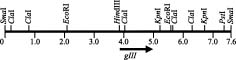
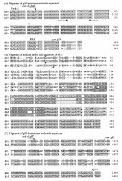
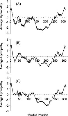
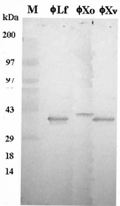
Abstract. fXo, Xf, fLf, fXv, and Cf are filamentous bacteriophages isolated in Taiwan. Both fXo and Xf specifically infect Xanthomonas oryzae pv. oryzae, and fLf, fXv, and Cf specifically infect X. campestris pv. campestris, X. campestris pv. vesicatoria, and X. campestris pv. citri, respectively. In this study, the fXo gene III (gIII) encoding the adsorption protein (pIII) was cloned by probing with the fLf gIII. Sequence analysis revealed that the fXo gIII is 1,023-nt long and able to encode a pre-protein of 340 aa (35,337 Da), with structural features typical of filamentous phage adsorption proteins: an N-terminal signal sequence (18 aa), a central region (90 aa) containing 38 glycine, 29 aspartic acid and 19 histidine residues, and a C-terminal membrane-anchoring domain (17 aa). The fXo pIII purified from the phage particles migrated as a 42-kDa band in SDS-polyacrylamide gel, which is substantially larger than that deduced from the nucleotide sequence, presumably due to the presence of the long stretch of charged residues in the central region. The fXo pIII could cross-react with the antiserum specific to fLf pIII, which also cross-reacts with fXv pIII. Like the situations in fLf and fXv, the pIIIs of fLf and fXo are also interchangeable. The gIII and the flanking regions of fLf, fXv and fXo are highly homologous and similar in size, but fXo has a genome (7.6 kb) larger than that of fLf (6.0 kb) and fXv (6.4 kb), suggesting that fXo is able to accommodate more genes and/or have longer intergenic regions in the remaining part of the genome. Difference in sizes between the pIIIs indicates that fXo and Xf, which has a predicted pre-pIII of 488 aa (51,036 Da), are distinct phages.

Keywords: Adsorption protein; Aspartic acid and histidine; Gene III; High content of glycine; Membrane-anchoring domain; Signal sequence; Xanthomonas.

Introduction

Several members of Xanthomonas, a genus of gram-negative phytopathogenic bacteria, are known to carry filamentous phages, e.g., fLf, fXv and Cf specifically infecting X. campestris, X. vesicatoria and X. citri, respectively, and Xf and fXo both infecting X. oryzae (Dai et al., 1980; Kuo et al., 1969; Lin et al., 1994; Tseng et al., 1990). Among them, all isolated in Taiwan, the nucleotide sequence has been determined for the fLf and Cf genomes (Kuo et al., 1991; Wen, 1992), whereas the amino acid sequence has been described in an abstract for the Xf gene III coding for the adsorption protein (pIII) (Yang and Yang, 1998), although the sequence was not yet available in the

database. Like other filamentous phages, they possess a circular single-stranded DNA (ssDNA) genome, produce replicative form (RF) during DNA replication, and propagate without lysis of the host cells (Dai et al., 1980; Kuo et al., 1969; Lin et al., 1994; Model and Russel, 1988; Tseng et al., 1990). Several interesting properties of these Xanthomonas phages have also been noticed, including: i) they hold restrictive host specificity, each phage being able to infect only its own host; however, they can propagate in the non-host Xanthomonas cells upon electroporation with RF or ssDNA, and the electroporated cells are capable of releasing authentic phage particles (Lin et al., 1994; Yang and Yang, 1997) indicating that host specificity is determined by the early steps of infection, i.e., adsorption and/or penetration, and ii) the genomes of Cf (7.8 kb), Xf (7.4 kb), and fXo (7.6 kb) are similar in size, but substantially larger than that of fLf (6.0 kb) and fXv (6.4 kb) (Kuo et al., 1991; Lin et al., 1994; Wen, 1992; Yang and Yang, 1998). Although Xf and fXo infect the same host, comparative study of these two phages has not been performed.

In filamentous phages, such as the best studied Ff phages (the closely related M13, f1 and fd), a phage particle contains about 2,700 copies of the major coat protein (pVIII) and three to five copies each of the four minor coat proteins (pIII, pVI, pVII and pIX), with pIII and pVI located at one end and pVII and pIX located at the other

1Present address: Food Industry Research & Development Institute, Hsinchu 300, Taiwan.

2Present address: Pesticide Toxicology Department, Taiwan Agricultural Chemicals and Toxic Substances Research Institute, Wu-Feng, Taichung 413, Taiwan.

3Present address: Center for Biophysics and Computational Biology, University of Illinois at Urbana-Champaign, Urbana, IL 61801, U.S.A.

4Present address: Department of Microbiology, Tzu-Chi University, Hualien 970, Taiwan.

*Corresponding author. Tel: 886-4-285-1885; Fax: 886-4-287-4879; E-mail: yhtseng@dragon.nchu.edu.tw


Botanical Bulletin of Academia Sinica, Vol. 42, 2001

(Model and Russel, 1988). pIII (adsorption protein) mediates phage adsorption to pilus (receptor recognition) and is necessary for phage uncoating and DNA penetration into the host cell (Endemann et al., 1992; Endemann and Model, 1995; Stengele et al., 1990), which also requires the function of host proteins TolQ, R and A (Levengood and Webster, 1989; Sun and Webster, 1987; Webster, 1991). In Xanthomonas filamentous phages, adsorption and penetration have not been studied in detail. It was only recently that we reported the purification of fLf and fXv pIIIs and the sequences for the corresponding genes (gIIIs) and demonstrated that they determine host specificity (Liu et al., 1998; Lin et al., 1999). In addition, interchangeability has been demonstrated between the pIIIs of fLf and fXv and between the pIIIs of Cf and Xf, which resulted in the change of host specificity (Liu et al., 1998; Lin et al., 1999; Yang and Yang, 1997). In this study, we purified the pIII from fXo phage particles and cloned and sequenced the corresponding gene. Western blotting demonstrated that the purified fXo pIII could cross-react with the antiserum specific to fLf pIII. Sequence analysis revealed that the fXo pIII possessed structural features typical of filamentous phage pIIIs. The fXo pre-pIII was similar to that of the fLf and fXv both in size and amino acid sequence, but 148 amino acid residues smaller than that of the Xf pre-pIII.

Materials and Methods

Bacterial Strains, Plasmids and Cultivation Conditions

Xanthomonas campestris pv. campestris strain P20H (Yang et al., 1988) and X. oryzae pv. oryzae strain Xo21 (Lin et al., 1994) were the hosts of fLf and fXo, respectively, and were used separately for phage propagation and as the indicator cells in plaque assay. Escherichia coli strain DH5a was used for gene cloning, and strain JM101 was the host for propagating M13 derivatives. LB broth and L agar (Miller, 1972) were the media for growing Xanthomonas (28°C) and E. coli (37°C). Plasmid pRKG3 (Liu et al., 1998) was constructed previously by cloning the fLf gIII, within a PCR-amplified 1,147-bp fragment, into the broad-host-range vector pRK415 (Keen et al., 1988), which contained the RK2 origin. To select for plasmids, the media were supplemented with antibiotics, ampicilin (50 µg/ml), kanamycin (50 µg/ml), or tetracycline (15 µg/ml).

Phage and DNA Techniques

Phages were propagated and purified as described by Lin et al. (1999). Double-layer bioassay (Eisenstark, 1967) was performed to determine phage titers. A spot test (Tseng et al., 1990) was carried out to verify phage sensitivity. Restriction endonucleases, T4 ligase and other enzymes were purchased from New England BioLabs and used in accordance with the instructions supplied. Preparation of plasmid and RF DNA, gene cloning, preparation of 32P-labeled probes, Southern hybridization, and trans

formation of E. coli were performed as described by Sambrook et al. (1989). Xanthomonas strains were transformed by electroporation (Wang and Tseng, 1992).

Sequence Analysis

Signal sequence and membrane anchoring domain were predicted with the PSORT program (Nakai and Kanehisa, 1991). Hydropathy plot was done by the program of Kyte and Doolittle (1982). Sequence of both strands was determined by the dideoxy chain termination method of Sanger et al. (1977).

Protein Techniques

The fXo pIII was purified with a fast protein liquid chromatography (FPLC) system as described for purification of the fLf pIII (Liu et al., 1998). Protein was separated by SDS-polyacrylamide gel electrophoresis (Laemmli, 1970). Western blotting was performed as described by Sambrook et al. (1989), using the antiserum prepared by immunizing a rabbit with the FPLC-purified fLf pIII (Liu et al., 1998).

Nucleotide Sequence

The nucleotide sequence reported here has been registered in GenBank under accession number AF162859.

Results and Discussion

Cloning and Sequencing of the fXo Gene III

To clone the fXo gIII, we used several restriction endonucleases to cut the fXo RF DNA, and the gIII-containing fragments were probed with the labeled pRKG3 containing the fLf gIII (Liu et al., 1998). The smallest fragment showing a hybridization signal was the 1.4-kb HindIII-KpnI fragment. The position of this fragment was near the middle of the restriction map of the linearized fXo RF DNA (Figure 1). The HindIII-KpnI fragment was subcloned into M13mp18 and mp19, and then the nucleotide sequence on both strands was determined. This fragment contained 1,386 bp with a G+C content of 56.5%, similar to that of the fLf genome (Wen, 1992), but deviating from that of the X. oryzae chromosome (64.6%) (Bradbury, 1984). The gIII-coding region spanned for 1,023 nt, starting with GTG at nt 146 and terminating with TGA at nt 1,168. Eleven nt upstream from the predicted translation initiation codon was a possible ribosome-binding site, 5'-AAGG-3', which was complementary to the 3'-end of the X. campestris pv. campestris 16S rRNA (Lin and Tseng, 1997; Figure 2A). This gIII was able to encode a polypeptide of 340 aa with a predicted molecular weight of 35,337, a size similar to that of the fLf pre-pIII (333 aa, 32,857) and the fXv pre-pIII (328 aa, 31,937 Da), but much smaller than that of the Xf pre-pIII (488 aa, 51,036 Da) (Yang and Yang, 1998).

Previously, nucleotide sequence analysis has shown that the upstream flanking regions of fLf gIII and fXv gIII are highly homologous, with an identity of 93% (Lin et al.,


Liu et al. — Adsorption protein of filamentous phage

1999). With a HindIII site at their left ends, they are 145 nt in length containing the C-terminal 5 codons (16 nt) of the major coat protein gene (gVIII) and the 129-bp gVIII/gIII intergenic region. Within these intergenic regions are the inverted repeats, resembling a transcription terminator for the upstream cistron (gVIII) and the gIII promoter (Lin et al., 1999). The gIII upstream region of fXo gIII was also 145-nt long, which shared 93% and 95% identity with the upstream regions of fLf and fXv gIIIs, respectively (Figure 2A). Consequently, sequences identical to the 16-nt C-terminus of gVIII and the putative transcriptional terminator present in the corresponding regions of fLf and fXv were all found in the upstream region of fXo gIII (Figure 2A). The nucleotide G at 74 nt upstream from the fLf gIII was identified as the transcriptional start site (Lin et al., 1999). Based on analogy, the same nucleotide G at 74 nt upstream from the fXo gIII start codon was predicted to be the transcriptional start site (Lin et al., 1999; Figure 2A).

The fLf gIII is followed by gVI, the gene coding for one of the minor coat proteins, after an intergenic region of 42 nt; it initiates with GTG situated 4 nt downstream from a possible ribosome binding site (Liu et al., 1997). In fXv, gIII is also followed by gVI, with a larger intergenic region of 129 nt (Wang, W.-H. and Tseng, Y.-H., unpublished results). In the downstream flanking sequence of fXo gIII (218 nt), which possessed 86.7% identity to the corresponding region of fLf, there was an incomplete open reading frame of 59 codons (Figure 2C). Its deduced amino acid sequence possessed 80.2% identity to that of the fLf gVI, although it was 40 nt instead of 42 nt behind gIII and initiated with ATG instead of GTG. These results indicated that, in gIII and the flanking regions, fXo had a genome organization identical to that of fLf and fXv, gVIII-gIII-gVI (Lin et al., 1999; Wang, W. -H. and Tseng Y.-H., unpublished results).

The size of the fXo RF DNA (7.6 kb) is similar to that of Xf (7.4 kb) and Cf (7.8 kb), but 1.6 kb and 1.2 kb larger than that of fLf and fXv, respectively (Kuo et al., 1991; Lin et al., 1994; Wen, 1992; Yang and Yang, 1998). However, the gIIIs and the flanking regions of the fLf, fXv and fXo are similar in length and contain the analogous genes, gVIII-gIII-gVI. Therefore, it seems safe to predict that fXo may accommodate more genes than fLf and fXo do or have larger intergenic regions in the remaining part of the fXo genome. In contrast, differences were noticed between the fXo gIII and the gIIIs of Cf and Xf: i) no homology was found between the fXo gIII and any open reading frame from the complete sequence of the Cf genome (Kuo et al., 1991), and ii) the fXo gIII was 444 nt less than the Xf gIII (Yang and Yang, 1998). These differences suggest fXo to be phylogenetically closer to fLf and fXv than to Cf, and that fXo and Xf, although infecting the same host, are distinct phages.

Amino Acid Sequence Analysis of the fXo pIII

The deduced amino acid sequence of the fXo pre-pIII shared 52% and 60% identities with that of the fLf pre-

Figure 1. Restriction map of the fXo RF DNA and the position of gene III. The 7.6-kb, double-stranded, circular DNA molecule is linearized at the unique SmaI site. The position of gene III, within the 1,386-bp HindIII-KpnI fragment, is indicated by an arrow.

Figure 2. (A) Alignment of the fXo gIII upstream region with that of the fLf and fXv. Shown are the left-most region of 163 nt from the sequenced HindIII-KpnI fragment, including the C-terminus of gVIII (16 nt), the intergenic region (145 nt), and the N-terminus of gIII (18 nt). Identical nucleotides are shadowed. The inverted arrows indicate the sequences having the potential to form a stem-loop structure resembling the transcription terminator. +1 is the transcription start site G determined for fLf gIII (Lin et al., 1999). RBS stands for the predicted ribosome-binding site, the Shine-Dalgarno sequence. (B) Alignment of the deduced amino acid sequences of the fXo, fLf and fXv pIIIs. Spaces are introduced for optimal alignments. Identical amino acid residues are shadowed. Vertical arrows indicate the computer-predicted sites for signal peptidase cleavage. The glycine-rich regions are blocked. (C) Alignment of the fXo gIII downstream region with that of the fLf. The end of gIII and the start of gVI are shown.


Botanical Bulletin of Academia Sinica, Vol. 42, 2001

pIII and fXv pre-pIII, respectively. The highest degree of identity was found in the C-terminal 100 aa, about 90.5 % among the three pre-pIIIs. In contrast, only 21.8% and 22.9% identity to the pre-pIIIs of fLf and fXv, respectively, were found in the N-terminal 100 aa. Computer analysis of the fXo pre-pIII predicted an 18-residue signal sequence in the N-terminus and a membrane-anchorage region of 17 residues in the C-terminus. In the central region was a 90-aa sequence rich in glycine, aspartic acid, and histidine (Figure 2B). These are structural features typical of filamentous phage pIIIs (Bross et al., 1988; Davis et al., 1985; Endemann and Model, 1995; Hillet al., 1991; Stengele et al., 1990). It is worth noting that similar to the situation in nucleotide sequence comparison, no homology was found between the fXo pre-pIII and the amino acid sequence deduced from the complete sequence of the Cf genome (Kuo et al., 1991).

The N-terminal 18 aa of the deduced fXo pre-pIII was highly hydrophobic; it had an arginine at position 3 which was followed by 11 hydrophobic residues and ended with an alanine (Figure 2B). This primary structure in the N-terminus of a protein molecule is typical of a signal sequence (Pugsley, 1993). After cleavage between Ala-18 and Ala-19, a mature protein with a MW of 33,158 would be produced. In the fLf and fXv pre-pIIIs, signal sequences have also been predicted. These sequences are different from one another in amino acid composition but similar in properties of the residues (Figure 2B).

The fLf pIII and fXv pIII each possesses a region rich in glycine and aspartic acid (Wen and Tseng, 1996; Lin et al., 1999). The fLf protein has the amino acid residues repeating as GD (7 times) and GGGD (11 times), whereas the fXv protein possesses the repeats appearing as GD (11 times), GGSD (5 times), and GGGD (4 times). As shown in Figure 2B, the central region in the deduced fXo pIII was 90-aa long, containing 38 glycine, 29 aspartic acid, and 19 histidine residues. They were clustered mainly as GD, GDGH and GDDH, repeating for 2, 12 and 7 times, respectively, between aa 156-245 (Figure 2B). This region was longer than the corresponding regions of fLf pIII (70 residues) and fXv pIII (75 residues) (Lin et al., 1999; Wen and Tseng, 1996). Comparison of the three central regions in amino acid composition indicated that the fLf pIII contains only glycine and aspartic acid, whereas in addition to these amino acids, the fXv protein contains serine (5 residues) and the fXo protein contains histidine. Alignment of the three pIIIs revealed that the insertion of three consecutive GDDHs caused the fXo central region to be longer than the corresponding regions in the other pIIIs (Figure 2B). In hydropathy plot, the glycine-rich region of the fXo pIII is strongly hydrophilic, similar to the property of the glycin-rich regions in the fLf and fXv pIIIs but a little stronger in hydrophilicity (Figure 3). This is consistent with the higher content of the charged amino acid residues in the fXo pIII central region than in the other pIIIs .

The predicted membrane-embedding domain at C-terminus was 17-residue long spanning aa 315 to 331 in the de

duced fXo pre-pIII (Figure 2B). A similar domain has also been predicted for fLf pIII (aa 308 to 324) and fXv pIII (aa 303 to 319) (Lin et al., 1999; Figure 2B), presumably required for anchoring the pIII proteins into the inner membrane of host cell. Within these domains, fLf and fXv have the same sequence, and only one amino acid residue was found to be different in the fXo pIII (Figure 2B).

The pIIIs of Ff and IKe possess a very low degree of overall homology (15%), with the highest homology (43%) being found in the regions required for penetration and the non-homologous regions being essential for receptor recognition to recognize and bind specifically to their respective receptor pilus. Therefore, in these two phages, one pIII can not replace the functionally analogous protein of the other phage (Bross et al., 1988; Endemann et al., 1992; Endemann et al., 1993). In contrast to these cases, the deduced pIIIs of fLf, fXv, and fXo possess a high degree of identity. In addition, the three pIIIs are interchangeable (Liu et al., 1998; Lin et al., 1999; see below). These findings suggest that these three phages share the same sequence information required for assembling the pIII

Figure 3. Hydropathy plots for the deduced pIIIs of fXo (A), fLf (B) and fXv (C). A window of 21 amino acids was used with the hydrophobicity scales of Kyte and Doolittle (1982).


Liu et al. — Adsorption protein of filamentous phage

into phage particles. The required sequence information in pIII is most likely located in the C-terminus of the polypeptide, since the highest degree of identity is concentrated in this region. On the other hand, to hold restrictive host specificity, specific sequences must be required for receptor recognition and binding. Thus, with a low degree of homology, the N-terminal regions of ca.100 aa of the three Xanthomonas phage pIIIs are the most probable candidates for the receptor recognition.

Purification and Western Blot Analysis of pIIIs

The fXo pIII was purified from the viral particles by two passages through the gel filtration column (Suprose 12) in fast protein liquid chromatography (FPLC) as described for the purification of the fLf and fXv pIIIs (Liu et al., 1998; Lin et al., 1999). The same peak patterns as those in the purification of the fLf and fXv pIIIs were observed, and the fXo pIII was recovered from the second peak from the second chromatography (data not shown). In SDS-PAGE, a single band with a molecular mass of ca. 42 kDa was visualized upon staining the proteins with Coomassie brilliant blue (data not shown). This size was substantially larger than the value calculated for the mature fXo pIII (322 residues, 33,158 Da), deduced from the nucleotide sequence determined in this study. Running in the same gel, the fLf and fXv pIIIs exhibited the same mobility corresponding to a molecular size of ca. 36 kDa, which is slightly larger than the sizes deduced for the mature pIIIs of fLf (311 aa, 30,497 Da) and fXv (304 aa, 29,463 Da) (Lin et al., 1999; Figure 4). A similar discrepancy has also been observed in Ff phages, in which the mature pIII (424 aa) with a calculated MW of 42,675 exhibits a mobility corresponding to sizes of 59-70 kDa in SDS-PAGE (Endemann and Model, 1995; Goldsmith and Konigsberg, 1977; Woolford et al., 1977). Several explanations have been proposed, one of which attributes the discrepancy to the presence of unusual clustering of glycine and serine in the protein molecule (Endemann and Model, 1995; Woolford et al., 1977). This proposal seems to explain the discrepancy observed in pIIIs of the Xanthomonas phages. In addition, the stronger effect on the discrepancy observed in fXo pIII than in the pIIIs of fLf and fXv, presumably due to the presence of three more GDDH repeats, may give further support to this proposal.

It has been shown that the antiserum specific to fLf pIII can cross-react with the fXv pIII (Lin et al., 1999). In this study, the purified fXo pIII was electrophoresed in SDS-polyacrylamide gel, then transferred onto a nylon membrane and subjected to Western blot analysis using the same antiserum, with the pIIIs of fLf and fXv as the controls. As shown in Figure 4, each of the pIIIs had the same mobility in the SDS-polyacrylamide gel as that described above, and the anti-fLf pIII serum was able to cross-react with fXo pIII. These results indicate that a high degree of identity in amino acid sequence is shared not only between fLf and fXv pIIIs, but also between fLf and fXo pIIIs.

Figure 4. Western blot analysis of the pIIIs from fLf, fXo and fXv. The pIIIs were purified by FPLC, subjected to SDS-polyacrylamide gel electrophoresis, transferred onto nylon membrane and reacted with the antiserum raised against the FPLC-purified fLf pIII.

Interchangeability of pIIIs

We have previously demonstrated that the fXv and fLf pIIIs are interchangeable (Liu et al., 1998; Lin et al., 1999). In this study, we electroporated the fXo RF DNA into X. campestris pv. campestris strain P20H carrying pRKG3, a plasmid containing cloned fLf gIII. The results showed that the culture supernatant contained a mixture of authentic fXo, which could infect Xo21, and a chimeric phage which could infect P20H. These results indicated that the pIIIs of fLf and fXo are also interchangeable.

Acknowledgments. We thank Ming-Ren Yen and David Hsia for the help in preparation of this manuscript. This study was supported by National Science Council of Republic of China, grant number NSC86-2311-B-005-038

Literature Cited

Bradbury, J.F. 1984. Genus II. Xanthomonas. In N. R. Kreig and J. G. Holt (eds.), Bergey's Manual of Systematic Bacteriology. Vol. 1. The Williams & Wilkins Co., Baltimore, Md., pp. 199.

Bross, P., K. Bussmann, W. Keppner, and I. Rasched. 1988. Functional analysis of the adsorption protein of two filamentous phages with different host specificities. J. Gen. Microbiol. 134: 461-471.

Dai, H., K.-S. Chiang, and T.-T. Kuo. 1980. Characterization of a new filamentous phage Cf from Xanthomonas citri. J. Gen. Virol. 46: 277-289.

Davis, N.G., J.D. Boeke, and P. Model. 1985. Fine structure of a membrane anchor domain. J. Mol. Biol. 181: 111-121.

Eisenstark, A. 1967. Bacteriophage Techniques. In K. Maramorosch and H. Koprowski (eds.), Methods in


Botanical Bulletin of Academia Sinica, Vol. 42, 2001

Virology. Academic Press, New York.

Endemann, H., P. Bross, and I. Rasched. 1992. The adsorption protein of phage IKe. Localization by deletion mutagenesis of domains involved in infectivity. Mol. Microbiol. 6: 471-478.

Endemann, H., V. Gailus, and I. Rasched. 1993. Interchangeability of the adsorption proteins of bacteriophages Ff and IKe. J. Virol. 67: 3332-3337.

Endemann, H. and P. Model. 1995. Location of filamentous phage minor coat proteins in phage and in infected cells. J. Mol. Biol. 250: 496-506.

Goldsmith, M.E. and W.H. Konigsberg. 1977. Adsorption protein of the bacteriophage fd: isolation, molecular properties, and location in the virus. Biochemistry 16: 2686-2694.

Hill, D.F., N.J. Short, R.N. Perham, and G.B. Petersen. 1991. DNA sequence of the filamentous bacteriophage Pf1. J. Mol. Biol. 218: 349-364.

Keen, N.T., S. Tamaki, D. Kobayashi, and D. Trollinger. 1988. Improved broad-host-range plasmids for DNA cloning in gram-negative bacteria. Gene 70: 191-197.

Kuo, T.-T., T.-C. Huang, and T.-Y. Chow. 1969. A filamentous bacteriophage from Xanthomonas oryzae. Virology 39: 548-555.

Kuo, T.-T., M.-S. Tan, M.-T. Su, and M.-K. Yang. 1991. Complete nucleotide sequence of filamentous phage Cf1c from Xanthomonas campestris pv. citri. Nucleic Acids Res. 19: 2498.

Kyte, J. and R.F. Doolittle. 1982. A simple method for displaying the hydropathic character of a protein. J. Mol. Biol. 157: 105-132.

Laemmli, U.K. 1970. Cleavage of structural proteins during the assembly of the head of bacteriophage T4. Nature 227: 680-685.

Levengood, S.K. and R.E. Webster. 1989. Nucleotide sequences of the tolA and tolB genes and localization of their products, components of a multistep translocation system in Escherichia coli. J. Bacteriol. 171: 6600-6609.

Lin, N.-T., B.-Y. You, C.-Y. Huang, C.-W. Kuo, F.-S Wen, J.- S. Yang, and Y.-H. Tseng. 1994. Characterization of two novel filamentous phages of Xanthomonas. J. Gen. Virol. 75: 2543-2547.

Lin, N.-T. and Y.-H. Tseng. 1997. Sequence and copy number of the Xanthomonas campestris pv. campestris gene encoding 16S rRNA. Biochem. Biophys. Res. Commun. 235: 276-280.

Lin, N.-T., T.-J Liu, T.-C. Lee, B.-Y. You, M.-H. Yang, F.-S. Wen, and Y.-H. Tseng. 1999. The adsorption protein genes of Xanthomonas campestris filamentous phages determining host specificity. J. Bacteriol. 181: 2465-2471.

Liu, T.-J., F.-S. Wen, T.-T. Tseng, M.-T. Yang, N.-T. Lin, and Y.-H. Tseng. 1997. Identification of gene VI of filamentous phage fLf coding for a 10-kDa minor coat protein. Biochem. Biophys. Res. Commun. 239: 752-755.

Liu, T.-J., B.-Y. You, N.-T. Lin, M.-T. Yang, and Y.-H. Tseng. 1998. Purification and expression of the gene III protein from filamentous phage fLf. Biochem. Biophys. Res. Commun. 242: 113-117.

Miller, J.H. 1972. Expriments in Molecular Genetics. Cold Spring Harbor, New York.

Model, P. and M. Russel. 1988. Filamentous Bacteriophage. In R. Calender (ed.), Filamentous Bacteriophage. Plenum Press, New York.

Nakai, K. and M. Kanehisa. 1991. Expert system for predicting protein localization sites in gram-negative bacteria. Proteins 11: 95-110.

Pugsley, A.P. 1993. The complete general secretory pathway in gram-negative bacteria. Microbiol. Rev. 57: 50-108.

Sambrook, J., E.F. Fritsch, and T. Maniatis. 1989. Molecular Cloning: A Laboratory Manual. 2nd ed. Cold Spring Harbor Laboratory Press, Cold Spring Harbor, New York.

Sanger, F., S. Nicklen, and A. R. Coulson. 1977. DNA sequencing with chain-terminating inhibitors. Proc. Natl. Acad. Sci. U S A 74: 5463-5467.

Stengele, I., P. Bross, X. Garces, J. Giray, and I. Rasched. 1990. Dissection of functional domains in phage fd adsorption protein. Discrimination between attachment and penetration sites. J. Mol. Biol. 212: 143-149.

Sun, T.P. and R.E. Webster. 1987. Nucleotide sequence of a gene cluster involved in entry of E colicins and single-stranded DNA of infecting filamentous bacteriophages into Escherichia coli. J. Bacteriol. 169: 2667-2674.

Tseng, Y.-H., M.-C Lo, K.-C Lin, C.-C Pan, and R.-Y. Chang. 1990. Characterization of filamentous bacteriophage fLf from Xanthomonas campestris pv. campestris. J. Gen. Virol. 71: 1881-1884.

Wang, T.-W. and Y.-H. Tseng. 1992. Electrotransformation of Xanthomonas campestris by RF DNA of filamentous phage fLf. Lett. Appl. Microbiol. 14: 65-68.

Webster, R. E. 1991. The tol gene products and the import of macromolecules into Escherichia coli. Mol. Microbiol. 5: 1005-1011.

Wen, F.-S. 1992. Genomic organization of filamentous phage fLf of Xanthomonas campestris pv. campestris. Ph. D.Dissertation, National Chung University.

Wen, F.-S. and Y.-H. Tseng. 1996. Nucleotide sequence of the gene presumably encoding the adsorption protein of filamentous phage fLf. Gene 172: 161-162.

Woolford, J.L., Jr., H.M. Steinman, and R.E. Webster. 1977. Adsorption protein of bacteriophage fl: solubilization in deoxycholate and localization in the fl virion. Biochemistry 16: 2694-2700.

Yang, B.-Y., H.-F. Tsai, and Y.-H. Tseng. 1988. Broad host range cosmid pLAFR1 and non-mucoid mutant XCP20 provide a suitable vector-host system for cloning genes in Xanthomonas campestris pv. campestris. Chin. J. Microbiol. Immunol. 21: 40-49.

Yang, M.-K. and Y.-C. Yang. 1997. The A protein of the filamentous bacteriophage Cf of Xanthomonas campestris pv. citri. J. Bacteriol. 179: 2840-2844.

Yang, Y.-C. and M.-K. Yang. 1998. The identification of coat protein genes of filamentous bacteriophage Xf from Xanthomonas campestris pv. oryzae. Abstract 98th Gen. Meet., Amer. Soc. Microbiol., pp. 364, M-15.


Liu et al. — Adsorption protein of filamentous phage

¤ô½_¥Õ¸­¬\¯fµß½uª¬¾½µßÅé fXo ¤§§lªþ³J¥Õ

¼B¯ª§g ´åºÑ®K ´¿¦b¥Ð ªL©Àæ¦ ·¨©ú¼w ´¿¸q¶¯

°ê¥ß¤¤¿³¤j¾Ç¤À¤l¥Íª«¾Ç¬ã¨s©Ò

fXo¡BXf¡BfLf¡BfXv »P Cf ¬°¦b¥xÆW¤ÀÂ÷¤§½uª¬¾½µßÅé¡C fXo »P Xf ±M¤@·P¬V Xanthomonas oryzae pv. oryzae¡AfLf¡BfXv »P Xf «h¤À§O±M¤@·P¬V X. campestris pv. campestris¡BX. campestris pv. vesicatoria »P X. campestris pv. citri¡C¥»¬ã¨s¥H fLf ¤§°ò¦] III (gIII) (¥D¾É§lªþ³J¥Õ pIII) ¬°±´¤l¿ï´Þ¨ì fXv ¤§ gIII¡C ®Ö»Ä§Ç¦C¤ÀªR¤§µ²ªGÅã¥Ü¸Ó gIII ¥þªø 1,023 nt¡A¥D¾É¤@­Ó 340 ­ÓÓi°ò»Ä (¤À¤l¶q 35,337) ¤§«e³J¥Õ¡C¸Ó ³J¥Õ¨ã¦³½uª¬¾½µßÅé§lªþ³J¥Õ¤§¯S¼x¡G¦b N-ºÝ¦³¤@«H¸¹§Ç¦C (18 ­ÓÓi°ò»Ä)¡A¦b¤¤¬q (90 ­ÓÓi°ò»Ä) ¦³°ª§t¶qªº¥ÌÓi»Ä (38 ­Ó)¡A¤Ñªù¥VÓi»Ä (29 ­Ó) ¤Î²ÕÓi»Ä (19 ­Ó)¡A¤Î¦b C-ºÝ¦³¤@­Ó¨Ñ´O¤J²Ó­M½¤ªº¤æ ªù (17 ­ÓÓi°ò»Ä)¡C¥Ñ¾½µßÅé fXo Áû²É¯Â¤Æ¥X¨Óªº pIII ¦b SDS-»E¤þ²mÓi¾®½¦¹qªa®É¡Aªa°Êªº¶ZÂ÷»P 42 kDa ³J¥Õ¤§ªa°Ê±¡§Î¬Û·í¡A¦Ó»·¤j©ó¸g¥Ñ gIII ©Ò±Àºâªº¤À¤l¶q¡C¦¹¤@±¡§Î¥i¯à»P fXo pIII ªº¤¤¬q§t¦³°ª ¶qªº±a¹q²üÓi°ò»Ä°ò¸s¦³Ãö¡C¯Â¤Æ¤§ fXo pIII ¯à»P§Ü fLf pIII ¦å²M°_¥æ¤e¤ÏÀ³¡C fXo ¤§ pIII ¯à»P fLf ¬Û¥æ´«¯Ç¤J¾½µßÅéÁû²É¡A±¡§Î»P fLf ªº pIII »P fXv ªº pIII ¯à¬Û¤¬¥æ´«Ãþ¦ü¡C¦b°ò¦]Åé¤j¤p¤è­±¡A fXo (7.6 kb) »P Xf (7.4 kb) ¨âªÌ¬Û¦ü¡A¦ý¤ñ fLf (6.0 kb) »P fXv (6.4 kb) ¤§°ò¦]Åé³£¤j¡F¤£¹L¡A fXo¡BfLf »P fXv ªº gIII ¤Î¤W¤U´åÃä¬Éªº¬q¸¨¤£¶È°ò¦]²Õ¦¨¬Û¦P¡Aªøµu»P®Ö»Ä§Ç¦C¤]³£°ª«×¬Û¦ü¡C¦¹¤@¨Æ¹êÅã¥Ü¡A fXo ¥i¯à¤ñ fLf ¤Î fXv ±a¦³§ó¦h°ò¦]¡A¤Î / ©Î fXo ¦b°ò¦]Å骺¨ä¥L³¡¤À¥i¯à¦³¸ûªøªº°ò¦]¶¡°Ï°ì¡C ¥Ñ©ó Xf ªº pIII ªø¹F 448 ­ÓÓi°ò»Ä¡A¥i¨£ fXo »P Xf ¬° X. oryzae pv. oryzae ªº¨âºØ¤£¦P½uª¬¾½µßÅé¡C

ÃöÁäµü¡G §lªþ³J¥Õ¡F¤Ñªù¥VÓi»Ä»P²ÕÓi»Ä¡F°ò¦] III ¡F°ª§t¶q¤§¥ÌÓi»Ä¡F´O¤J²Ó­M½¤ªº¤æªù¡F«H¸¹§Ç¦C¡C