Bot. Bull. Acad. Sin. (2001) 42: 207-214

Liu and Lin — Ultrastructural study and lipid formation of Isochrysis sp.

Ultrastructural study and lipid formation of Isochrysis sp. CCMP1324

Ching-Piao Liu and Liang-Ping Lin*

Graduate Institute of Agricultural Chemistry, National Taiwan University, Taipei 106, Taiwan, Republic of China

(Received May 22, 2000; Accepted December 12, 2000)

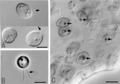
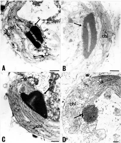
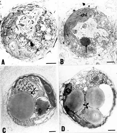
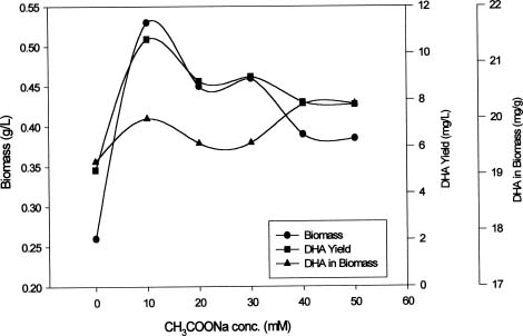
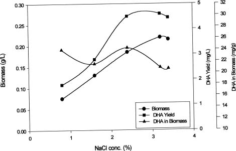
Abstract. This study investigates methods for extracting lipids from microalgae and analyzes the effects of culture media as well as culture conditions on PUFA yields and total fatty acid contents. Experimental results of an optimal culturing of Isochrysis spp. were based on a 3.2% salinity culture medium. These microalgae were cultured in a 1-2 L Roux's flat-flask and a 5 L jar fermentor. The optimum culture temperature and initial pH for DHA production were 25°C and 8.0, respectively. Pigments included chlorophylls a and c. The DHA yield increased with cultivation time until the eighth day. Optimum DHA amounts in the cells were reached under aeration with 10% CO2 and with continuous illumination of 10 klux. The biomass dry weight reached 4 g per liter of culture, and the DHA production reached 16 mg per liter of culture. Lipid bodies in Isochrysis spp. and related genera were observed during culture by light and transmission electron microscopy; 0.5~3.0 µm sized lipid bodies were confirmed by staining with Sudan Black B in cells from log stage to stationary stage cultures. These results demonstrated that DHA-containing lipid bodies in cells can be produced and accumulated in marine Isochrysis spp.

Keywords: Docosahexaenoic acid; Isochrysis sp.; Lipid formation; Polyunsaturated fatty acids (PUFA); Ultrastructure.

Introduction

Marine microalgae such as Isochrysis have received increasing interest because of their ability to produce the polyunsaturated fatty acid docosahexaenoic acid (DHA), one of the n-3 fatty acids believed to provide health benefits associated with the consumption of certain marine fish and their oils. DHA, a C22-polyunsaturated fatty acid, and its derivatives help prevent and treat pathologies such as coronary heart disease and atherosclerosis (Norday and Hansen, 1994), inflammatory problems, and some cancers, and are believed to play a role in infant nutrition (Conner and Neuringer, 1987). DHA accumulates in the membranes of nervous, visual, and reproductive tissues (Dratz and Deese, 1986). Polyunsaturated fatty acids are especially helpful in preventing heart and circulatory disease and facilitating brain development in infants (Yongmanitchai and Ward, 1991). Fish oils may not be an ideal source of n-3 PUFAs due to their scarcity and odor, as well as geographical and seasonal variations in quality (Varela et al., 1990).

Isochrysis has been widely used as a mariculture feed due to its high content of long chain polyunsaturated fatty acids (PUFAs) (Jeffrey et al., 1994). However, the lipid class and fatty acid compositions of microalgal cells at different growth phases can differ significantly (Emdadi and Berland, 1989), and can change with variations in culture conditions e.g. nutrient status, temperature, salinity, pH,

photoperiod, light intensity and light quality (reviewed by Yongmanitchai and Ward, 1989; Roessler, 1990). The cell structure of Isochrysis has attracted the attention of many investigators. Earlier studies (Green and Pienaar, 1977; Hori and Green, 1985; 1991) on Isochrysis galbana have mainly focused on its flagellar root system.

In this study, EM technologies were employed to survey this alga since previous papers have lacked detailed investigations of lipid formation in marine Isochrysis. This work also examines marine microalgae Isochrysis spp. as an alternative source of PUFAs and analyzes the culture medium and culture conditions that affect yields of PUFAs and their content in the total fatty acids. Lipid bodies in Isochrysis spp. and related genera are observed by light and transmission electron microscopy. Lipid granules are confirmed by staining with Sudan Black B in cells from the stationary cultures. The results suggest that DHA-containing lipid bodies in cells can be produced by marine Isochrysis. The possible commercial production of biomass and DHA-rich oil for use as food and feed ingredients is also predicted.

Materials and Methods

Cell Growth

Microalgal strains CCMP 463, 1324, 1325 and Pavlova salina were obtained from the Provasoli-Guillard Center for Culture of Marine Phytoplankton (West Boothbay Harbor, Maine USA). Isochrysis galbana TK1, TK2, were originally isolated by the Tungkang Marine Laboratory (Pingstung, Taiwan). Nannochloropsis oculata and Chlo

*Corresponding author. Fax: 886-2-23626455; E-mail: m046@ccms.ntu.edu.tw


Botanical Bulletin of Academia Sinica, Vol. 42, 2001

rella minutussima UTEX 2341 were obtained from our laboratory's previously collected strains. The cultures were grown under constant illumination in f/2 medium (Guillard and Ryther, 1962) at 25°C, pH 8, and an air-specific supply rate of 250 mL min-1. Artificial seawater was sterilized in an autoclave at 120°C for twenty minutes. Microalgae were cultured in 1-2 L Roux's flat-flasks and 5 L jar fermentors. The different salinities (NaCl concentrations) examined were 0.8, 1.6, 2.4 and 3.2%. Sodium acetate 10~50 mM was applied to the mixotrophic culture of microalgae. All cultures were harvested by centrifugation during the stationary growth phase for subsequent analysis. The algal biomass was lyophilized and stored at -30°C, and the lyophilized cells were utilized for analysis within two weeks. A lipid analysis was performed after the saponification and methylation. The extracted pigment concentrations of chl a and chl c were estimated by spectrophotometry (Jeffrey and Humphrey, 1975).

Light and Electron Microscopies

The lipid bodies in cells from the stationary cultures were stained with Sudan Black B (Weete et al., 1997) and observed under a Normarski differential interference contrast light microscope (LM). The algal cells were collected by centrifugation at a higher concentration for viewing by a Normarski DIC on a Nikon E-600 microscope.

The algal cells for electron microscopy (EM) were collected by centrifugation at 3,000 g, and fixed with 2.5% (v/v) glutaraldehyde in 0.2 M sodium cacodylate buffer at pH 7.2. The cells were postfixed for two hours with 1% osmium tetroxide in a cacodylate buffer. The fixed material was washed once in a cacodylate buffer prior to dehydration. The samples were dehydrated in a series of 30, 50, 70, 85, 90, 95 and 100% (v/v) acetone solutions for ten minutes each. The dehydrated cells were suspended in a 50:50 mixture of Spurr's resin and acetone for one hour and then embedded in 100% Spurr's resin. The embed

ded samples were polymerized at 65°C for twenty-four hours and sectioned using an LKB ultramicrotome. Thin sections were picked up on 300-mesh copper grids and post-stained with uranyl acetate for thirty minutes. After rinsing with distilled water, the ultra-sections were post-stained with lead citrate for four minutes and finally rinsed with distilled water. The sections were examined under a transmission electron microscope (JEOL JEM 1200 EXII) at an accelerating voltage of 80 kV.

Total Lipid and Fatty Acid Analysis

The total lipid was extracted from dry cells (ca. 20 mg) using the Bligh and Dyer (1959) procedure. The dry cells were re-extracted two or three times with small portions of CHCl3-MeOH (2:1, v/v) and purified by removing nonlipid contaminants (Folch et al., 1957). The total amount of lipid was calculated from the gas chromatographic data. The methyl ester derivatives of the fatty acid methyl esters were directly prepared from the cells, after adding pentadecanoic acid FAME (fatty acid methyl ester) as an internal standard. All FAME were analyzed by FID-GC, using a capillary column (RTX 225, 30 m length, 0.32 mm in diameter) in a Hewlett Packard 5890 gas chromatograph. The initial oven temperature was set at 150°C, followed by a temperature program of 4°C min-1 to a final oven temperature of 210°C. The injector and detector temperature were set at 230°C, and the flow rates for hydrogen and air were 30 and 400 mL min-1, respectively. Fatty acid contents were determined by comparing their peak areas with that of the internal standard.

Results

The growth and biomass production of Isochrysis spp. (in Roux's flat flask cultures) were examined after eight days of growth, the cells were harvested and the solvent extractable lipid content of cells was determined. The DHA

Table 1. The fatty acid composition of several marine microalgae (% total fatty acids).

Fatty Pavlova Isochrysis sp. Isochrysis sp. Isochrysis Isochrysis Pavlova lutheri Nannochloropsis Chlorella

acid salina CCMP 463 CCMP 1324 galbana galbana CCMP 1325 oculata minutussima

TK1 TK2 UTEX2341

14:0 10.1±0.2 10.4±0.9 10.2±0.5 17.5±1.0 16.3±0.9 10.3±0.5 5.1±0.4 4.5±0.4

16:0 23.4±0.4 14.9±0.7 17.6±0.6 14.3±0.4 12.9±0.7 20.8±1.2 32.1±1.4 33.9±1.6

16:1n-7 6.2±0.1 4.5±0.2 3.9±0.1 6.3±0.5 4.0±0.1 18.4±0.4 24.9±1.7 23.2±1.0

18:0 0.9±0.1 N.D. N.D. N.D. N.D. 0.4±0.1 2.7±0.2 2.9±0.1

18:1n-9 16.9±0.3 29.8±1.2 32.0±1.4 15.1±0.7 28.1±1.0 3.3±0.5 16.5±0.9 20.4±1.1

18:2n-6 7.8±0.4 5.7±0.4 4.1±0.3 8.8±0.4 3.0±0.1 1.9±0.1 1.9±0.3 3.4±0.1

18:3n-3 3.1±0.1 6.4±0.5 6.4±0.3 8.2±0.3 5.5±0.2 1.5±0.2 N.D. N.D. 18:4n-3 5.9±0.1 17.5±0.9 15.1±0.6 24.9±1.4 18.9±0.3 6.8±0.5 N.D. N.D.

20:4n-6 1.6±0.1 N.D. N.D. N.D. N.D. N.D. 2.8±0.2 1.9±0.1

20:5n-3 11.8±0.3 N.D. N.D. N.D. N.D. 21.0±0.5 9.4±0.7 8.7±0.2

22:6n-3 4.4±0.4 10.7±0.5 10.9±0.3 8.2±0.6 11.1±0.4 6.2±0.3 N.D. N.D.

SUn-3 25.2 34.6 32.3 41.4 31.8 35.5 12.2 8.7

SUn-6 9.4 5.7 4.1 8.8 3.0 1.9 4.7 5.2

n-3/n-6 2.7 6.1 7.9 4.7 10.6 18.7 2.6 1.7

N.D. = None detected.


Liu and Lin — Ultrastructural study and lipid formation of Isochrysis sp.

yield increased with cultivation time until the eighth day (data not shown). Eight species of marine microalgae were examined, including Pavlova lutheri CCMP1325, Pavlova salina, Isochrysis galbana TK1, Isochrysis galbana TK2, Isochrysis sp. CCMP1324, Isochrysis sp. CCMP463, Nannochloropsis oculata, and Chlorella minutussima UTEX 2341. Each species was cultured and analyzed for its fatty acid composition (Table 1). The fatty acid distribution of Isochrysis spp. and P. lutheri (Haptophyceae) revealed the dominance of 14:0, 16:0, 16:1n-9 or 18:1n-9 and 22:6n-3. In addition, Isochrysis spp. had a higher content of 18:1n-9 and 18:4n-3, whereas P. lutheri had a higher level of 20:5n-3. The abundance of PUFAs demonstrated a pronounced variation between algal species and classes. A similar finding was reported previously (Kjell et al., 1994). According to Table 1, 22:6 n-3 composition of Isochrysis sp. CCMP1324 and Isochrysis sp. CCMP463 were 10.9% and 10.7%, respectively, found to be higher in 22:6 contents and 20:5n-3 was absent. They were then subjected to further culture tests, and to test similarities in growth rates. Typical growth curves showed a lag phase of 4 days, a log phase of 4 to 8 days, and 8 to 14 days in a stationary phase under these conditions.

The extracted pigment concentration of chl a and chl c were estimated by spectrophotometry; chl a and c content were 2.8 µg/mL and 1.4 µg/mL in the culture periods (data not shown). Optical and electron microscopic photographs, confirmed the presence of lipid bodies (or osmiophilic droplets) in Isochrysis sp. CCMP 1324 (Figures

1-3). Many oil droplets accumulated in the late phase of culture, and the lipid granules in cells from the stationary cultures were confirmed by staining with Sudan Black B (Figure 1). The TEM photomicrographs demonstrate the lipid body formation occurs in the thylakoid space of the chloroplast structure. Their sizes vary with growth phase stage, and they finally form a rounded shape (Figure 2). The accumulated dense lipid granules were partially dissolved and diffused into cytosol and form less dense, large lipid globules. Lipid bodies from 0.5 to 3.0 µm were detected in samples harvested from the four to eleven day culture. The oil droplet accumulation, surveyed at each growth stage and illustrated in the EM photographs (Figure 3), was similar to that in a previous report (Weete et al., 1997). The Isochrysis sp. cells have no distinct cell wall, as confirmed by Zhu et al. (1997), and only possess a plasma membrane covering. Cells are generally solitary, motile, 5-6 µm long, 2-4 µm wide, and 2.5-3 µm thick in ellipsoid forms. There are two flagella, more or less equal, smooth, approximately 7 µm long, cells inserted with abbreviated haptonema; normally plastid usually single, parietal, yellow-brown with an immersed fusiform pyrenoid, the latter traversed by a pair of thylakoids, resembled that described for Isochrysis galbana previously (Green and Pienaar, 1977). The cells were fragile, and plasmolysis occurred when the naked cells were exposed to a sudden change of osmotic pressure. Some of the TEM micrographs are similar to those in a previous report (Goldman and Dennet, 1985). The ultrastructural morphological

Figure 1. Nomarski differential interference contrast microscopic photograph of Isochrysis sp., cells, showing two flagella at one end of a cell (A) (arrows). The photograph of phase contrast microscope, was shown in (B). After staining with Sudan Black B, showing the lipid accumulation inside the cells (C) (arrows). Scale bar = 5 µm.


Botanical Bulletin of Academia Sinica, Vol. 42, 2001

Figure 2. Transmission electron micrographs of Isochrysis sp. CCMP 1324, showing the lipid body (arrow) formation in chloroplast (A~C), their size from small to large and finally rounded in spherical form (D) at early log phase. Scale bar = 200 nm.

Table 2. Influence of different salinities on the fatty acid composition of Isochrysis sp. CCMP 1324 (% total fatty acids).

Fatty acids NaCl conc

0.8% 1.6% 2.4% 3.2%

14:0 18.4±0.9 18.6±0.9 16.9±1.2 16.3±0.6

16:0 15.5±0.7 13.4±0.7 13.4±0.7 12.9±0.4

16:1n-7 5.6±0.3 5.5±0.4 4.3±0.2 4.0±0.2

18:0 N.D. N.D. N.D. N.D.

18:1n-9 27.7±1.1 27.9±1.7 28.0±1.4 28.1±1.5

18:2n-6 4.3±0.2 6.2±0.7 5.0±0.4 3.0±0.3

18:3n-3 5.1±0.2 4.8±0.2 4.9±0.3 5.5±0.1

18:4n-3 14.9±0.9 14.6±0.7 17.1±0.8 18.9±1.1

20:5n-3 N.D. N.D. N.D. N.D.

22:6n-3 9.4±0.9 8.9±0.7 10.5±0.9 11.2±0.9

Total n-3 29.4 28.4 32.4 35.6

PUFA 33.6 34.9 37.4 38.6

N.D. = None detected.


Liu and Lin — Ultrastructural study and lipid formation of Isochrysis sp.

Figure 3. Transmission electron micrographs of Isochrysis sp. CCMP 1324 vegetative cell. After the fourth day of growth, no oil drop was observed (A), but oil droplets (arrows) could be observed in the stationary phase (6~11th day) (B~D). Scale bar = 500 nm.

Table 3. Variation in fatty acid composition of Isochrysis sp. CCMP 1324 in different sodium acetate concentration (% total fatty acids).

Fatty acids CH3COONa

10 mM 20 mM 30 mM 40 mM 50 mM

14:0 17.8±0.9 16.5±0. 8 16.4±0.4 15.9±0.7 16.9±0.5

16:0 12.8±0.6 11.9±0.5 12.0±0.4 11.7±0.4 12.7±0.4

16:1n-7 4.7±0.2 4.5±0.2 3.7±0.1 4.6±0.1 4.8±0.1

18:0 N.D. N.D. N.D. N.D. N.D.

18:1n-9 30.7±1.2 31.8±1.4 32.9±1.2 32.4±1.5 33.5±1.0

18:2n-6 5.7±0.2 4.8±0.1 4.7±0.2 4.9±0.2 3.8±0.1

18:3n-3 5.1±0.3 4.4±0.2 4.7±0.1 4.7±0.4 3.9±0.1

18:4n-3 12.8±0.5 13.1±0.4 13.6±0.7 13.0±0.5 12.7±0.3

20:5n-3 N.D. N.D. N.D. N.D. N.D.

22:6n-3 10.3±0.6 13.1±0.5 11.9±0.5 12.7±0.8 12.1±0.1

Total n-3 28.3 30.8 30.2 30.4 28.7

PUFA 34.0 35.4 34.9 35.3 32.2

N.D. = None detected.


Botanical Bulletin of Academia Sinica, Vol. 42, 2001

changes appear to be associated with lipid synthesis in these microalgae.

The fatty acid analysis, from the GC profile of Isochrysis sp. CCMP1324, indicated that 22:6n-3 ranged from approximately 9-11%. The acid 18:1n-9, was the dominant fatty acid, and 20:5n-3 was below detection. The fatty acid composition of microalgal cells can vary in different growth phases and with changes in culture conditions such as nutrient status, temperature, salinity, pH, light intensity, and aeration rate (Yongmanitchai and Ward, 1989). In spite of these variations 18:1n-9 was the main fatty acid at all growth rates. Environmental factors were altered to enhance PUFA production, especially the DHA content. The optimum culture temperature and initial pH for DHA production were 25°C and 8.0, respectively. Cells did not grow well when deprived of suitable illumination, but optimum growth was achieved by continuous illumination at 10 klux light.

The lipid contents of Isochrysis sp. increased with an increase of salinity. Lipid content was higher (DHA and PUFA were 11.2% and 38.6%, respectively) at 3.2% salinity (Table 2). The biomass dry weight reached 0.23 g per liter of culture, and the DHA production reached 4.6 mg per liter of culture (Figure 4). From the data, the microalgae growth well in higher salinity, and attempt to reduce the salinity, the cell growth and lipid composition were not good than in higher salinity. For mixotrophic culture, the DHA in the cells increased when sodium acetate was added to the Isochrysis sp. CCMP1324 culture. The appropriate concentration of sodium acetate for mixotrophic culture of Isochrysis sp. was 10 mM (Figure 5) and the DHA content was 20 mM (Table 3). Optimum DHA amounts in the cells were reached under aeration with 10% CO2, and continuous illumination at 10 klux in our study. The dry weight of cells reached 4 g per liter of culture, and the DHA production reached 16 mg per liter of culture in a fermentor experiment (data not shown).

Discussion

Isochrysis is a commonly used marine algal feed for aquaculture. The main pigments are chlorophyll a, c1, and c2, but c3 is not present (Zapata and Garrido, 1997), and there is more chl a than chl c. Isochrysis galbana Parke and clone T-ISO strains, both members of the Haptophyceae, are often referred to simply as Isochrysis galbana despite some obvious differences (Whyte, 1987). While DHA is present in both, the Isochrysis galbana clone T-ISO lacks EPA. In addition, the optimum growth temperature of T-ISO is 27.5°C, while for Isochrysis galbana Parke it is 20°C (Molina et al., 1994). The strains employed herein were Isochrysis spp. CCMP 463 and 1324. These strains also lack EPA, which correlates with data in a report by Jeffrey et al. (1994), and we believe that they belong to Isochrysis galbana clone T-ISO and the strain CCMP1324, which both originate from Tahiti. The original CCMP report also notes this Isochrysis sp. is the clone synonym : TISO : NEPCC 601. Thus, Isochrysis sp. CCMP 1324 species is termed herein as Isochrysis galbana clone T-ISO. Since the strain grows at relatively high temperature (30°C in the present study) and contains DHA, it could be cultivated in the tropics as planktonic feed for mariculture.

Fatty acid synthesis may be qualitatively and quantitatively affected by environmental conditions such as media composition, temperature, light intensity, and the age of the culture (Bajpai and Bajpai, 1993). For Isochrysis sp. CCMP 1324, this study demonstrated that the highest total DHA amounts in cells were attained under aeration with 10% CO2. The optimum illumination was achieved by continuous illumination at 10 klux light. The GC profile revealed that Isochrysis sp. CCMP 1324 had DHA as a major, and 18:1n-9 as the dominant, fatty acid. Previous reports by Saoudi-Helis et al. (1994) and Kjell et al. (1994) have suggested that oleic acid (18:1n-9) prevents heart and cir

Figure 4. The biomass and DHA yield of Isochrysis sp. CCMP 1324 in each liter from cultures grown at different NaCl concentrations.

Figure 5. The biomass and DHA yield of Isochrysis sp. CCMP 1324 in each liter from different sodium acetate concentration in cultures.


Liu and Lin — Ultrastructural study and lipid formation of Isochrysis sp.

culatory diseases, and also has general health benefits. However, Isochrysis sp. (clone T-ISO) contained the unusual very-long-chain unsaturated methyl and ethyl alkenones, hydrocarbons, and methyl and ethyl esters of a 36:2 fatty acid (Volkman et al., 1980; Marlowe et al., 1984). The role of these compounds in Isochrysis sp. is not known, but it seems likely that they are associated with membrane structure (Dunstan et al., 1993). These alkenones are significant components in stationary phase cultures of Isochrysis (25.6% of total lipid), a fact which must be taken into account when calculating fatty acid content from total lipid (Lopez Alonso et al., 1992). It may also affect the lipid formation in this microalga, especially for the distribution of lipid body's density.

The EM photographs illustrate that several oil drops accumulated during the late phase of growth. The lipid body formed in the inner thylakoid spaces of the chloroplast structure. Their sizes ranged from small to large in different growth phase stages, and they finally formed a rounded shape. The accumulated dense lipid granules were partially dissolved and diffused into cytosol and formed less dense large lipid globules. The oil droplet accumulation was surveyed at each growth stage and is illustrated in the EM photographs (Figure 3). These show prominent lipid bodies similar to those in a previous report (Weete et al., 1997). Lipid bodies of 0.5 to 3.0 µm existed in cells containing 3~7 granules. These structures seem to be closely associated with lipid synthesis. The concentrations of polyunsaturated fatty acids increased significantly as the culture reached stationary stage, which correlates with data in a report by Moreton (1987). LM and EM technologies were employed to survey this alga since no investigations of the marine Isochrysis sp. CCMP 1324 lipid formation have previously been done. The DHA in algal oil exhibited a greater degree of oxidative stability than that in fish oil. It also lacks the fishy odor or taste present in fish oils (Varela et al., 1990). The oil from Isochrysis sp. CCMP 1324, which contains various polyunsaturated fatty acids, has an advantage in the DHA purification process. The haptophyte Isochrysis sp. is a common marine unicellular algae for aquaculture (Sukenik and Wahnon, 1991). The developed strain can be cultivated in the tropics as a planktonic feed for mariculture since it can grow at relatively high temperatures and contains a high quantity of n-3 PUFAs (Zhu and Lee, 1997). Haptophytes are also rich in B, C, D, and K vitamins. The cells are easily assimilated by larval animals because of their small size and absence of a tough cell wall. Other attributes include fast growth rates, easy mass-culture, wide temperature and salinity tolerance, and absence of toxins (Jeffrey et al., 1994). Therefore, Isochrysis spp. are a potential source of DHA where marine microalgae are used. Isochrysis sp. CCMP 1324 appears be an another promising source for microbial DHA production since it has a simple polyunsaturated fatty acid profile and is quite productive. We believe that DHA-containing lipid bodies in cells can be produced and accumulated in sufficient quantity for mass culturing and DHA production.

Acknowledgements. We are especially grateful to Ms. Ji, S. J. for technical assistance. The material in this paper is part of the dissertation submitted to the National Taiwan University, Taipei, Taiwan, in partial fulfillment of the requirements for the degree of Doctor in Agricultural Chemistry.

Literature Cited

Bajpai, P. and P.K. Bajpai. 1993. Eicosapentaenoic acid (EPA) production from microorganism: a review. J. Biotechnol. 30: 161-183.

Bligh, E.G. and W.J. Dyer. 1959. A rapid method of total lipid extraction and purification. Can. J. Biochem. Physiol. 37: 913-917.

Conner, W.E. and M. Neuringer. 1987. Importance of Dietary Omega-3 Fatty Acids in Retinal Function and Brain Chemistry, in Nutritional Modulation of Neural Function. Academic Press, New York, pp.191-201.

Dratz, E.A. and A.J. Deese. 1986. The Role of Docosahexaenoic Acid in Biological Membranes: Examples from Photoreactors and Model Membrane Bilayers, in Health Effects of Polyunsaturated Fatty Acids in Sea Foods. Academic Press, Orlando, pp. 319-351.

Dunstan, G.A., J.K. Volkman, S.M. Barrett, and C.D. Garland. 1993. Changes in the lipid composition and maximization of the polyunsaturated fatty acid content of three microalgae grown in mass culture. J. Appl. Phycol. 5: 71-83.

Emdadi, D. and B. Berland. 1989. Variation in lipid class composition during batch growth of Nannochloropsis salina and Pavlova lutheri. Mar. Chem. 26: 215-225.

Folch, J., M. Lee, and G.H. Sloan-Stanly. 1957. A simple method for the isolation and purification of total lipids from animal tissue. J. Biol. Chem. 226: 497-509.

Goldman, J.C. and M.R. Dennet. 1985. Susceptibility of some marine phytoplankton species to cell breakage during filtration and post filtration rinsing. J. Exp. Mar. Biol. Ecol. 86: 47-58.

Green, J.C. and R.N. Pienaar. 1977. The taxonomy of the order Isochrysidales (Prymnesiophyceae) with special reference to the genera Isochrysis Parke, Dictateria Parke and Imantonia Reynolds. J. Mar. Biol. Ass. UK 57: 7-17.

Guillard, R.L. and J.H. Ryther. 1962. Studies on marine planktonic diatoms. I. Cyclotella nana Hustedt and Detinula confervacea (Cleve) Gran. Can. J. Microbiol. 18: 229-239.

Hori, T. and J.C. Green. 1985. The ultrastructure of mitosis in Isochrysis galbana Parke (Prymnesiophyceae). Protoplasm 125: 140-151.

Hori, T. and J.C. Green. 1991. The ultrastructure of the flagellar root system of Isochrysis galbana (Prymnesiophyta). J. Mar. Biol. UK 71: 137-152.

Jeffrey, S.W. and G.F. Humphrey. 1975. New spectrophotometric equations for determining chlorophylls a, b, c and c in higher plants, algae and natural phytoplankton. Biochem. Physiol. Pflanz. 167: 191-194.

Jeffrey, S.W., M.R. Brown, and J.K. Volkman. 1994. Haptophyte as feedstocks in mariculture. In J.C. Green and B.S.C. Leadbeater (eds.), The Haptophyte Algae, Clarendon Press, Oxford, pp. 287-302.

Kjell, I.R., R. Jose, and O. Yngvar. 1994. Effect of nutrient limi


Botanical Bulletin of Academia Sinica, Vol. 42, 2001

tation of fatty acid and lipid content of marine microalgae. J. Phycol. 30: 972-979.

Lopez Alonso, D., G.E. Molina, P.E. Sanchez, S.J.L. Garcia, and C.F. Garcia. 1992. Isolation of clones of Isochrysis galbana rich in eicosapentaenoic acid. Aquaculture 102: 363-371.

Marlowe, I.T., J.C. Green, A.C. Neal, S.C. Brassel, G. Eglinton, and P.A. Course. 1984. Long chain (n-C37-C39) alkenones in the Prymnesiophyceae. Distribution of alkenones and other lipids and their taxonomic significance. Br. Phycol. J. 19: 203-216.

Molina, G.E., P.J.A. Sanchez, C.F. Garcia, S.J.M. Fernandez, and A.F.G. Fernandez. 1994. Effect of growth rate on the eicosapentaenoic acid and docosahexaenoic acid content of Isochrysis galbana in chemostat culture. Microbiol. Biotechnol. 41: 23-27.

Moreton, R.S. 1987. "Single cell oil" Longman Scientific & Technical. New York, pp. 1-32.

Norday, A. and J.B. Hansen. 1994. n-3 fatty acids and cardiovascular risk factors. World Rev. Nutr. Diet. 76: 51-54.

Roessler, P.G. 1990. Environmental control of glycerolipid metabolism in microalgae: commercial implications and future research directions. J. Phycol. 26: 393-399.

Saoudi-Helis, L., J.P. Dubacq, Y. Marty, F.J. Asmain, and C. Gudin. 1994. Influence of growth rate on pigment and lipid composition of the microalga Isochrysis aff. galbana clone T. ISO. J. Appl. Phycol. 6: 315-322.

Sukenik, A. and R. Wahnon. 1991. Biochemical quality of marine unicellular algae with special emphasis on lipid composition. I. Isochrysis galbana. Aquaculture 97: 61-72.

Varela, G., M. Perez, and R.B. Ruiz. 1990. Changes in the quan

titative and qualitative composition if fat from fish, due to seasonality and industrial and culinary processing. In J.C. Somogyi and D. Hotzel (eds.), vol. 40. Marine Foods. Biol. Nutr. Dieta. Basel, Karger., pp. 104-109.

Volkman, J. K., G. Eglington, E.D. Corner, and J.R. Sargent. 1980. Novel unsaturated straight chain C37-C39 methyl and ethyl ketones in marine sediments and a cocolithophore Emiliania huxleyi. In A.G. Douglas and J.R. Maxwell (eds.), Advances in Organic Geochemistry 1979. Pergamon Press, Oxford, pp. 219-277.

Whyte, J.N.C. 1987. Biochemical composition and energy content in six species of phytoplankton used in mariculture of bivalves. Aquaculture 60: 231-241.

Weete, J.D., H. Kim, S.R. Gandhi, Y. Wang, and R. Dute. 1997. Lipids and ultrastructure of Thraustochytrium sp. ATCC 26185. Lipids 32: 839-845.

Yongmanitchai, W. and O.P. Ward. 1989. Omega-3 fatty acids: alternative sources of production. Process Biochem. 24: 117-125.

Yongmanitchai, W. and O.P. Ward. 1991. Screening of algae for potential alternative sources of eicosapentaenoic acid. Phytochemistry 30: 2963-2967.

Zapata, M. and J.L. Garrido. 1997. Occurrence of phytylated chlorophyll c in Isochrysis galbana and Isochrysis sp. (clone T. ISO) (Prymnesiophyceae). J. Phycol. 33: 209-214.

Zhu, C.J. and Y.K. Lee. 1997. Determination of biomass dry weight of marine microalgae. J. Appl. Phycol. 9: 189-194.

Zhu, C.J., Y.K. Lee, and T.M. Chao. 1997. Effects of temperature and growth phase on lipid and biochemical composition of Isochrysis galbana TK1. J. Appl. Phycol. 9: 451-457

µ¥Ã@ª÷Ħªº¯×½è§Î¦¨¹Lµ{»P¶W·Lµ²ºc¤§¬ã¨s

¼B²M¼Ð ªL¨}¥­

°ê¥ß¥xÆW¤j¾Ç¹A·~¤Æ¾Ç¬ã¨s©Ò

§Q¥Î¥ú¾Ç¤Î¬ï³z¦¡¹q¤lÅã·LÃè§Þ³N¡AÆ[¹î®ü¬v·LĦ Isochrysis sp. CCMP 1324 ¨ä§ÎºA»P¯×ªÕ§Î¦¨¤§¹L µ{¡Aµo²{¨ä¯×ªÕ§t¦³¦h¤¸¤£¹¡©M¯×ªÕ»Ä¡A¤×¨ä¬O¤Ü¤GºÒ¤»²m»Ä (DHA)¡A¬°®É¤U¬y¦æ¤§°·±d­¹«~¡A¹ï©ó¤H Åé°·±dºûÅ@¤Î¤ô²£¾i´Þ¤§»ç®Æ¡A´£¨Ñ¬Û·í¤j¤§§U¯q¡C¦b 10% CO2 ¤§³q®ð¤Î 10 klux ¤§³sÄò·Ó©ú¤U¡A²Ó­M ¤¤¤Ü¤GºÒ¤»²m»Ä¤§§t¶qÄ~Äò¼W¥[¦Ü¤K¤Ñ¤§°ö¾i¡A³Ì²×§t¶q¬°¨C¤É§t 16 ­½§J¡C¹êÅ礤¨Ã±´°Q¨ä¯×ªÕ¦b¥Íªø ¹Lµ{¤§§Î¦¨»PÅܤơA¬°­º¦¸§Q¥Î¹qÅã±´°Qµ¥Ã@ª÷Ħªo¯×§Î¦¨¤§¬ã¨s³ø§i¡C

ÃöÁäµü¡G ¤Ü¤GºÒ¤»²m»Ä¡Fµ¥Ã@ª÷Ħ¡Fªo¯×§Î¦¨¡F¦h¤¸¤£¹¡©M¯×ªÕ»Ä¡F¶W·Lµ²ºc¡C