Bot. Bull. Acad. Sin. (2002) 43: 13-20

Chen et al. — Sequencing and protein expression of TMMV

Molecular characterization of Tuberose mild mosaic virus and preparation of its antiserum to the coat protein expressed in bacteria

Chin-Chih Chen1, Tom Hsiang2, Fen-Lan Chiang1, and Chin-An Chang1,*

1Department of Plant Pathology, Taiwan Agricultural Research Institute, Wu-feng, Taichung 413, Taiwan, Republic of China

2Environmental Biology, University of Guelph, Guelph, Ontario, Canada

(Received April 3, 2001; Accepted September 12, 2001)

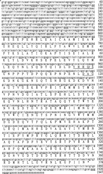
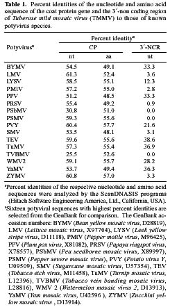
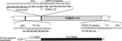
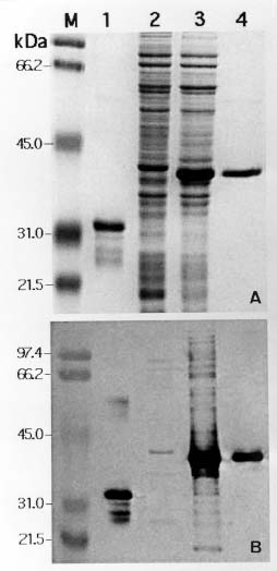
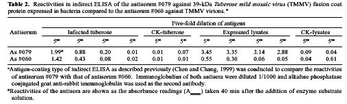
Abstract. A 2-kb DNA product was amplified from purified Tuberose mild mosaic virus (TMMV) virions as well as from infected tissues of tuberose by the use of degenerate primers for potyvirus. The PCR product was subsequently cloned and its sequence analyzed. It was found comprised of 1947 nucleotides (nts) corresponding to the 3´-terminal region of potyviruses. The deduced amino acid sequence contained 598 residues encoding part of the 3´-terminal region of NIb gene (319 residues) and the complete sequence of coat protein (CP) gene (279 residues). A 136 nts of non-coding region (NCR) was found located at the 3´-terminal region of the DNA. A genetic code for aphid transmissibility of potyviruses, DAG triplet, was found at the 19-21 residues from the N-terminus of CP gene. Compared to the known sequences of potyviruses, the percent of nucleotide identities of the CP gene and the NCR were less than 62% and 39%, respectively. Similarly, percent identities of TMMV's CP amino acid sequence to those of other known potyviruses were all below 58%, confirming our previous finding that TMMV is a new species of Potyvirus. Using directional cloning technology, a 39-kDa fusion protein containing a complete CP sequence of TMMV and a partial sequence encoded by the expression vector plasmid (pET-30b, Novagene) was highly expressed and purified from E. coli cell cultures. The antigenicity of the fusion protein was determined to be indistinguishable from the viral CP. Antiserum prepared against this fusion protein showed comparable reactivities in the serological detection of TMMV with the conventional antibodies against purified virus particles.

Keywords: 3´-terminal region; Antiserum; Expression vector; Fusion protein; Potyvirus; Sequences; Tuberose mild mosaic virus.

Introduction

Tuberose (Polianthes tuberosa L.) has been cultivated in Taiwan for over 300 years and has become an economically important ornamental bulb crop (Shen et al., 1987, 1993). Tuberose mild mosaic virus (TMMV), a newly recognized potyvirus, based on its serological and biological distinction to other known potyviruses, was recorded in 1998 (Chen and Chang, 1998). However, the molecular characteristics of this tuberose virus such as the coat protein (CP) gene sequence, the key taxonomic factor, have not been described (Chen and Chang, 1998). One of the major objectives of this study is to clone and analyze the sequences of the CP gene and the 3´-non coding region (3´-NCR) of TMMV. This sequence information is provided as molecular evidence for finalizing its actual taxonomic status. Secondly, due to generations of vegetative propagation, all of the major tuberose varieties in Taiwan have been found infected by TMMV. Preparation of specific antiserum was hampered by the difficulties in obtain

ing sufficient quantities of purified TMMV (Horner and Person, 1988). Recently an antiserum against TMMV was successfully prepared and applied in virus identification and indexing in Taiwan (Chen and Chang, 1998). Using the antiserum, a complete virus indexing and bulb certification program for tuberose has been developed (Chen et al., 1998; Chen and Chang, 1999). In addition, the clone of a virus-free multi-petal tuberose variety was rescued by meristem-tip tissue culture and identified by indexing with this antiserum (Chen et al., 1999). As a result, an industrialized virus-free tuberose bulb propagation program is becoming established. However, its success will depend upon a consistent supply of antiserum to TMMV. The efficient production of good quality antiserum to TMMV is not an easy task under conventional methods due to their instability and extremely low yield in tuberose tissue. In this study, we took the approach of cloning the complete coat protein (CP) gene and expressing it in a bacterial culture system. The bacteria-expressed viral CP was then efficiently purified in sufficient quantity for antiserum preparation. Serological experiments showed that antiserum prepared by this approach was equivalent to traditional antiserum against purified virions in the detection of TMMV in tuberose tissue.

*Corresponding author. Tel: 886-4-23302803; Fax: 886-4-23331089; E-mail: cachang@wufeng.tari.gov.tw


Botanical Bulletin of Academia Sinica, Vol. 43, 2002

into expression vector plasmid. Using this primer pair to react with purified TMMV RNA in PCR, DNA fragment corresponding to the estimated size of TMMV CP gene was amplified, cloned into pET-30b vector (Novagen, Inc., Madison, WI, USA) by a site-specific directional cloning procedure, and subsequently transformed into the E. coli strain DH5a . Bacterial clones containing inserted pET DNA were identified by PCR using the same primer pair (CP1/CP2) followed by subcloning into the E. coli strain BL21(DE3)pLysS (Novagen) for protein expression. The inducer, isopropyl b-D-thiogalactopyranoside (IPTG), was added to a bacterial culture in LB broth to make a final concentration of 1 mM for extra protein expression (Li et al., 1998).

Analysis of the Bacteria-Expressed Coat Protein of TMMV

About 4 h after adding IPTG, an aliquot of 5 ml of bacterial culture was sampled from the main culture, centrifuged at 8,000 rpm for 5 min, resuspended in TE buffer (10 mM Tris-HCl, pH 8.0, containing 1 mM EDTA) and treated with an equal volume of protein denaturing solution (0.25 M Tris-HCl, pH 6.8, containing 2% [w/v] of SDS, 4% [w/v] of 2-mercaptoethanol, and 10% [w/v] of sucrose). The sample was then analyzed by SDS-polyacrylamide gel electrophoresis (SDS-PAGE) (Laemmli, 1970). The size and expression level of viral CP was determined by western blotting analysis using an antiserum (#060) against traditionally purified TMMV virions (Chen and Chang, 1998). Antigenecity of the expressed viral CP relative to the native protein was determined by the SDS-immunodiffusion test (Chang et al., 1988).

Purification of Expressed Viral Coat Protein

Since the complete CP gene of TMMV was inserted into the pET-30b, the N-terminus of the protein translation cassette of which contained a histidine-tag (Figure 3), we were enabled to efficiently purify the expressed protein by histidine affinity column chromatography. One of the bacterial clones expressing a fusion protein that positively reacted to TMMV antiserum was selected and grown in modified M9 medium (Li et al., 1998). In order to purify the expressed TMMV CP, a 1,000 ml of bacterial culture was pooled 4 h after IPTG induction and centrifuged at 8,000 rpm for 10 min to collect the bacterial cells. The cell pellet was resuspended in 100 ml of cell-lysis buffer (50 mM NaH2PO4, 10 mM Tris-HCl, 100 mM NaCl, 8 M Urea, pH 8.0) and was frozen at -20°C overnight. After thawing in a 37°C water bath, the bacterial cells were further disrupted by a sonicater (VCX 600, Sonics & Materials Inc., CT) followed by centrifuging at 10,000 rpm for 15 min to remove the cell debris. The supernatant was then mixed with histidine resin (Talon Resin, Clontech, Inc., CA) and subjected to affinity chromatography purification according to the manufacturer's instruction. The eluted protein solution was adjusted to a concentration of 1.0 OD280 per ml for immunological studies.

Materials and Methods

Virus Isolate and Maintenance

Tbr1 isolate, the original culture for identification, of TMMV was used in this study (Chen and Chang, 1998). Tbr1 infected tuberose bulbs were routinely stored at 5°C and planted in a 25°C growth chamber to propagate infected tissue for virion purification (Chen and Chang, 1998).

Viral RNA Extraction and Reverse Transcription-Polymerase Chain Reaction (RT-PCR)

RNA template for RT-PCR was isolated from TMMV virions purified as described previously (Chen and Chang, 1998). The purified virion (1 mg/ml) was treated with 1/10 volume of disruption buffer (40 mM Tris-HCl, pH 7.8, containing 20 mM sodium acetate, 1 mM EDTA and 1% [w/v] SDS), extracted with phenol/chloroform (1:1), precipitated with ethanol, and resuspended in 20 µl of sterilized water. The first strand of cDNA was synthesized from the viral RNA by a cDNA synthesis kit (Strategene, La Jolla, CA) with the addition of an oligo dT primer. Subsequently, a set of potyvirus degenerate primers (Zerbini et al., 1995) was used to PCR-amplify the 3´-terminal region of TMMV genome. The exTaq polymerase (TaKaRa, Shuzo Co., Shiga, Japan) was used in the amplification reaction for 30 cycles (Perkin-Elmer GeneAmp model 2400, Norwalk, CT) as follows: denaturing at 94°C for 1 min, annealing at 60°C for 30 s, and DNA synthesis at 72°C for 2 min. An elongation step at 72°C for 8 min was conducted during the last cycle. The amplification products were analyzed by electrophoresis in a 1% agarose gel.

Cloning and Sequencing

The amplified DNA product in a total of 200 µl of PCR reaction mixture was precipitated with ethanol, resuspended in 20 µl of sterilized water, and cloned into pCRII-TOPO vector (Invitrogen, California, USA) according to manufacturer's instruction. Plasmid clones with expected size DNA inserts were identified and used for sequence analyses (Sambrook et al., 1989). Sequencing of the target insert DNA was done by an automatic DNA sequencer (ABI PRISM 377, Perkin-Elmer, CA, USA) with the BigDye Terminator Cycle Sequencing Ready Reaction Kit (Perkin-Elmer Applied Biosystems, CA, USA). Sequence data was analyzed by the ScanDNASIS program (Hitachi Software Engineering America, Ltd., California, USA).

Expression of Viral Coat Protein Gene

Based on the sequence data obtained from the previous experiment, a primer pair was designed for the site-specific amplification of the putative complete CP gene of TMMV. The restriction enzymes NcoI and NotI digestion sites (underlined) were created at the 5´-end of the upstream (CP1: 5´- CATCATCCATGGCTAGCGGGAATCC ACCACCA-3´) and downstream (CP2: 5´-TAATGT GCGGCCGCGAGGGAACACTA-3´) primer, respectively, to facilitate subsequent directional cloning of the CP gene


Chen et al. — Sequencing and protein expression of TMMV

Antiserum Preparation and Serological Test

An antiserum (#079) against the TMMV CP expressed in bacteria was prepared by immunizing New Zealand white rabbit intramuscularly. The immunization protocol was the same as in those described previously for preparing antisera against native viral antigens (Chang et al., 1988; Chen and Chang, 1998). SDS-immunodiffusion, ELISA, and direct tissue blotting tests (Chen and Chang, 1998, 1999) were carried out to compare the reactivity of this antiserum (#079) with the antiserum (#060) prepared against TMMV virion purified by traditional protocols.

Results

Cloning the 3´-Terminal Region of TMMV Genome and Analysis of its Sequence

A DNA fragment 2-kb in size was consistently amplified from TMMV RNA or from total RNA of TMMV-infected tissue by running a potyvirus-specific RT-PCR (Figure 1, lane 1, 2). The PCR product was cloned into pCRII-TOPO plasmid and subsequently sequenced. The DNA fragment was 1947 nucleotides (nts) long, corresponding to the 3´-terminal region of potyvirus genome

Figure 2. The nucleotide and deduced amino acid sequences of the cloned 3´-terminal region of Tuberose mild mosaic virus (TMMV) genome. The putative protease cleavage site (Q/S) between nuclear inclusion protein b (NIb) and coat protein (CP) and the DAG triplet, a genetic indication for aphid transmissibility, are underlined. The cloned 1947 nucleotides of TMMV are shown starting from the first nt. The termination codon is indicated by an asterisk followed by 136 nts of non-translated region and 17 nts of poly-A tail. The sequence has been submitted to GenBank with the accession no. AF062926.

including a part of the NIb gene (957 nts), the CP gene (837 nts) and a 3´ non-coding region (NCR) of 136 nts followed by a poly-A tail of 17 nts (Figure 2). The deduced amino acid sequences of the partial NIb and the complete CP gene contained 319 and 279 residues, respectively (Figure 2). As with other potyviruses, the predicted protease recognition site for cleaving NIb and CP was between Q/S (Shukla et al., 1991). The DAG triplet needed for aphid transmissibility (Atreya et al., 1991) was found 19-21 amino acid residues from the N-terminus of CP (Figure 2). With the help of the ScanDNASIS program, the percent identities between nucleotide sequences of the CP gene and the 3´-NCR of TMMV to those of other known potyviral se

Figure 1. Agarose gel electrophoresis of DNA products derived by amplification of Tuberose mild mosaic virus (TMMV) RNA with potyvirus specific degenerate and TMMV coat protein specific primers by reverse-transcription polymerase chain reaction (RT-PCR). Lane M, molecular markers; lanes 1 and 2, DNA product amplified with potyvirus degenerate primers (Zerbini et al., 1995) from the RNA templates purified from TMMV virion and TMMV-infected tissue, respectively; lane 3, DNA product amplified from purified TMMV RNA with TMMV coat protein specific primers (CP1/CP2) as described in the text; lane 4, no product could be amplified from the RNA extracted from healthy tuberose tissue with CP1/CP2 primers.


Botanical Bulletin of Academia Sinica, Vol. 43, 2002

quences documented in the GenBank were less than 62% and 39%, respectively (Table 1). In addition, the percent identities of the amino acid sequence of TMMV CP to other known potyviruses were below 58% (Table 1). These results provided strong evidence that TMMV was a unique member of the genus Potyvirus.

Expression of the CP Gene of TMMV in E. coli

Based on the derived 3´-terminal sequence of TMMV (Figure 2), two site-specific primers (CP1 and 2) were designed, and a 837 bp DNA product equivalent to the estimated size of the full length CP gene of TMMV was amplified by PCR (Figure 1, lane 3). This 837 bp DNA was subsequently constructed in expression vector pET-30b by directional cloning (Figure 3) and cloned in E. coli strain DH5a. After confirmation by PCR using CP1 and CP2 primers, six clones with correct DNA insert sizes were obtained, and the plasmids were then transformed into E. coli strain BL21 for protein expression. A protein about 39 kDa in size was detected by electrophoresis in bacterial lysates of transformed BL21 clones induced with IPTG, but not in lysates of control clones (Figure 4, A). Western blotting analyses showed that the 39-kDa protein as well as the TMMV CP purified from infected tissue both strongly reacted with TMMV antiserum #060 (Figure 4, B), indicating the expressed protein was antigenically related to the native TMMV coat protein. Furthermore, when compared in an SDS-immunodiffusion test they were antigenically indistinguishable as no spur reaction formed between their precipitation lines (Figure 5). These serological tests clearly showed that the cloned TMMV CP gene in pET-30b expressed a fusion protein antigenically indistinguishable from the native viral TMMV CP. In order to mass-produce the fusion protein for immunization, a high expressing clone, TM29, was selected for large-scale culture and the protein was purified by affinity chromatography. Routinely, about 4 mg of expressed protein was purified from 1,000 ml of bacterial culture.

Figure 3. Schematic representation of the construction of full-length coat protein gene of Tuberose mild mosaic virus (TMMV-CP gene) in the expression vector pET-30b. The upper map shows the detail sequence composition in the linkage between TMMV-CP (£), and the pET-30b (£) by NcoI and NotI restriction enzyme sites. Protein translation is done starting from the ATG codon of the NdeI site from the expression vector. After reading through a sequence encoded by the vector, translation is terminated at the TAG codon provided by the viral CP sequence. The lower map indicates the relative size of the translated protein with a 5.1 kDa vector-encoded fragment from the N-terminus and followed by the expressed 31.4 kDa of TMMV CP. The molecular size of the expressed fusion protein is calculated by ScanDNASIS program based on the amino acid sequence.


Chen et al. — Sequencing and protein expression of TMMV

Serological Reactivity of the Antiserum Prepared Against Viral CP Expressed in Bacteria

An antiserum (#079) against the TMMV CP expressed in bacteria was successfully prepared, and its reactivities in ELISA, SDS-immunodiffusion, and direct tissue blotting tests as compared to the antiserum #060 against purified virions (Chen and Chang, 1998) were analyzed. In indirect ELISA, both antisera reacted with TMMV-infected tuberose tissue and the bacteria lysates containing expressed fusion protein, but did not react with healthy tuberose and the bacteria lysates without an insert of the TMMV CP gene (Table 2). Although ELISA tests implicated slight differences in the optimum reaction titers of these two antisera, both were successful in the detection of TMMV from tuberose tissue. On the other hand, the reactivities of antiserum #079 and #060 against the fusion protein expressed in bacteria were significantly different (Table 2). The dilution end point of the fusion protein in reacting with antiserum #060 was only 1/125, while that

Figure 5. Reactivity of the antiserum prepared against coat protein (CP) of Tuberose mild mosaic virus (TMMV) expressed in bacteria as compared to the antiserum against purified TMMV virion in sodium dodecyl sulfate (SDS) immunodiffusion tests. The central wells BT and T were charged with antiserum #079 against bacteria expressed TMMV CP and antiserum #060 against purified TMMV virions, respectively. The peripheral wells were filled with SDS-treated antigens of TMMV-infected tuberose tissue (1), IPTG-induced lysate of bacteria (E. coli BL21(DE3)pLysS) clone TM29 containing TMMV CP gene inserted pET-30b (2), IPTG-induced bacterial lysate with pET-30b without TMMV CP gene insert (3), the same bacterial lysate as (3) but non-induced by IPTG (4), and healthy tuberose tissue (H), respectively.

Figure 4. Sodium dodecyl sulfate-polyacrylamide gel electrophoresis (SDS-PAGE) of the coat protein of Tuberose mild mosaic virus (TMMV) expressed in bacteria (A) and western blotting (B) with the antiserum against purified TMMV virion. Lane M, protein marker; lane 1, purified TMMV virion; lane 2, IPTG-induced bacteria (E. coli BL21(DE3)pLysS) lysate containing expression vector pET-30b without TMMV CP gene insert; lane 3, IPTG-induced bacteria lysate of clone TM29 containing TMMV CP gene inserted pET-30b; lane 4, bacteria-expressed TMMV CP purified by His-Tag affinity chromatography.


Botanical Bulletin of Academia Sinica, Vol. 43, 2002

against antiserum #079 was higher than 1/57. This result indicated that antiserum #079 contained antibodies specific to the antigenic determinants residing in the vector-encoded protein sequence of the fusion protein. These antibodies however were not present in antiserum #060. Nevertheless, apparently these antibodies in antiserum #079 did not interfere with the detection of TMMV.

In SDS-immunodiffusion tests, antiserum #079 reacted strongly with IPTG induced bacterial lysates of clone TM29 and TMMV-infected tuberose tissues, and no discernible spur formed between them. It did not react with non-infected tuberose antigen. Unlike antiserum #060, #079 reacted with IPTG-induced control bacterial lysate (well 3, Figure 5) but not with the non-IPTG-induced culture (well 4, Figure 5). This result further confirmed the previous ELISA finding that antiserum #079 did contain antibodies specific to the vector-encoded fragment of the expressed fusion protein so that it reacted with the control bacterial lysate only after induction by IPTG. Without IPTG induction, the control lysate contained no vector protein and thus did not react with antiserum #079. Based on the intensity of the precipitation lines against TMMV-infected tuberose tissue, the reactions of antiserum #079 and #060 under the same dilution factor were indistinguishable (Figure 5).

In direct tissue blotting tests, both antisera #079 and #060 readily detected TMMV in infected tuberose bulbs or leaves (Figure 6). As in the results of SDS-immunodiffusion tests, no differences in the color intensity of positive reactions were observed between the two antisera.

However, antiserum #060 reacted slightly with control blots containing proteins from non-infected tissues, indicating a low background reaction still associated with it. Although this background reaction could easily be removed by cross absorption with non-infected tuberose antigen as shown in Figure 6, this procedure was unnecessary for antiserum #079.

Discussion

Based on our previous studies of serological and biological distinction (Chen and Chang, 1998), TMMV was proposed as a new species of the genus Potyvirus. It is currently the only documented potyvirus known to infect tuberose. In this study, we presented molecular evidence showing that the CP gene and 3´-NCR of TMMV has less than 62% identity with other known potyviruses at the nucleotide level. Similarly, the amino acid sequence identity of CP gene of TMMV with other known potyviruses was less than 58%. It is now widely accepted that amino acid sequence identities of CP among species of potyviruses ranged from 38 to 71% (average 54%) while those among strains of the same species ranged from 90 to 99% (average 95%) (Shukla and Ward, 1988, 1989; Ward et al., 1992). Therefore, our results of sequence comparison provide conclusive evidence that TMMV is a new species of Potyvirus.

TMMV was found in nearly every tuberose plant examined in Taiwan but it did not cause evident damage on cut flower production during the summer season. However, during other growing seasons when the temperature is lower than 25°C infected tuberoses tend to express pronounced foliar symptoms and usually their growth vigor is significantly reduced (unpublished data). Therefore, implementation with TMMV-indexed bulbs as growing materials is proposed as a solution to this problem. In order to eliminate the infection of TMMV from tuberoses, a constant supply of specific antiserum for indexing is necessary. However, tuberose is the only host plant found to be infected by TMMV, and the virus concentration in infected tissue is so low that purification of virus particle is difficult (Chen and Chang, 1998). It has been shown that plant virus antiserum can be prepared by immunizing the viral coat protein expressed in bacteria (Li et al., 1998; Nagel and Hiebert, 1985; Nikolaeva et al., 1995). In this study, we also successfully introduced a molecular technique to overcome the obstacle encountered in the purification of TMMV. A 39-kDa fusion protein containing complete CP sequence of TMMV (Figure 4) was efficiently expressed in bacterial culture and subsequently purified. It was shown that the bacteria expressed protein preserved an antigenicity indistinguishable from the native viral CP, although the antiserum prepared against the fusion protein did contain some antibodies specific to the amino acid sequence originated from the expression vector. However, ELISA, tissue blotting and SDS-immunodiffusion tests showed that the prepared antiserum was equivalent in reactivities and detected TMMV antigens to the traditionally made antiserum against TMMV virions.

Figure 6. Reactivity of the antiserum prepared against coat protein (CP) of Tuberose mild mosaic virus (TMMV) expressed in bacteria as compared to the antiserum against purified TMMV virion in direct tissue blotting tests. Cross-sectioned TMMV-infected tuberose bulbs (Diseased) and healthy tuberose leaves (H-CK) were blotted on separated Nylon membrane (Duralon-UV membranes, Stratagene, La Jolla, CA) and reacted with the antiserum #079 against TMMV CP expressed in bacteria (BT), with antiserum #060 against purified TMMV virion (T), and with antiserum #060 but pre-absorbed with healthy tuberose tissue (T/H). The treated membranes were further reacted with alkaline phosphatase conjugated goat anti-rabbit immunoglobulin followed by treating with (BCIP/NBT) substrate solution as previously described (Chen and Chang, 1999). Positive reactions were shown by the dark bluish color on the blots.


Chen et al. — Sequencing and protein expression of TMMV

Therefore, our results confirm that cloning and expressing the viral CP gene in a bacterial system can be applied in the purification of viral antigens for antiserum preparation or for other possible studies. Furthermore, we would particularly like to point out that the directional cloning approach in the construction of the CP gene expression cassette is more efficient than those applied by previous workers (Li et al., 1998; Nagel and Hiebert, 1985). We believe that the protocol developed in this study can easily and efficiently produce viral antigens in good quantities for advanced studies.

Literature Cited

Atreya, P.L., C.D. Atreya, and T.P. Pirone. 1991. Amino acid substitutions in the coat protein result in the loss of insect transmissibility of a plant virus. Proc. Natl. Acad. Sci. USA 88: 7887-7891.

Chang, C.A., D.E. Purcifull, and E. Hiebert. 1988. Purification, characterization, and immunological analysis of nuclear inclusions induced by bean yellow mosaic and clover yellow vein potyviruses. Phytopathology 78: 1266-1275.

Chen, C.C. and C.A. Chang. 1999. The improvement for the detection of tuberose mild mosaic potyvirus in tuberose bulbs by temperature treatment and direct tissue blotting. Plant Pathol. Bull. 8: 83-88.

Chen, C.C., F.L. Chiang, and C.A. Chang. 1998. Distribution of tuberose mild mosaic potyvirus in tuberose (Polianthes tuberosa L.) and its influence on virus detection. Plant Pathol. Bull. 40: 199-207.

Chen, C.C. and C.A. Chang. 1998. Characterization of a potyvirus causing mild mosaic on tuberose. Plant Dis. 82: 45-49.

Chen, C.C., C.A. Chang, and F.L. Chiang. 1999. Elimination of tuberose mild mosaic potyvirus from tuberose and its influence on plant growth. Plant. Pathol. Bull. 8(4): 180.

Horner, M.B. and M.N. Person. 1988. Purification and electron microscopy studies of a probable potyvirus from Polianthes tuberosa L. J. Phytopathol. 122: 261-266.

Laemmli, U.K. 1970. Cleavage of structural proteins during the assembly of the head of bacteriophage T-4. Nature 227: 680-685.

Li, R.H., F.W. Zettler, D.E. Purcifull, and E. Hiebert. 1998. The

nucleotide sequence of the 3´-terminal region of dasheen mosaic virus (Caladium isolate) and expression of its coat protein in Escherichia coli for antiserum production. Arch. Virol. 143: 2461-2469.

Nagel, J. and E. Hiebert. 1985. Complementary DNA cloning and expression of the papaya ringspot potyvirus sequences encoding capsid protein and a nuclear inclusion-like protein in Escherichia coli. Virology 143: 435-441.

Nikolaeva, O.V., A.V. Karasev, D.J. Gumpf, R.F. Lee, and S.M. Garnsey. 1995. Production of polyclonal antisera to the coat protein of citrus tristeza virus expressed in Escherichia coli: Application for immunodiagnosis. Phytopathology 85: 691-694.

Sambrook, J., E.F. Fritsch, and T. Maniatis. 1989. Molecular Cloning: A Laboratory Manual. Cold Spring Harbor Laboratory, New York.

Shen, T.M., K.L. Huang, and T.S. Huang. 1987. Study on hybridization and sexual propagation of tuberose. In Symposium on the Improvement for Flower Production. Tau-Yuan Dis. Agric. Improv. Sta. Taiwan, R.O.C., pp. 167-175.

Shen, T.M., T.S. Huang, B.S. Du, and K.L Huang. 1993. Breeding of tuberose for cut flower. J. Chin. Soc. Hort. Sci. 39(1): 23-29.

Shukla, D.D. and C.W. Ward. 1988. Amino acid sequence homology of coat proteins as a basis for identification and classification of the potyvirus group. J. Gen. Virol. 69: 2703-2710.

Shukla, D.D. and C.W. Ward. 1989. Structure of potyvirus coat proteins and its application in the taxonomy of the potyvirus group. Adv. Virus Res. 36: 273-314.

Shukla, D.D., M.J. Frenkel, and C.W. Ward. 1991. Structure and function of the potyvirus genome with special reference to the coat protein coding region. Can. J. Plant Pathol, 13: 178-191.

Ward, C.W., N.M. McKern, M.J. Frenkel, and D.D. Shukla. 1992. Sequence data as the major criterion for potyvirus classification. In O.W. Barnett (ed.), Potyvirus Taxonomy. Springer, Wien and New York [Archives of Virology (Supplement) 5], pp. 283-297.

Zerbini, F.M., S.T. Koike, and R.L. Giberston. 1995. Biological and molecular characterization of lettuce mosaic potyvirus isolates from the Salinas Valley of California. Phytopathology 85: 746-752.


Botanical Bulletin of Academia Sinica, Vol. 43, 2002