Bot. Bull. Acad. Sin. (2002) 43: 21-29

Liou et al. — DNA fingerprint analysis of Phytophthora parasitica

Analysis of Phytophthora parasitica by retrotransposon-derived DNA fingerprinting

Ruey-Fen Liou1,*, Jent-Turn Lee1, Hsiao-Ching Lee1, and Pao-Jen Ann2

1Department of Plant Pathology, National Taiwan University, Taipei 106, Taiwan

2Department of Plant Pathology, Taiwan Agricultural Research Institute, Wu Fong 413, Taiwan

(Received August 2, 2001; Accepted September 14, 2001)

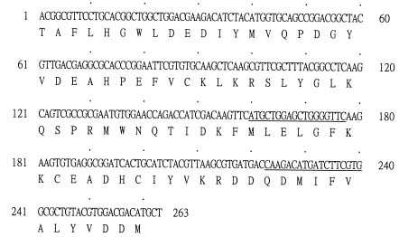
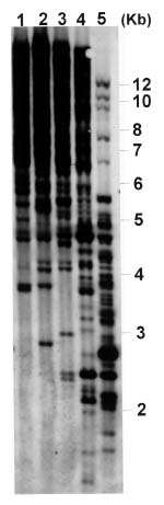
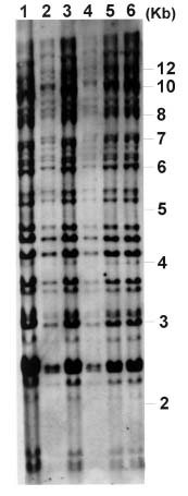
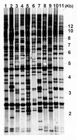
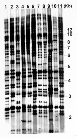
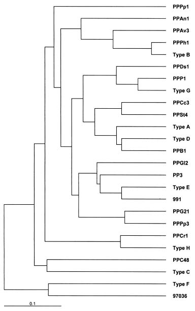
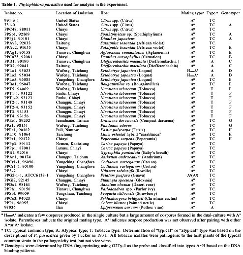
Abstract. Phytophthora parasitica, being able to attack a wide variety of plants, is a very important plant pathogen in Taiwan. It has been shown that isolates of P. parasitica from tobacco, dieffenbachia, and loquat differed significantly from other isolates in morphology and pathogenicity and were recognized as "atypical" types of the fungus. In the current study, a partial sequence of the reverse transcriptase gene of a Ty1-copia retrotransposon was cloned from P. parasitica and used as the probe, designated herein as G2Ty-1, for genomic Southern hybridization analyses of P. parasitica. It was demonstrated that G2Ty-1 existed as a moderately repetitive sequence in the genome of P. parasitica. The banding pattern of G2Ty-1 was mitotically stable for at least five consecutive asexual generations. DNA fingerprint analysis of P. parasitica using G2Ty-1 as the probe demonstrated that the banding patterns of G2Ty-1 were highly polymorphic among the isolates analyzed. "Atypical" strains from tobacco, dieffenbachia, and loquat, nonetheless, displayed host-specific banding patterns. Phylogram generated by cluster analysis demonstrated that isolates from tobacco and dieffenbachia were well separated from each other and from all other isolates of P. parasitica. These results provided a genetic basis for distinguishing these isolates from others and validate the use of retrotransposon as a genetic marker to study phylogeny in Phytophthora parasitica.

Keywords: DNA fingerprinting; Genotypes; Host specificity, Phytophthora parasitica; Retrotransposon.

Introduction

Phytophthora parasitica Dastur (= Phytophthora nicotianae Breda de Haan), an oomyceteous fungus, is the most widespread and important species of Phytophthora in Taiwan (Ho et al., 1995). It causes root rot, foot rot, leaf blight, and fruit rot in a variety of economically important crops (Ann et al., 1992; Ann, 1992, 2000a, 2000b; Erwin and Ribeiro, 1996). The taxonomy of Phytophthora is based mainly on morphological and physiological characteristics (Waterhouse, 1963). Although isolates morphologically identified as P. parasitica are pathogenic to a wide range of plant species, considerable evidence shows that some isolates are specifically pathogenic to a few hosts or a single host, such as tobacco (Bonnet et al., 1978, 1994; Matheron and Matejka, 1990; Erwin and Ribeiro, 1996). In support of this view, it was demonstrated in a recent study that isolates from dieffenbachia (Dieffenbachia maculata Don) were nonpathogenic or weakly pathogenic to other hosts of P. parasitica, nor were isolates from these plants pathogenic to dieffenbachia (Ann, 1992). Besides, some isolates from loquat (Eriobotrya japonica (Thunb.) Lindl.), known as "atypical" strains of P. parasitica, were found to be highly

pathogenic to loquat while displaying reduced virulence toward eggplant fruits, sweet orange, and tomato seedlings as compared to typical strains in the pathogenicity test (Chern et al., 1998). It is not clear if atypical characteristics in pathogenicity observed in these isolates result from differentiation of the fungi at the genetic level. In past years, molecular markers such as isozyme patterns (Oudemans and Coffey, 1991) or mitochondrial restriction fragment length polymorphism (RFLP) (Föster et al., 1990; Lacourt et al., 1994) have been used for analysis of P. parasitica. These studies, however, revealed only a low level of polymorphism among the P. parasitica isolates analyzed, and no correlation with pathogenicity was observed. As a first attempt to seek molecular support for host specialization in P. parasitica isolates, retrotransposon-derived DNA fingerprinting was used to analyze the genotypes of various P. parasitica isolates in the current study.

Retrotransposons are mobile genetic elements, which replicate through an RNA intermediate prior to integration into the chromosomal DNA of their host. As a result, they may constitute a substantial portion of the repetitive sequences in an organism, and provide useful markers for analyses of the plant and fungal genomes (Hamer et al., 1989; Purugganan and Wessler, 1995; Flavell et al., 1998). According to the structural organization and the amino acid sequences of the encoded reverse transcriptase and

*Corresponding author. Fax: 886-2-23620271; E-mail: rfliou@ccms.ntu.edu.tw


Botanical Bulletin of Academia Sinica, Vol. 43, 2002

integrase, retrotransposons can be classified into three major groups: the Ty1-copia, the Ty3-gypsy, and LINE (long interspersed nuclear elements)-like retrotransposons (Boeke and Sandmeyer, 1991, and references therein). The Ty1-copia and Ty3-gypsy retrotransposons are characterized by the flanking long terminal repeats (LTR). LINE, on the contrary, does not have LTRs. In this report, partial reverse transcriptase sequence of a Ty1-copia retrotransposon was cloned and used as the probe for DNA fingerprint analyses of P. parasitica. It was demonstrated that, although the DNA banding patterns of most isolates are highly polymorphic, P. parasitica isolated from tobacco, dieffenbachia, and loquat exhibited host-specific DNA banding patterns. These results provide a genetic basis for separating these P. parasitica isolates from others.

Materials and Methods

Phytophthora Isolates and Culture Conditions

Phytophthora parasitica isolates were from one of us (P. J. Ann) (Table 1; Ann, 1992, 1995; Chern et al., 1998; Ann, 2000a, 2000b). Cultures were grown on 5% V-8 juice agar (5% Campbell's V-8 juice, 0.02% CaCO3, and 2% Bacto agar) plates for 3-5 days, cut into small pieces (5×3×2 mm), and soaked in test tubes containing sterile distilled water at 24°C for maintenance. For preparation of DNA, fungal isolates were grown on 5%V-8 juice medium (5% Campbell's V-8 juice and 0.02% CaCO3) at 25°C in darkness for 10 days.

The method described by Hwang et al. (1976) was used for production and isolation of single-zoospore isolates. A few pieces of 3-5-day-old culture blocks (3×3×2 mm) grown on 5% V-8 agar were soaked in a small petri dish (6 cm in diameter) containing 5 ml sterile distilled water at room temperature for releasing zoospores. Single-zoospore isolates obtained from the "parent" isolates were designated as Z1. Subsequently, the second asexual generation was isolated from Z1 by the same method and designated as Z2. A total of five asexual generations (Z1~Z5) were collected from four different "parent" isolates (731-0, 991-3-1, PPPr 1-1, and PPT 2-1), respectively.

Polymerase Chain Reaction and DNA Sequence Analysis

General DNA manipulations including extraction of fungal DNA, preparation of plasmid DNA, DNA ligation, bacterial transformation, and agarose gel electrophoresis were performed as described by Sambrook et al. (1989). Nuclear DNA of P. parasitica was further purified by CsCl gradient centrifugation according to Garber and Yoder (1983).

For amplification of the reverse transcriptase domain of the Ty1-copia retrotransposon, two sets of degenerate oligonucleotides, ty1A (5´-ACNGCNTTYYTNCAYGG-3´) and ty1B (5´-ARCATRTCRTCNACRTA-3´) (Flavell et al., 1992), were used to prime the DNA polymerization reaction. The reaction mixture contained 100 ng of fungal

genomic DNA, 2.5 µM of oligonucleotide primers, 0.2 mM dNTP, 1X PCR buffer, and 1 U of DyNazymeTM II DNA polymerase (Finnzymes). PCR was performed in a thermocycler (GeneAmp PCR System 2400) programmed for denaturation at 94°C for 5 min, followed by 30 cycles at 94°C for 15 sec, 50°C for 15 sec, 72°C for 15 sec, and a final extension at 72°C for 10 min. Following analysis of the amplified products by 1.5% agarose gel electrophoresis, DNA bands of expected size were collected from the agarose gel using the Geneclean III kit (Bio101, Inc.), and cloned into pGemT-easy (Promega). DNA sequencing was performed on both strands of DNA by the dideoxy termination method (Sanger et al., 1977), using the BigDye terminator cycle sequencing ready reaction kit and an automated Applied Biosystem 373 instrument (Applied Biosystems). Sequence was analyzed using programs in the GCG software package (Genetics Computer Group, Wisconsin, Package Version 10.0).

Genomic Southern Hybridization

Phytophthora parasitica DNA was digested with 10 units of restriction enzyme and then separated on a 0.8% agarose gel in 0.5X TBE buffer at 1.2 V cm-1 overnight. Blotting was performed according to the standard protocol (Sambrook et al., 1989). DNA probe was labeled with digoxigenin (DIG)-11-dUTP by PCR using the PCR DIG probe synthesis kit (Roche). PCR was primed with oligonucleotides 1S and 1A (Figure 1) in order to exclude the conserved reverse transcriptase sequences and thereby to avoid cross hybridization with those of other retrotransposons. This probe was herein designated as G2Ty-1. Prehybridization, hybridization with DIG-labeled probe, and detection of the hybridization signal were performed according to the manufacturer's protocol (Roche).

Figure 1. Partial nucleotide and deduced amino acid sequence of the reverse transcriptase gene of a Ty1-copia retrotransposon from Phytophthora parasitica. The sequence (GeneBank accession number AF130855) was cloned by PCR with primers ty1A and ty1B (Flavell et al., 1992). Underlines indicate positions of PCR primers used for synthesis of the DIG-labeled G2Ty-1 probe: 1S (the upstream primer) and 1A (the downstream primer).


Liou et al. — DNA fingerprint analysis of Phytophthora parasitica

Statistical Analysis

Presence (1) or absence (0) of a band at a particular position in the Southern blot (ranging from 2 to 6 kb) were treated as discrete characters, and banding patterns were converted into binary matrices. The total banding number of each isolate and the number of bands shared by every pair of isolates were counted. "Similarity index" (S) was calculated according to the formula: Sab = 2 x nab/ (na+nb), where na and nb represent the total number of bands present in the DNA fingerprint patterns of fungal

isolates a and b, respectively, and nab is the number of bands shared by isolates a and b (Nei and Li, 1979). Subsequently, pairwise distance (Dab) between isolates a and b was obtained by the following formula: Dab = 1- Sab. The resulting distance values were then used as the basis of cluster analysis. Dendrograms were produced by cluster analysis using the UPGMA (unweighted pair group method using arithmetic average) (Sneath and Sokal, 1973) option of the Neighbor-joining program in the PHYLIP (Phylogeny Inference Package) (Version 3.5c; J. Felstein,

Figure 2. Southern hybridization analysis of the G2Ty-1 sequence in Phytophthora parasitica. Fungal DNA (isolate number PPAv3) was cut with XhoI (lane 1), SacI (lane 2), HindIII (lane 3), EcoRI (lane 4), or BamHI (lane 5), and subjected to Southern hybridization analysis using DIG-labeled G2Ty-1 as the probe. Positions of size standards are indicated in kilobases to the right.

Figure 3. Somatic stability of the G2Ty-1 banding pattern of Phytophthora parasitica. DNA from single-zoospore isolates representing five successive asexual generations (Z1 to Z5) (lanes 2~6) from a parent isolate (991-3-1) (lane 1) was cut with EcoRI and analyzed by Southern hybridization using DIG-labeled G2Ty-1 as the probe. Positions of size standards are indicated in kilobases to the right.


Botanical Bulletin of Academia Sinica, Vol. 43, 2002

to DNA sequence analysis. Of the ten recombinant clones being analyzed, one contained a full-length ORF which was obviously irrelevant to the reverse transcriptase sequence of the copia retrotransposon, and three contained short ORFs and stop codons. Thus, they were neglected in the subsequent analysis. The remaining six clones contained ORFs with an identical deduced amino acid sequence. Analysis of the amino acid sequence revealed the presence of TAFLH and YVDDM on the N- and C-termini of each ORF, which corresponded to the sequences of the PCR primers (Figure 1). Analysis by BLAST indicated that it is very similar to the reverse transcriptase sequences of Phytophthora infestans (AF262230), Alstroemeria inodora (AJ223608), and Picea abies (AJ290661), as well as to other Ty1-copia retrotransposons from plants. Moreover, a conserved reverse transcriptase

Dept. Genetics, University of Washington, Seattle). The TREEVIEW program was used to draw the resulting phylogenetic tree (Page, 1996).

Results

Cloning and Analysis of Partial Reverse Transcriptase Sequence of Ty1-Copia Retrotransposons from P. parasitica

PCR with ty1A and ty1B, which were designed according to the conserved reverse transcriptase sequence of Ty1-copia retrotransposons (Flavell et al., 1992), gave rise to a DNA fragment of the expected size, approximately 260 bp in length (data not shown). This fragment was eluted from the agarose gel, cloned into a T-vector, and subjected

(A)

(B)

Figure 4. Nuclear polymorphisms of Phytophthora parasitica based on the G2Ty-1 probe hybridization patterns. After digestion with EcoRI, fungal DNA was subjected to Southern hybridization analysis using DIG-labeled G2Ty-1 as the probe. Positions of size standards are indicated in kilobases to the right. Fungal isolates demonstrated in panel A included PPFa1 (lane 1), PPT1-1 (lane 2), PP3-2 (lane 3), PPEe1 (lane 4), PPPj1 (lane 5), PPPp1 (lane 6), PPPp3 (lane 7), PPL10 (lane 8), PPCc3 (lane 9), PPCr1-1 (lane 10), PPCr1-5 and (lane 11). Panel B included 731-0 (lane 1), 991-3-1 (lane 2), PPPr1 (lane 3), PPD1 (lane 4), PPD2 (lane 5), PPLo5 (lane 6), PPLo1 (lane 7), PPLo2 (lane 8), PPT1-2 (lane 9), PPT4 (lane 10), and PPT5 (lane 11).


Liou et al. — DNA fingerprint analysis of Phytophthora parasitica

motif for the Ty1-copia retrotransposons, SLYXLKQAXRXW (Flavell et al., 1992; Flavell et al., 1998; Xiong and Eickbush, 1990), was also identified in the sequence, indicating that it was amplified from the reverse transcriptase gene of a Ty1-copia retrotransposon.

Hybridization Patterns of the G2Ty-1 Probe

To better understand the nature of the cloned sequence, genomic Southern hybridization was performed following digestion of fungal DNA (isolate PPAV3-1) with restriction enzymes XhoI, SacI, HindIII, EcoRI or BamHI, using G2Ty-1 as the probe. As shown in Figure 2, the DIG-labeled G2Ty-1 probe hybridized to multiple DNA fragments in the genome of P. parasitica. The best resolution of banding pattern was obtained with the EcoRI digests (lane 4, Figure 2). Thus, genomic Southern hybridization was performed with EcoRI digest in the following analysis.

In order to verify the somatic stability of the banding patterns produced by G2Ty-1, single-zoospore isolates were prepared from four different "parent" fungal isolates and analyzed by genomic Southern hybridization using fungal DNA cut with EcoRI. The results indicated that, no matter which isolate was analyzed, the hybridization patterns of the single-zoospore progeny from Z1 to Z5, which encompassed a total of five consecutive asexual generations, were identical to their parent isolate (Figure 3). Thus, the DNA banding patterns produced by the G2Ty-1 probe were mitotically stable in P. parasitica.

Analysis of P. parasitica by G2Ty-1 DNA Fingerprinting

A total of 42 isolates of P. parasitica (Table 1) were studied. Among them, isolates from tobacco, dieffenbachia, and loquat, as well as one isolate from pothos vine (97036) were of special interest due to their characteristics in morphology and/or pathogenicity (Ann, 1995; Chern et al., 1998). Other isolates, on the contrary, were collected from a wide variety of host plants and were recognized as "typical common" strains of P. parasitica (Ann, 1995, 2000a, 2000b). Nuclear DNA of each isolate was digested with EcoR1, blotted, and hybridized with DIG-labeled probes. As shown in the representative Southern blots (Figure 4A and 4B), the banding patterns lit up by G2Ty-1 were highly polymorphic among the P. parasitica isolates being analyzed. Interestingly, some isolates displayed identical DNA banding patterns, and were classified into eight groups designated as types A~H. They included 731-0, PPSp1, and PPPj1 of type A, PPAv2, PPAg1, and PPCa75 of type B, PPLo5, PPBo1 and PPT5 of type E, PPEe1 and PPx1 of type G, and PPFa1, PPL10, and PPPr1 of type H (Table 1). In addition, of the seven tobacco isolates analyzed, six (PPT1-1, PPT1-2, PPT2-1, PPT2-4, PPT3, and PPT4) displayed an identical banding pattern and were designated as type F (Figure 4B). The banding pattern of PPT5 placed it in type E, the same as PPLo1 and PPBo1, isolates from loquat and bougainvillea (Bougainvillea sp.), respectively. Isolates from dieffenbachia, collected from Taitong (PPD1) and

Figure 5. Phylogram generated by UPGMA cluster analysis of the distance values. Types A~H indicated genotypes of Phytophthora parasitica as defined in the text (Table 1). Data were compiled as a (0/1) matrix from the DNA fingerprint patterns revealed by the G2Ty-1 probe. Pairwise genetic distance was calculated and cluster analysis was performed as described in Materials and Methods.

Changhua (PPD2), respectively, also displayed a unique banding pattern, designated as type C (Figure 4B, lanes 4 and 5). Two isolates from loquat, PPLo1 and PPLo2, known as `atypical' strains of P. parasitica (Chern et al., 1998), exhibited a characteristic banding pattern designated as type D (Figure 4B, lanes 7 and 8).

To analyze the relationship between the fungal isolates based on the G2Ty-1 banding patterns, a phylogram was derived from the fingerprints of the fungal isolates by cluster analysis using the UPGMA option of the Neighbor-joining program in PHYLIP. As shown in Figure 5, the 42 isolates tested were divided into three groups. Isolate 97036 was clustered with type F, which represented most of the tobacco isolates (Table 1), while isolate PPC48 was clustered with type C, which represented isolates from dieffenbachia. These two clusters were widely separated from each other and from all other fungal isolates. Type


Botanical Bulletin of Academia Sinica, Vol. 43, 2002

Kalendar et al., 1999). In our study, partial reverse transcriptase sequence of a Ty1-copia group retrotransposon was cloned by genomic PCR. This is the first indication that the genome of P. parasitica contains Ty1-copia retrotransposon. Molecular cloning and characterization of the full-length retrotransposon is now being performed in our laboratory.

Genomic Southern hybridization using G2Ty-1 as the probe revealed that this sequence exists as moderately repetitive sequences in the genome of P. parasitica. Furthermore, the G2Ty-1 hybridization patterns are highly polymorphic among the fungal isolates being analyzed. Since the presence of a reverse transcriptase sequence is

D, which represented "atypical" strains isolated from loquat was, in contrast, closest to isolate PPB1 and comprised part of the main cluster.

Discussions

Retrotransposons are mobile genetic elements, which replicate by successive transcription, reverse transcription, and insertion of the new cDNA copies back into the genome. Insertion of retrotransposon in a new site may be detected by the appearance of a new DNA banding in a genomic Southern blot, and thus is useful for establishment of phylogenies (Ellis et al., 1998;


Liou et al. — DNA fingerprint analysis of Phytophthora parasitica

It is intriguing to ask why characteristics of P. parasitica, such as host specialization, are correlated with the insertion site polymorphism of a retrotransposon and why type C is clustered most closely with PPC48, a "typical common" strain isolated from citrus. Since activity of transposable elements may cause mutation and thereby lead to pathogenic specialization in plant pathogenic fungi (McHale et al., 1992; Shull and Hamer, 1995; He et al., 1996), it is not surprising that transposition of a retrotransposon in the genome of P. parasitica can result in changes of its phenotypes as well. What is really relevant is the insertion sites of the retrotransposon, namely the genes that have been disrupted by the retrotransposon in individual fungal isolate. In plants, retrotransposons are frequently found in the regions flanking functional genes (White et al., 1994). Nothing is known, however, about their insertion sites in P. parasitica. Analysis of sequences flanking the retrotransposons may help elucidate the mechanisms underlying host specialization as well as other characteristics of this fungus.

Acknowledgements. This research was supported by a grant from the National Science Council, Taiwan, ROC.

Literature Cited

Ann, P.J. 1992. Phytophthora diseases of ornamental plants in Araceae in Taiwan. Plant Pathol. Bull. 1: 79-89.

Ann, P.J. 1995. Studies on the Pathogenicity and Biology of Phytophthora parasitica. National Science Council Research Report. Taipei, Taiwan.

Ann, P.J. 2000a. New diseases and records of flowering potted plants caused by Phytophthora species in Taiwan. Plant Pathol. Bull. 9: 1-10.

Ann, P.J. 2000b. Phytophthora diseases of some ornamental foliage plants as new records in Taiwan. Plant Pathol. Bull. 9: 47-52.

Ann, P.J., C.T. Lo, and T.F. Hsieh. 1992. Phytophthora blight of Lilium spp. in Taiwan. Plant Prot. Bull. 34: 64-69.

Boeke, J.D. and S.B. Sandmeyer. 1991. Yeast transposable elements. In R. Broach, J.R. Pringle, and E.W. Jones (eds.), The Molecular and Cellular Biology of the Yeast Saccharomyces, vol. 1. Genome Dynamics, Protein Synthesis, and Energetics, Cold Spring Harbor Laboratory Press, Cold Spring Harbor, USA, pp. 193-261.

Bonnet, P., I. Lacourt, P. Venard, and P. Ricci. 1994. Diversity in pathogenicity to tobacco and in elicitin production among isolates of Phytophthora parasitica. J. Phytopathol. 141: 25-37.

Bonnet, P., N. Maia, J. Tello-Marquina, and P. Venard. 1978. Pathogenic capacity of Phytophthora parasitica [Dastur]: Factors on variability and concept of parasitic specialization. Ann. Rev. Phytopathol. 10: 15-29.

Chern, L.L., P.J. Ann, and H.R. Young. 1998. Root and foot rot of loquat in Taiwan caused by Phytophthora. Plant Dis. 82: 651-656.

Colas, V., I. Lacourt, P. Ricci, F. Vanlerberghe-Masutte, P. Venard, A. Poupet, and F. Panabičres. 1998. Diversity of virulence in Phytophthora parasitica on tobacco, as reflected by nuclear RFLPs. Phytopathology 88: 205-212.

indicative of the existence of retrotransposons, it is possible that the DNA polymorphism observed in our study was caused by active transposition of a retrotransposon. Analysis of single-zoospore progeny, which encompass five successive asexual generations from four different "parent" isolates, however, indicates that the G2Ty-1 banding patterns are mitotically stable, at least to the extent genomic Southern hybridization analysis may detect. Analysis by Northern blot and reverse transcriptase-PCR also confirmed absence of the corresponding RNA message in P. parasitica (data not shown). It is thus very likely that the highly polymorphic banding patterns of G2Ty-1 revealed in the current study resulted from a once very active retrotransposon which is unable to transpose anymore. This may provide an excellent basis for the development of marker systems in P. parasitica. Previous studies using mitochondrial RFLP (Föster et al., 1990; Lacourt et al., 1994) or isozyme patterns (Oudemans and Coffey, 1991) as genetic markers revealed only a low level of polymorphism, and thus lower intraspecific diversity in P. parasitica than other Phytophthora species. Analysis by a retrotransposon-derived marker system, as demonstrated in this study, may provide new insights into the genetic diversity of intraspecific taxa of P. parasitica isolates.

In our study, P. parasitica isolates were analyzed by G2Ty-1-based DNA fingerprinting. The results demonstrated that, although the fingerprints of most isolates were highly polymorphic, some isolates displayed identical banding patterns. In particular, "atypical" strains from loquat, dieffenbachia, and tobacco displayed host-specific DNA fingerprints. These isolates are known to differ significantly from others in regard to morphology, virulence, and host range (Ann, 1992, 1995; Chern et al., 1998). "Atypical" strains of P. parasitica isolated from loquat (PPLo1 and PPLo2) displayed characteristic banding patterns of type D, which was distinct from that of PPLo5, also an isolate from loquat but known as a "typical common" strain of the fungus. Dieffenbachia isolates from Changhwa (Tianwei) or Taitung, known to be nonpathogenic or weakly pathogenic to other hosts of P. parasitica, showed identical banding patterns of type C. Isolates from tobacco, with one exception, displayed host-specific banding patterns of type F. Furthermore, a phylogram generated by cluster analyses of the G2Ty-1 banding patterns demonstrated that isolates from tobacco (type F) and dieffenbachia (type C) were widely separated from each other and all other fungal isolates. These results provide a genetic basis for distinguishing these fungal isolates from others and illustrate the potential of retrotransposon-derived DNA fingerprints for phylogeny analysis in Phytophthora parasitica. In support of our result, a recent study by Colas et al. (1998) also demonstrated that black shank isolates could be distinguished from other P. parasitica isolates on the basis of nuclear RFLP. It is thus very likely that the tobacco and dieffenbachia isolates have evolved from separate clonal origins in the course of fungal evolution, and host specialization as observed in these isolates may have a genetic basis.


Botanical Bulletin of Academia Sinica, Vol. 43, 2002

Ellis, T.H.N., S.J. Poyser, M.R. Knox, A.V. Vershinin, and M.J. Ambrose. 1998. Polymorphism of insertion sites of Ty1-copia class retrotransposons and its use for linkage and diversity analysis in pea. Mol. Gen. Genet. 260: 9-19.

Erwin, D.C. and O.K. Ribeiro. 1996. Phytophthora diseases worldwide. APS Press, the American Phytopathological Society, Minnesota, USA. Exp. Sta. Res. Bull. 153, 208 p.

Flavell, A.J., E. Dunbar, R. Anderson, S.R. Pearce, R. Hartley, and A. Kumar. 1992. Ty1-copia group retrotransposons are ubiquitous and heterogeneous in higher plants. Nucl. Acids Res. 20: 3639-3644.

Flavell, A.J., M.R. Knox, S.R. Pearce, and T.H.N. Ellis. 1998. Retrotransposon based insertion polymorphisms (RBIP) for high throughput marker analysis. Plant J. 16: 643-650.

Föster, H., P. Oudemans, and M.D. Coffey. 1990. Mitochondrial and nuclear DNA diversity within six species of Phytophthora. Exp. Mycol. 14: 18-31.

Garber, R.C. and O.C. Yoder. 1983. Isolation of DNA from filamentous fungi and separation into nuclear, mitochondrial, ribosomal and plasmid components. Anal. Biochem. 135: 416-422.

Hamer, J.E., L. Farrall, M.J. Orbach, B. Valent, and F.G. Chumley. 1989. Host species-specific conservation of a family of repeated DNA sequences in the genome of a fungal plant pathogen. Proc. Natl. Acad. Sci. USA 86: 9981-9985.

He, C., J.P. Nourse, S. Kelemu, J.A.G. Irwin, and J.M. Manners. 1996. CgT1: a non-LTR retrotransposon with restricted distribution in the fungal phytopathogen Colletotrichm gloeosporioides. Mol. Gen. Genet. 252: 320-330.

Ho, H.H., P.J. Ann, and H.S. Chang. 1995. The Genus Phytophthora in Taiwan. Acad. Sin. Mon. Ser. 15. Taipei, Taiwan, ROC. 86 pp.

Hwang, S.C., W.H. Ko, and M. Aragki. 1976. A simplified method for sporangial production by Phytophthora cinnamomi. Mycologia 68: 1233-1234.

Kalendar, R., T. Grob, M. Regina, A. Suoniemi, and A. Schulman. 1999. IRAP and REMAP: two new retrotransposon-based DNA fingerprinting techniques. Theor. Appl. Genet. 98: 704-711.

Lacourt, I., F. Panabieres, A. Marais, P. Venard, and P. Ricci. 1994. Intraspecific polymorphism of Phytophthora parasitica revealed by analysis of mitochondrial DNA restriction fragment length polymorphism. Mycol. Res. 98: 562-568.

Matheron, M.E. and J.C. Matejka. 1990. Differential virulence of Phytophthora parasitica recovered from citrus and other plants to rough lemon and tomato. Plant Dis. 74: 138-140.

McHale, M.T., I.N. Roberts, S.M. Nobel, C. Beaumont, M.P. Whitehead, D. Seth, and R.P. Oliver. 1992. CfT1: an LTR-retrotransposon in Cladosporium fulvum, a fungal pathogen of tomato. Mol. Gen. Genet. 233: 337-347.

Nei, M. and W.H. Li. 1979. Mathematical model for studying genetic variation in terms of restriction endonuclease. Proc. Natl. Acad. Sci. USA 76: 5269-5273.

Oudemans, P. and M.D. Coffey. 1991. A revised systematics of twelve papillate Phytophthora species based on isozyme analysis. Mycol. Res. 95: 1025-1046.

Page, R.D.M. 1996. TREEVIEW: An application to display phylogenetic tees on personal computers. Comput. Appl. Biosci. 12: 357-358.

Purugganan, M.D. and S.R. Wessler. 1995. Transposon signature: species-specific molecular markers that utilize a class of multiple-copy nuclear DNA. Mol. Ecol. 4: 265-269.

Sambrook, J., E.F. Fritsch, and T. Maniatis. 1989. Molecular Cloning: A Laboratory Manual, 2nd ed. Cold Spring Harbor Laboratory Press, USA.

Sanger, F., S. Nicklen, and A.R. Coulson. 1977. DNA sequencing with chain terminating inhibitors. Proc. Natl. Acad. Sci. USA 74: 5463-5468.

Shull, V. and J.E. Hamer. 1995. Genome structure and variability in Pyricular grisea. In R.S. Zeigler, S.A. Leong, and P.S. Teng (eds.), Rice Blast Disease. CAB International, Oxford, pp. 65-86.

Sneath, P.H.A. and R.R. Sokal. 1973. Numerical Taxonomy: The Principles and Practice of Numerical Classification. W.H. Freeman and Company, San Francisco.

Tucker, C.M. 1931. Taxonomy of the Genus Phytophthora de Bary. Mo. Agric. Exp. Sta. Res. Bull. 153, 208 pp.

Waterhouse, G.M. 1963. Key to the Species of Phytophthora de Bary. Mycol. Pap. 92. Commonwealth, Mycol. Inst. Kew, UK, 22 pp.

White, S.E., L. Habers, and S.R. Wesseler. 1994. Retrotransposons in the flanking regions of normal plant genes. A role for copia-like elements in the evolution of gene structure and expression. Proc. Natl. Acad. Sci. USA 91: 11792-11796.

Xiong, Y. and T.H. Eickbush. 1990. Origin and evolution of retroelements based upon their reverse transcriptase sequences. EMBO J. 9: 3353-3362.


Liou et al. — DNA fingerprint analysis of Phytophthora parasitica