Bot. Bull. Acad. Sin. (2002) 43: 227-230

Ho et al. — Phytophthora insolita on Hainan Island

Phytophthora insolita on Hainan Island

H.H. Ho1,*, H.C. Zeng2, and F.C. Zheng2

1Department of Biology, State University of New York, New Paltz, New York 12561, USA

2Plant Protection Research Institute, Chinese Academy of Tropical Agricultural Sciences, Danzhou City, Hainan 571737, People's Republic of China

(Received July 17, 2001; Accepted September 27, 2001)

Abstract. Phytophthora insolita was isolated from forest soil, streams, and ponds on southern China's Hainan Island. This is the first documented report of its occurrence outside Taiwan, and the significance of the finding is discussed.

Keywords: China; Hainan Province; Phytophthora; P. insolita.

Introduction

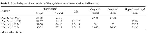
Phytophthora insolita Ann & Ko is an interesting but little known fungus, distinguished from all other species of Phytophthora in the production of abundant oospores in single cultures without the presence of antheridia (Stamps et al., 1990). Although parthenogenic "oospores" have been reported occasionally in P. infestans de Bary and P. fragariae Hickman (Pethybridge and Murphy, 1913; Savage et al., 1968), they are rare and produced only under certain cultural conditions. Phytophthora insolita was first reported from citrus soil in Changhua, Taiwan (Ann and Ko, 1980) and later from diseased poinsettia (Euphorbia pulcherrima Willd.), also in Changhya. Later, the common occurrence of asexual isolates of P. insolita was discovered in soil, ditch water, and diseased plant tissues in southern and central Taiwan (Ann and Ko, 1994). In a recent survey of Phytophthora species on southern China's Hainan Island we have obtained isolates of P. insolita and are reporting them herewith as new findings.

Materials and Methods

In the summers of 1998 and 1999, samples of soil, stream and pond water were collected from natural mountain forests and other localites and brought back to the Plant Pathology Laboratory in Danzhou City for the isolation of Phytophthora species. Samples were placed in 9-inch specimen dishes, with the soil samples flooded with an equal amount of deionized water. Citrus leaves were used as the bait, submerged or partially submerged in water. When lesions started to develop within a few days at room temperature (27-30°C), they were removed, scrubbed slightly with domestic bleaching powder and washed clean under running tap water. Small pieces of plant tissues (ca 2×4 mm) were cut out from the margin the lesions, blotted

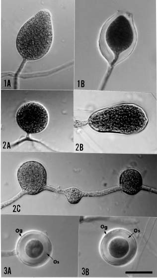
dry with paper towels and plated on selective medium in 9-cm petri dishes. The medium was made up by mixing 10 ml Campbell's V-8 juice, 90 ml deionized water, and 0.2 g CaCO3. This was filtered through two layers of cheescloth , diluted with deionized water to 1000 ml, boiled to dissolve 20 gm Bacto agar, and autoclaved at 121°C for 15 min. Prior to pouring the agar medium into sterilized petri dishes, benomyl (150 ppm), hymexazol (50 ppm), rifampicin (100 ppm), nystatin (50 ppm), and ampicillin (100 ppm) were added. The inoculated plates were incubated in dark at 25°C. When mycelial colonies appeared in 2-3 days, they were examined under light microscope to determine their identity as species of Phytophthora based on the colony morphology and the characteristics of the hyphae and branching characteristics (Ho et al., 1995). The culture was then transferred onto 10% V-8 agar plates for further studies. To induce sporangial production, small mycelial agar discs (ca 2×2 mm) were cut from the edge of a growing colony and transferred to sterile distilled water in 6-cm petri dishes, left under regular indoor light and at room temperature.

The pathogenicity of Phytophthora insolita to fruits was tested by inoculating apple, avocado, cucumber, eggplant, mango, green pepper and tomato with the fungus. The fruit was washed clean, surface sterilized with 90% ethanol, and a slit was made with a flamed scalpel. A small mycelial disc from a young colony of isolate Bds 1-18 was inserted into the crevice, which was then sealed with Scotch tape. Alternatively, a small mycelial agar disc (ca 5×5 mm) was placed on an unwounded surface, kept moist with wet cotton, and held in place by Scotch tape. The inoculated fruits were kept in sealed plastic bags and incubated at 28°C for 5 days.

Results

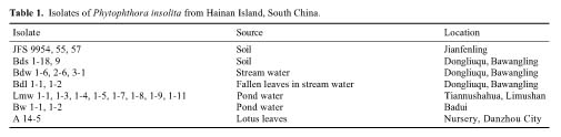
Of the numerous isolates of Phytophthora recovered, those that have been identified as belonging to P. insolita are presented in Table 1. They were all similar in producing appressed and petaloid colonies on V-8 agar plates,

*Corresponding author. Tel: 845-257-3780; Fax: 845-257-3791; E-mail: hoh@newpaltz.edu


Botanical Bulletin of Academia Sinica, Vol. 43, 2002

Figures 1-3. Figure 1A-B, Sporangia of Phytophthora insolita. Figure 2A-C, Hyphal swellings of Phytophthora insolita. Figure 3A-B, Oospores of Phytophthora insolita. Note the thin oogonial wall (Og) and the thick oospore wall (Os). Scale bar: 20 µm. All at the same magnification.


Ho et al. — Phytophthora insolita on Hainan Island

with a maximal temperature for growth at 40°C. Sporangia were not found on agar plates but readily produced overnight in water. They were oval to obpyriform, nonpapillate, nondeciduous and terminal on unbranched or irregularly branched sporangiophore, which displayed internal proliferation to form new sporangia nested within or outside the sporangium (Figure 1A-B). Laterally biflagellate zoospores were produced within the sporangium proper. The sporangia measured (27-)36-51(-62) × (22-)27-39(-51) µm with length/breadth ratio of (1.1)1.3-1.6(-1.9). Hyphal swellings (Figure 2A-C) were commonly found, terminal or intercalary, single or catenuous, often lateral and sessile. The shape of hyphal swellings ranged from spherical (12-)21-30(-31) µm diam to oval or obpyriform. Oogonia with oospores were produced in 1-5 month old cultures, well sealed with parafilm and kept in dark at 25°C. They were found only in isolates BDW 2-6, Bds 1-18, Bdl 1-2, Bds 9, JFS 9954, 9955 and 9957, especially around the inocula. The oogonia were yellowish, spherical, measuring (27-)29-33(-37) µm diam, and the spherical oospores, (26-)27-30(-32) µm diam almost filled up the oogonia (Figure 3A-B). The oospore wall was (2.5-)3-4(-6) µm thick. No antheridia could be found.

By artificial inoculation, P. insolita could not infect unwounded fruit but was pathogenic to wounded apple, avocado, cucumber, eggplant, mango, green pepper and tomato, causing fruit rots. The fungus was re-isolated from the diseased plant tissues in all cases.

Discussion

In addition to the absence of antheridia, P. insolita is also characterized by its appressed, chrysanthemum growth pattern, high maximal temperature for growth (39-40°C), nondeciduous, nonpapillate, internally proliferating sporangia with low length/breadth ratio and hyphal swellings of medium sizes (Ann and Ko, 1994; Ho et al., 1995). For comparison, the morphological characteristics of P. insolita reported in literature are summarized in Table 2. The production of oospores only in old cultures of some isolates of this species on Hainan Island is of special interest. Ann and Ko (1994) confirmed the identity of the asexual isolates of P. insolita by comparing the electrophoretic patterns of soluble proteins of these isolates with the sexual isolates. They suggested that "in nature, the asexual isolates of P. insolita probably originated from sexual isolates of P. insolita by losing their ability to pro

duce oospores." The discovery of P. insolita isolates on Hainan Island, which produce oospores only in aged cultures, tends to support their hypothesis. These sexual isolates may be in the process of losing their ability to produce oospores. The thickness of the oospore wall may be influenced by the age and the condition of the culture. Whereas the thickness of oospores from 5-day old cultures was 2.4 µm (Ann and Ko, 1980), 4-6 µm from 2-week old cultures (Ho et al., 1995), oospore wall from 1-5 month cultures in present study measured (2.5-)3-4(-6) µm thick.

The distribution of P. insolita is also intriguing. Ann and Ko (1994) concluded that the "high temperature variant of P. megasperma Drechsler" from alfalfa in Southern California (Ribeiro et al., 1978) should be re-classified as P. insolita. Otherwise, all isolates of this species have been reported from Taiwan only. Taking into consideration the occurrence of P. insolita on Hainan Island, the fungus is now known to exist on both sides of the Pacific Basin. Whereas Hainan Island and Taiwan are similar in having tropical to subtropical maritime climate and a central mountainous region surrounded by flat agricultural plains around the edge of the islands, southern California is different in having hot and dry weather and flat topography. We should determine if P. insolita exists in other parts of the world before speculating on the origin of this species. Neverthless, since most of the isolates on Hainan Island were obtained from relatively undisturbed and protected forests on high mountains, P. insolita is possibly indigenous to the island. It is not clear how important P. insolita is as a plant pathogen in nature. It is pathogenic to alfalfa root (Ribeiro et al., 1978), causes damage to poinsettia stem base (Ann and Ko, 1990), and is associated with strawberry fruit rot (Chang, 1988). The strawberry isolate was initially identified as a "high temperature variant of P. fragariae" but was reassigned to P. insolita (Ann and Ko, 1994). Although P. insolita was pathogenic to various wounded fruits by artificial inoculation in the present study, no report on plant diseases on Hainan Island attributable to P. insolita has appeared.

Literature Cited

Ann, P.J. and W.H. Ko. 1980. Phytophthora insolita, a new species from Taiwan. Mycologia 72: 1180-85.

Ann, P.J. and W.H. Ko. 1990. New Phytophthora diseases of flowers and ornamental plants in Taiwan. Plant Prot. Bull. 32: 341.


Botanical Bulletin of Academia Sinica, Vol. 43, 2002

Ann, P.J. and W.H. Ko. 1994. An asexual variant of Phytophthora insolita. Can. J. Microbiol. 40: 810-815.

Chang, H.S. 1988. Phytophthora species associated with strawberry fruit rot in Taiwan. Bot. Bull. Acad. Sin. 29: 61-67.

Ho, H.H., P.J. Ann, and H.S. Chang. 1995. The genus Phytophthora in Taiwan. Monogr. Ser., Inst. Bot., Acad. Sin. 15: 1-86.

Pethybridge, G.H. and P.A. Murphy. 1913. On pure cultures of Phytophthora infestans de Bary, and the development of oospores. Proc. Roy. Dublin Soc. 13: 566-588.

Ribeiro, O.K., D.C. Erwin, and R.A. Khan. 1978. A new high temperature Phytophthora pathogenic to roots of alfalfa. Phytopathology 68: 155-161.

Savage, E.J., C.W. Clayton, J.H. Hunter, J.A. Brenneman, C. Laviola, and M.E. Gallegly. 1968. Homothallism, heterothallism and interspecific hybridization in the genus Phytophthora. Phytopathology 58: 1004-1021.

Stamps, D.J., G.M. Waterhouse, F.J. Newhook, and G.S. Hall. 1990. Revised tabular key to the. species of Phytophthora. Mycol. Pap. 162. C.A.B. International, Kew, Surrey, England, 28 pp.