Bot. Bull. Acad. Sin. (2002) 43: 241-250

Wu et al. — Peniophora in Taiwan

A study of Peniophora species in Taiwan with clamped hyphae

Sheng-Hua Wu

Department of Botany, National Museum of Natural Science, Taichung, Taiwan 40419, Republic of China

(Received April 26, 2001; Accepted October 9, 2001)

Abstract. In this preliminary survey of Taiwanian Peniophora species with clamped hyphae five species are described: P. cinerea, P. ovalispora, P. pseudonuda, P. scintillans, and P. cf. septentrionalis. Apart from P. cinerea, all are newly recorded from Taiwan. Cultural studies are provided for the five species. Compatibility tests have been conducted for many specimens to confirm their specific identifications.

Keywords: Compatibility; Corticiaceae; Cultural studies; Peniophora; Taiwan; Taxonomy.

Introduction

This study is a preliminary survey of the Peniophora species in Taiwan with clamped hyphae. The genus Peniophora Cooke is generally regarded by mycologists as a member of the Corticiaceae s.l. (Basidiomycota). Peniophora is characterized by a combination of features: Presence of lamprocystidia or gloeocystidia or both; basidia clavate; pale-red spore deposits; smooth-walled, inamyloid and indextrinoid spores; tetrapolar sexuality and normal nuclear behavior or homothallism. Some species of Peniophora are capable of growing on living plants as parasites. Therefore, studies of this group may enhance our knowledge of plant pathology. Some species of Peniophora are associated with Tremella spp. (Chen, 1998). Species delimitation in Peniophora is more difficult than in most other corticioid genera, due to the similarity of the main morphological characters. Information concerning plant hosts, measurements of basidiospores based on spore deposits, and compatibility tests will be helpful for species identification. Before this study, Lin and Chen (1990) reported four species of Peniophora from Taiwan. They are P. aurantiaca, P. cinerea, P. incarnata, and P. spathulata. Their identification of P. cinerea was confirmed by my examination of voucher specimens.

Materials and Methods

Specimens provided for this study were collected from various localities in Taiwan during 1988-1997. Studied fungal specimens and cultures are deposited at the herbarium of the National Museum of Natural Science of ROC (TNM). Methods for examining specimens have been previously described (Wu, 1990). Cultural description and species code system are basically from Nobles (1965) with amendments by Boidin and Lanquetin (1983). Minor modifications to Nobles' code system have been presented by

some other mycologists. Nobles' code as detailed by Nakasone (1990) was adopted in this study. Methods of cultural studies, determination of sexuality, and compatible tests have been previously described (Wu, 1996).

Taxonomy

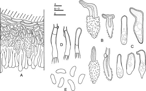
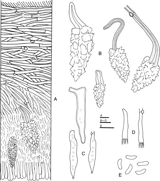
1. Peniophora cinerea (Pers.:Fr.) Cooke, Grevillea 8: 20. 1879. Figure 1

Basidiomes resupinate, effuse, adnate, membranaceous or subceraceous, 40-150 µm thick in section. Hymenial surface gray, purplish gray, pinkish gray, or yellowish gray, smooth, cracked; margin fairly determinate, concolorous.

Hyphal system monomitic; hyphae nodose-septate. Subiculum fairly uniform, composed of a thin basal layer, with compact texture; hyphae brownish, mainly horizonal, usually glued together, 2.5-5 µm diam., ± thick-walled. Hymenium ± thickening, of compact texture; hyphae brownish to colorless, mainly vertical, rather indistinct, slightly thick-walled. Lamprocystidia heavily encrusted, conical, brownish towards bases, 20-45 × 8-12 µm (encrustation included), with 1.5-4 µm thick walls. Gloeocystidia colorless or brownish especially towards bases, cylindrical, sometimes with acute apices, 25-60 × 6-11 µm, ± thick-walled towards bases, SA-. Basidia subclavate, 25-35 × 5-6.5 µm, 4-sterigmate. Basidiospores narrowly ellipsoid, adaxially slightly concave or flat, smooth, thin-walled, (6.3-) 7-9 × 2.5-4 µm, IKI-, CB-.

Specimens examined. TAIWAN. TAIPEI: National Taiwan University, on branch of Bridelia balansae, 31 May 1988, Wu 880531-2 (TNM); on branch of Liquidambar formosana, 19 Aug 1988, Wu 880819-3 (TNM); on branch of Ficus virgata, 15 Jan 1988, Wu 880115 (TNM); on branch of Elaeocarpus sylvestris, 15 May 1989, Wu 890515a (TNM). Yangminshan, alt. 600 m, on branch of angiosperm, 13 Dec 1990, Wu 901213-3 (TNM). HSINCHU: Kuanwu, alt. 1,900 m, on branch of angiosperm, 24 Aug 1988, Wu 880824-51, 880824-60 (TNM). MIAOLI: Takeshan, alt. 550 m, on branch of Prunus campanulata,

Fax: +886-4-3258684; E-mail: shwu@mail.nmns.edu.tw


Botanical Bulletin of Academia Sinica, Vol. 43, 2002

Figure 1. Peniophora cinerea (Wu 9310-63). A, Basidiome section; B, Lamprocystidia; C, Gloeocystidia; D, Basidia; E, Basidiospores. Scale bars = 10 µm.

9210-68 (TNM); alt. 1,950 m, on branch of angiosperm, 3 May 1994, Wu 9405-54 (TNM); alt. 1,650 m, on branch of angiosperm, 3 May 1994, Wu 9405-20 (TNM). Yushan National Park, Shenmu Forest Road, alt. 1,950 m, on branch of Alnus formosana, 7 Oct 1992, Wu 9210-72 (TNM). KAOHSIUNG: Liukuei, on branch of angiosperm, 13 Jul 1989, Wu 890713-4 (TNM). ILAN: Chilanshan, alt. 400 m, on branch of angiosperm, 18 Nov 1992, Wu 9211-1 (TNM).

Statistical analyses of basidiospores. Wu 911011-41: 6.5-8 × 2.8-3.8 µm (X = 7.39 ±0.52 × 3.01 ±0.24 µm, n = 30). Wu 9210-68: 7-8.2 × 3-4 µm (X = 7.61 ±0.44 × 3.45 ±0.36 µm, n = 30). Wu 9302-43: 7.8-8.9 × 3-4 µm (X = 8.26 ±0.34 × 3.34 ±0.28 µm, n = 30). Wu 9209-45: 7-8.8 × 2.8-3.1 µm (X = 7.61 ±0.48 × 2.93 ±0.10 µm, n = 30). Wu 9302-52: 7-8.6 × 2.5-3 µm (X = 7.89 ±0.53 × 2.77 ±0.19 µm, n = 30). Wu 9310-63: 7-9 × 2.5-3.6 µm (X = 8.15 ±0.55 × 3.01 ±0.24 µm, n = 30). Wu 9311-48: 6.3-8 × 2.5-3.5 µm (X=7.11 ±0.56 × 2.93 ±0.29 µm, n = 30). Wu 9403-17: 7.2-9.1 × 2.5-3.2 µm (X = 8.44 ±0.49 × 2.98 ±0.14 µm, n = 30). Wu 9403-18: 7.2-9.1 × 2.8-3.3 µm (X = 8.29 ±0.53 × 3.02 ±0.09 µm, n = 30).

Distribution. Cosmopolitan.

Cultural description (combined from PS-mycelia of Wu 9302-43, Wu 9405-5, and Wu 9405-20). 1 wk growth: Colony radius 27-35 mm. Advancing zone even. Mat whitish. Aerial mycelium absent or pellicular near inoculum, downy-cottony elsewhere. 2 wk growth: Colony radius 60-

25 Mar 1994, Wu 9403-17, 9403-18 (TNM). TAICHUNG: Pilu, alt. 2,000 m, on branch of Alnus formosana, alt. 1,900 m, 6 Dec 1991, Wu 911206-9 (TNM); on branch of angiosperm, Wu 911206-16 (TNM). Hsienpaotai, alt. 900 m, on branch of angiosperm, 6 Dec 1991, Wu 911206-46, 911206-54 (TNM). NANTOU: Sunlinhsi, alt. 1,700 m, on branch of angiosperm, 11 Oct 1991, Wu 911011-41 (TNM); on branch of Liquidambar formosana, 1 Jul 1992, Wu 9207-12 (TNM); 19 Sep 1992, Wu 9209-45, 9209-46 (TNM). Tsuifeng, alt. 2,300 m, on branch of angiosperm, 16 Feb 1993, Wu 9302-43, 9302-52, 9302-65 (TNM); 21 Sep 1993, Wu 9309-16 (TNM). Piluhsi, alt. 2,300 m, on branch of angiosperm, 13 Apr 1993, Wu 9304-8 (TNM); alt. 2,100 m, Wu 9304-43 (TNM). Yushan National Park, between Kuankao and Patungkuan, alt. 2,700-3,000 m, on branch of angiosperm, leg. Y.M. Ju, 8 Jan 1988, Wu 880108-15 (TNM); between Kuankao and Tuikuan, alt. 2,400 m, on branch of angiosperm, 26 Jul 1988, Wu 880726-53 (TNM). Huisun Forestry Station, alt. 770 m, on branch of angiosperm, 12 Dec 1997, Wu 9712-35 (TNM). CHIAYI: Yushan National Park, Lulinshan, alt. 2,650 m, on stem of Polygonum cuspidatum, 13 Oct 1993, Wu 9310-63 (TNM). Yushan National Park, entrance of Salihsienhsi Forest Road near Tungpu, alt. 1,300 m, on branch of angiosperm, 24 Nov 1993, Wu 9311-48 (TNM); 7 Dec 1994, Wu 9412-1 (TNM). Yushan National Park, Nanhsi Forest Road, alt. 2,100 m, on branch of Alnus formosana, 7 Oct 1992, Wu


Wu et al. — Peniophora in Taiwan

70 mm. Mat whitish. Aerial mycelium pellicular near inoculum, downy-cottony elsewhere. Advancing hyphae colorless, nodose-septate, 1.5-4.5 µm diam., thin-walled. 3 wk growth: Plates covered. 6 wk growth: Mat whitish near inoculum, brownish elsewhere. Aerial mycelium sparse and slightly pellicular near inoculum, downy towards growth margins. Hyphal system monomitic; hyphae nodose-septate. Aerial hyphae colorless, yellow or brownish, moderately ramified, 1.5-5 µm diam., thin-walled, sometimes irregularly swollen, usually guttulate, occasionally covered with brownish excreted materials; gloeoplerous hyphae or gloeocystidia occasionally present, SA-. Submerged hyphae colorless, moderately ramified, usually guttulate, 1.5-6.5 µm diam., Crystals present, thin-walled. No distinct odor. Not fruiting.

Oxidase reacitons. TAA: ++++, 30-40; ++++, 40-65. GAA: ++++, tr; ++++, tr to 12. TyA: - (slightly brownish), 35-50; - (slightly brownish), 90+.

Sexuality. Tetrapolar (A1B1: 1, 8; A1B2: 4, 6, 7, 9; A2B1: 3, 5, 9; A2B2: 2, 10) for Wu 9302-52.

Species code. 2a, 3c, 15b, 32, 37, 39, 43, 54, 60, 61.

Nuclear behavior. Normal. Spores uninucleate, MS-mycelium uninucleate, PS-mycelium dikaryotic (studied from Wu 9302-52).

Remarks. This species complex has cosmopolitan distribution, from temperate to tropical regions. Hallenberg and Larsson (1992) performed compatibility tests mating three specimens from Taiwan (Wu 890515, Wu 880726-53,Wu 880824-51) with foreign strains. All three have high degree of intercompatibility with European and Canadian specimens. The following Taiwanian collections of P. cinerea were compatible in this study with the tester strain Wu 9302-52. Wu 880531-2, Wu 880726-53, Wu 880824-51, Wu 890515, Wu 890713-4, Wu 911011-41, Wu 911206-9, Wu 911206-54, Wu 9207-12, Wu 9210-68, Wu 9210-72, Wu 9211-1, Wu 9302-43, Wu 9304-8, Wu 9304-43, Wu 9309-16, Wu 9310-63, Wu 9311-48, Wu 9403-17, and Wu 9405-54.

2. Peniophora ovalispora Boidin et al., Bull. Soc. Mycol. France 107: 108. 1991. Figure 2

Basidiome resupinate, effuse, adnate, membranaceous, 40-120 µm thick in section. Hymenial surface yellowish orange, smooth or slightly tuberculate, sometimes cracked; margin fairly determinate, concolorous.

Hyphal system monomitic; hyphae nodose-septate. Subiculum fairly uniform, composed of a fairly thin basal layer, with compact texture; hyphae colorless, mainly horizonal, sometimes glued together, 1.5-4.5 µm diam., thin- or slightly thick-walled. Hymenium thickening, with compact texture; hyphae colorless, mainly vertical, dextrinoid, thin-walled. Lamprocystidia numerous, immersed or emergent, overlapping in hymenial layer, heavily encrusted, conical, colorless, 20-40 × 7-11 µm (encrustation included), with 1-2 µm thick walls. Gloeocystidia not abundant, colorless, cylindrical, usually tapering to nar

row apices, 25-40 × 6-9 µm, thin-walled, SA-. Basidia subclavate to almost cylindrical, sometimes slightly constricted in the middle, 14-20 × 4-4.5 µm, some with thickened walls toward bases, 4-sterigmate. Basidiospores broadly ellipsoid, smooth, thin-walled, (4-) 5-6 (-6.3) × 3-4 µm, IKI-, CB-.

Specimens examined. TAIWAN. TAIPEI: Yangminshan, alt. 400 m, on branch ofDuranta repens, 1 Jul 1988, Wu 880701-4 (TNM); alt. 600 m, on branch of angiosperm, 20 Nov 1992, Wu 9211-93 (TNM). Kungliao, alt. 200 m, on branch of Araliaceae, 25 Jul 1991, Wu 910725-7, Lin 566-1 (TNM). HSINCHU: Sanshui, on branch of angiosperm, 4 Apr 1988, Wu 880404-2, 880404-4 (TNM). ILAN: Jentse, alt. 380 m, on branch of angiosperm, 22 Nov 1988, Wu 881122-45 (TNM); Chilanshan, alt. 400 m, on branch of angiosperm, 18 Nov 1992, Wu 9211-15 (TNM); Fushan Nature Reserve, alt. 600 m, on branch of angiosperm, 9 Mar 1993, Chen 160 (TNM). NANTOU: Yushan National Park, between Kuankao and Tuikuan, alt. 2,300 m, on branch of living Pittosporum illicioides, 28 Jul 1988, Wu 880728-42 (TNM); Shenmu Forest Road, alt. 1,300 m, on rotten culm of bamboo, 7 Oct 1992, Wu 9210-87 (TNM). Tsuifeng, alt. 2,300 m, on branch of angiosperm, 16 Feb 1993, Wu 9302-62 (TNM 9306-37); Sunlinhsi, alt. 1,700 m, on branch of living? Acer serrulatum, 19 Jun 1993, Wu 9306-34, 9306-37, 9306-47 (TNM); Neimaopu, Dachieshan, alt. 750 m, on branch of angiosperm, 13 Oct 1994, Wu 9410-17 (TNM); Tungpu, alt. 1,300 m, on branch of angiosperm, 13 Oct 1994, Wu 9410-32 (TNM). KAOHSIUNG: Liukuei, Shanping, alt. 750 m, on branch of angiosperm, 7 Nov 1991, Wu 911107-26, 911107-41 (TNM).

Statistical analyses of basidiospores. Wu 9211-93: 4-5.5 × 3-4 µm (X = 4.83 ±0.46 × 3.23 ±0.33 µm, n = 30). Wu 9302-62: 5-6.3 × 3.3-4 µm (X = 5.75 ±0.40 × 3.73 ±0.22 µm, n = 30). Wu 911107-26: 5-6 × 3-4 µm (X = 5.60 ±0.36 × 3.48 ±0.38 µm, n = 30).

Distribution. Réunion (type locality) and Taiwan.

Cultural description (from PS-mycelia of Wu 9211-93). 1 wk growth: Colony radius 25 mm. Advancing zone even. Mat white. Aerial mycelium pellicular around inoculum, downy-cottony towards growth margins. 2 wk growth: Colony radius 57 mm. Advancing zone even. Mat white. Aerial mycelium pellicular around inoculum, downy towards growth margins. 3 wk growth: Plates covered. 6 wk growth: Mat whitish, slightly brownish-tinted. Aerial mycelium pellicular. Hyphal system monomitic; hyphae nodose-septate. Aerial hyphae colorless, sparsely ramified, usually guttulate, 2-3.5 µm diam., thin-walled. Submerged hyphae colorless, moderately ramified, 2-5 µm diam., thin-walled. Cuboid crystals present. Odor fragrant. Not fruiting.

Oxidase reactions. TAA: +++, 14-20; 37-43. GAA: ++++, tr; tr. TyA: -, 45-50; 90+.

Species code. 2a, 3c, 7, 32, 36, 37, 38, 43, 50, 54, 60, 61.

Sexuality. Tetrapolar (A1B1: 1, 8; A1B2: 4, 5, 10; A2B1: 2, 6, 7; A2B2: 3, 9) for Wu 9211-93.


Botanical Bulletin of Academia Sinica, Vol. 43, 2002

Nuclear behavior. Normal. Spores uninucleate, MS-mycelium uninucleate, SC-mycelium dikaryotic (studied from Wu 9211-93).

Remarks. Peniophora ovalispora is closely related to P. scintillans. Morphologically the former has smaller basidiospores. Boidin et al. (1991) proved incompatibility between specimens of these two species. Incompatibility was also obtained in this study by crossing Wu 9211-39 (P. ovalispora) with Wu 881026-21 (P. scintillans). In this study, specimens Wu 880404-2, Wu 880404-4, Wu 880701-4, Wu 881122-45, Wu 911107-26, and Wu 9211-15, were compatible with the tester Wu 9211-93.

3. Peniophora pseudonuda Hallenb., Mycotaxon 11(2): 459. 1980. Figure 3

Basidiome resupinate, effuse, adnate, membranaceous or subceraceous, 50-120 µm thick in section. Hymenial surface yellowish gray, smooth, cracked; margin thinning, concolorous, pruinose.

Hyphal system monomitic; hyphae nodose-septate. Subiculum fairly uniform, composed of basal layer, with compact texture; hyphae brownish, mainly horizonal, usually glued together, 2.5-5 µm diam., ± thick-walled. Hymenium ± thickening, with compact texture; hyphae colorless, mainly vertical, rather indistinct, thin-walled.

Figure 2. Peniophora ovalispora (Wu 9211-93). A, Basidiome section; B, Lamprocystidia; C, Gloeocystidia; D, Basidia; E, Basidiospores. Scale bars = 10 µm.


Wu et al. — Peniophora in Taiwan

Figure 3. Peniophora pseudonuda (Wu 9211-110). A, Basidiome section; B, Lamprocystidia; C, Gloeocystidia; D, Basidia; E. Basidiospores. Scale bars = 10 µm.

Lamprocystidia usually immersed, heavily encrusted, conical, colorless or slightly brown, 20-45 × 8-15 µm (encrustation included), with 1-2.5 µm thick walls. Gloeocystidia numerous, brownish towards bases, cylindrical with narrower apices, slightly flexuous, 40-90 × 7-14 µm, thick-walled towards bases, slightly SA+. Basidia subclavate, 24-33 × 5.5-7 µm, 4-sterigmate. Basidiospores ellipsoid, adaxially slightly concave, smooth, thin-walled, 8-10.2 × 3-4.2 µm (X = 9.15 ±0.64 × 3.56 ±0.36 µm, n = 30), IKI-, CB-.

Specimen examined. TAIWAN. TAIPEI: Yangminshan, alt. 600 m, on branch of angiosperm, 20 Nov 1992, Wu 9211-110 (TNM).

Distribution. Iran (type locality) and Taiwan.

Cultural description (from PS-mycelium of Wu 9211-110). 1 wk growth: Colony radius 20-25 mm. Advancing zone fairly even. Mat white. Aerial mycelium pellicular. 2 wk growth: Colony radius 40-46 mm. Advancing zone slightly fimbriate. Mat whitish. Aerial mycelium almost absent. 3 wk growth: Colony radius 63-75 mm. Advanc

ing hyphae colorless, nodose-septate, 1.5-3 µm diam., thin-walled. 4 wk growth: Plates covered. 6 wk growth: Mat white, slightly brownish-tinted. Aerial mycelium pellicular, irregularly reticulate-porous. Monomitic. Hyphae nodose-septate. Aerial hyphae colorless, moderately ramified, usually guttulate, occasionally irregularly swollen, 1.5-4.5 µm diam., thin-walled; gloeoplerous hyphae and gloeocystidia occasionally present, SA-. Submerged hyphae colorless, moderately ramified, occasionally swollen, usually guttulate, secondary septa occasionally present, 1.5-6.5 µm diam., thin-walled. Cuboid crystals present. No distinct odor. Not fruiting.

Oxidase reactions. TAA: ++++, tr; 23-37. GAA: ++++, tr; tr to 15. TyA: - (slightly brown), 12-15; 25-30.

Species code. 2a, 3c, 15b, 32, 36, 37, 38, 44, 54, 60, 61.

Sexuality. Tetrapolar (A1B1: 1, 3, 10; A1B2: 4, 6, 8; A2B1: 2; A2B2: 5, 7, 9) for Wu 9211-110.

Nuclear behavior. Normal. Spores uninucleate, MS-mycelium uninucleate, SC-mycelium dikaryotic (studied from Wu 9211-110).


Botanical Bulletin of Academia Sinica, Vol. 43, 2002

Figure 4. Peniophora scintillans (Wu 901125-16). A, Basidiome section; B, Quasi-binding hyphae; C, Lamprocystidia; D, Gloeocystidia; E, Basidia; F, Basidiospores. Scale bars = 10 µm.

Remarks. Peniophora pseudonuda differs from P. nuda (Fr.) Bres. in having thicker basal layer of the basidiomes and broader basidiospores, additionally has comparatively longer and narrower gloeocystidia. It also resembles P. fissilis Boidin et al., but differs in having obtuse apices of the gloeocystidia, and has slightly larger basidiospores.

4. Peniophora scintillans G.H. Cunn., Trans. Roy. Soc. New Zealand 83: 268. 1955. Figure 4

Basidiome resupinate, effuse, adnate, membranaceous, 50-150 µm thick in section. Hymenial surface grayish yellow or grayish pink, smooth, sometimes cracked; margin fairly determinate, concolorous.

Hyphal system essentially monomitic, some specimens with quasi-binding hyphae near substratum; generative hyphae nodose-septate. Subiculum fairly uniform, composed of a fairly thin basal layer, with compact texture; generative hyphae colorless or yellowish, glued together, 2-4 µm diam., usually slightly thick-walled; quasi-binding hyphae rarely present near substratum, colorless or yellowish, 0.5-1.5 µm diam. Hymenium thickening, with compact texture; hyphae colorless, mainly vertical, thin-

walled, dextrinoid. Lamprocystidia numerous, immersed or emergent, overlapping in subhymenium, heavily encrusted, conical, colorless, slightly yellow or rarely slightly brown, 25-45 × 8-12 µm (encrustation included), with 1-2.5 µm thick walls. Gloeocystidia fairly few, colorless, cylindrical, tapering to narrow apices, 25-50 × 6-10 µm, thin-walled, SA-. Basidia subclavate, 25-33 × 5-6 µm, 4-sterigmate. Basidiospores broadly ellipsoid, smooth, thin-walled, (5-) 5.7-7 (-7.5) × 3.7-4.8 µm, IKI-, CB-.

Specimens examined. NEW ZEALAND. Bay of Plenty, Mt. Te Aroha, on Laurelia novaezelandiae, leg. G.H. Cunn., Sep 1954 (holotype, PDD 13828). TAIWAN. TAIPEI: Kungliao, alt. 200 m, on branch of Lagerstroemia subcostata, 25 Nov 1990, Wu 901125-16 (TNM). Yangminshan, alt. 600 m, on branch of angiosperm, 13 Dec 1990, Wu 901213-13 (TNM). NANTOU: Sun-Moon Lake, on branch of angiosperm, alt. 750 m, 26 Oct 1988, Wu 881026-21 (TNM). TAICHUNG: Chiapaotai, alt. 900 m, on branch of angiosperm, 6 Dec 1991, Wu 911206-14 (TNM). PINGTUNG: Kengting National Park, Changle, alt. 250 m, on branch of angiosperm, 20 Feb 1992, Wu 9202-36 (TNM). TAITUNG: Taimali, alt. 600 m, on branch of angiosperm, 19 May 1989, Wu 890519-35 (TNM).


Wu et al. — Peniophora in Taiwan

Distribution. New Zealand (type locality), Madagascar, Réunion, and Taiwan.

Cultural description (FB-mycelium from Wu 901125-16). 1 wk growth: Colony radius 27 mm. Advancing zone fairly even. Mat white, slightly dirty-tinted. Aerial mycelium pellicular around inoculum, downy-cottony elsewhere. 2 wk growth: Colony radius 60 mm. Advancing zone some

what bayed. Mat white, slightly clay colored tinted. Aerial mycelium pellicular-downy around inoculum, downy-cottony elsewhere, ± zonate. 3 wk growth: Plates covered. 6 wk growth: Mat white, slightly brownish-tinted. Aerial mycelium pellicular, almost absent around inoculum. Hyphal system monomitic; hyphae nodose-septate. Aerial hyphae moderately or richly ramified, colorless or rarely

Figure 5. Peniophora cf. septentrionalis (Wu 9310-114). A, Basidiome section; B, Lamprocystidia; C, Gloeocystidia; D, Basidia; E, Basidiospores. Scale bars = 10 µm.


Botanical Bulletin of Academia Sinica, Vol. 43, 2002

brownish, usually guttulate, 1-3.5 µm diam., thin-walled; gloeoplerous hyphae or gloeocystidia colorless or rarely brownish, 3-6 µm diam., with walls up to ca. 1 µm thick, SA-. Submerged hyphae colorless, moderately or sparsely ramified, usually guttulate, 1-3.5 µm diam., thin-walled. Cubical crystals present. No distinct odor. Not fruiting.

Oxidase reactions. TAA: +++, 28-33; 45-53. GAA: ++++, tr; tr. TyA: -, 36-42; 90+.

Species code. 2a, 3c, 15b, 32, 36, 37, 38, 43, 54, 60, 61.

Sexuality. Tetrapolar (Boidin et al., 1991).

Nuclear behavior. Normal. Spores uninucleate, MS-mycelium uninucleate, PS-mycelium dikaryotic (Boidin et al., 1991).

Remarks. As mentioned above, P. scintillans has larger basidiospores than P. ovalispora. Cunningham (1963) reported spores for P. scintillans to be 6-7 × 3.5-4 µm. My measurements from the type specimen are similar, 6-7.5 × 3.5-4.5 µm. Stalpers and Buchanan (1991), based on the same specimen, found spores sizes to be 5.5-8 × 3.5-4.5 (-5) µm. The measurement provided by Boidin et al. (1991) for this species is slightly smaller (4.5-6(-7) × 3.2-4.2 µm). I found one of the specimens (LY 12415) studied by Boidin et al. (1991) as P. scintillans to be compatible with two Taiwanian specimens (Wu 890519-35 and Wu 901125-16) in this study.

5. Peniophora cf. septentrionalis Laurila, Ann. Bot. Soc. Zool.-Bot. Fenn. Vanamo 10 (4): 10. 1939. Figure 5

Basidiome resupinate, effuse, adnate, subceraceous, 150-500 µm thick in section. Hymenial surface pink or grayish pink, smooth, occasionally cracked; margin determinate, quite often separate from the substratum, concolorous or slightly paler.

Hyphal system monomitic; hyphae nodose-septate. Subiculum fairly uniform, composed of a rather thick basal layer, with compact texture; hyphae brownish next to substratum and hymenium, colorless elsewhere, mainly horizonal, sometimes glued together, 2.5-5 µm diam., with 0.6-1.2 µm thick walls. Hymenium ± thickening, with compact texture; hyphae colorless, mainly vertical, rather indistinct, thin-walled. Lamprocystidia variably abundant, mostly immersed, heavily encrusted, conical, colorless or slightly brown, 30-90 × 10-20 µm (encrustation included), with 1-2.5 µm thick walls. Gloeocystidia variably abundant, colorless, cylindrical with obtuse apices or fusiform with acute apices, 30-80 × 5-10 µm, thin- to thick-walled, SA-. Basidia subclavate, 25-30 × 4-5 µm, 4-sterigmate. Basidiospores suballantoid, adaxially slightly concave, smooth, thin-walled, 6.3-7.5 × 2-2.5 µm, IKI-, CB-.

Specimen examined. TAIWAN. NANTOU: Yushan National Park, Nanhsi Forest Road, alt. 2,200 m, on branch of angiosperm, 14 Oct 1993, Wu 9310-114 (TNM); Yushan National Park, between Kuankao and Tuikuan, alt. 2,400 m, on branch of Alnus formosanum, 26 Jul 1988, Wu 880726-55 (TNM).

Distribution. North Europe, North America, Russia, and Taiwan.

Cultural description (from PS-mycelium of Wu 9310-114). 1 wk growth: Colony radius 35 mm. Advancing zone even. Mat whitish or slightly brown. Aerial mycelium downy-cottony. 2 wk growth: Colony radius 70 mm. Advancing zone fairly even. Mat white. Aerial mycelium pellicular near inoculum, downy-cottony and with sparsely porose-reticulate morphology elsewhere. Advancing hyphae sparsely ramified, nodose-septate, 2.5-5 µm diam., thin-walled. 3 wk growth: Plates covered. 6 wk growth: Mat white. Aerial mycelium almost absent close to inoculum, slightly pellicular with porose-reticulate appearance elsewhere. Hyphal system monomitic; hyphae nodose-septate. Aerial hyphae colorless, sparsely or moderately ramified, usually guttulate, 1.5-3 µm diam., thin-walled. Submerged hyphae colorless, moderately ramified, 2-5 µm diam., thin-walled. No distinct odor. Not fruiting.

Oxidase reactions. TAA: +++, 30; 50-60. GAA: ++++, 25-40; 50-60. TyA: -, 30-50; 90+.

Species code. 2a, 3c, 7, 32, 36, 38, 43, 54, 60, 61.

Sexuality. Tetrapolar (A1B1: 1; A1B2: 3, 6, 7, 8, 10; A2B1: 4, 5; A2B2: 2, 9) for Wu 9310-114.

Nuclear behavior. Normal. Spores uninucleate, MS-mycelium uninucleate, SC-mycelium dikaryotic (studied from Wu 9310-114).

Remarks. General morphological characters of two specimens collected from temperate belt of Taiwan resemble those of P. septentrionalis. The only significant difference between Taiwanian specimens and those of other localities is in the host plants. Collections from the other regions of the world occur on gymnosperm (Eriksson et al., 1978; Ginns and Lefebvre, 1993) while two specimens from Taiwan were collected from angiosperm. Specimens Wu 9310-114 and Wu 880726-55 were intercompatible in this study.

Studies on Peniophora specimens reported from Taiwan by Lin and Chen (1990)

Lin and Chen (1990) described four species in Peniophora in their preliminary survey of corticioid fungi from Taiwan. All have clamped hyphae. I examined voucher specimens cited in their paper and conclude:

1. Peniophora aurantiaca (Bres.) Höhn. & Litsch.

Specimen examined. TAIWAN. TAITUNG: Pinglin, roadside between Orchid Valley and Mt. Suchuanglo, under the hardwood forest, alt. 400-500 m, 24 Jul 1975, S. Lin (NTU-2925).

Remarks. Specimen NTU-2925 is not Peniophora aurantiaca, but a species of Hypochnicium. Subiculum texture is loose. Hymenial surface is slightly grandinioid, due to the projecting encrusted cystidia. Subicular hyphae are nodose-septate, thick-walled. Spores are subglobose,


Wu et al. — Peniophora in Taiwan

Council of ROC (Nos. NSC 83-0211-B-178-002 and NSC 88-2311-B-178-001).

Literature Cited

Boidin, J. and P. Lanquetin. 1983. Basidiomycètes Aphyllophorales épithéloïdes étalés. Mycotaxon 16: 461-499.

Boidin, J., P. Lanquetin, and G. Gilles. 1991. Les Peniophoraceae de la zone intertropicale (Basidiomycetes, Aphyllophorales). A. Especes paleotropicales. Bull. Soc. Mycol. France 107: 91-147.

Chen, C.J. 1998. Morphological and molecular studies in the genus Tremella. Biblioth. Mycol. 174: 1-225.

Cunningham, G.H. 1963. The Thelephoraceae of Australia and New Zealand. New Zealand Dept. Sci. Ind. Res. Bull. 145: 1-359.

Eriksson, J., K. Hjortstam, and L. Ryvarden. 1978. The Corticiaceae of North Europe 5. Mycoaciella - Phanerochaete. Fungiflora, Oslo, pp. 889-1047.

Ginns, J. and M.N.L. Lefebvre. 1993. Lignicolous corticioid fungi (Basidiomycota) of North America. Mycol. Mem. 19:1-247.

Hallenberg, N. and E. Larsson. 1992. Mating biology in Peniophora cinerea (Basidiomycetes). Can. J. Bot. 70: 1758-1764.

Lin, S.H. and Z.C. Chen. 1990. The Corticiaceae and the resupinate Hydnaceae of Taiwan. Taiwania 35: 69-111.

Nakasone, K.K. 1990. Cultural studies and identification of wood-inhabiting Corticiaceae and selected Hymenomycetes from North America. Mycol. Mem. 15: 1-412.

Nobles, M.K. 1965. Identification of cultures of wood-inhabiting Hymenomycetes. Can. J. Bot. 43: 1097-1139.

Stalpers, J.A. and P.K. Buchanan. 1991. Type studies of the species of Pellicularia and Peniophora described by G.H. Cunningham. New Zealand J. Bot. 29: 331-340.

Wu, S.H. 1990. The Corticiaceae (Basidiomycetes) subfamilies Phlebioideae, Phanerochaetoideae and Hyphodermoideae in Taiwan. Acta Bot. Fennica 142: 1-123.

Wu, S.H. 1996. Studies on Gloeocystidiellum sensu lato (Basidiomycotina) in Taiwan. Mycotaxon 58: 1-68.

Wu, S.H. 1997. New species and new records of Hyphoderma (Basidiomycotina) from Taiwan. Bot. Bull. Acad. Sin. 38: 63-72.

smooth, 11.5-13 × 10-11.5 µm, with 0.5-1 µm thick walls, IKI-. This Hypochnicium species will be described in detail in a separate publication. Lin and Chen (1990) misspelt the specific epithet as "aurantica".

2. Peniophora cinerea (Pers.:Fr.) Cooke

Specimen examined. TAIWAN. TAIPEI: Chungho, Yuanntong Temple, roadside, under the broad-leaved forest, alt. 300 m, 25 Nov 1975, S. Lin (NTU-3903, 3396).

Remarks. General characters of these two specimens correspond with this species.

3. Peniophora incarnata (Pers.:Fr.) P. Karst.

Specimen examined. TAIWAN. TAITUNG: Lanyu, Yehyiu, in the ditch of mountain, under the broad-leaved primary forest, alt. 50-300 m, 22 Aug 1975, S. Lin (NTU-3649).

Remarks. The specimen is Hyphoderma variolosum Boidin et al. The basidia have a median-constriction, typical of Hyphoderma basidia. The spores are about 12.5 × 4.7 µm. This measurement fits H. variolosum well. Peniophora incarnata is a temperate species, has pink basidiome, not white-gray of this specimen. In addition, P. incarnata has shorter spores (8-12 × 3.5-5 µm, according to Eriksson et al., 1978). Hyphoderma variolosum has been recorded from Taiwan by Wu (1997), based on a specimen collected also from the tropical lowland in southern Taiwan.

4. Peniophora spathulata S.H. Lin & Z.C. Chen

Specimen examined. TAIWAN. TAIPEI: Mt. Seven-star, roadside, under the hardwood forest, alt. 700 m, 6 Dec 1975, S. Lin (NTU-4304, holotype).

Remarks. The "spathulate cystidia" in the type specimen diagnostic for this species, do not deviate from the ordinary conical lamprocystidia that are fairly common in many species of Peniophora. Various features of the holotype of Peniophora spathulata resemble P. cinerea. I herein treat P. spathulata as a synonym of P. cinerea.

Acknowledgments. The author is indebted to the curator of PDD and Dr. Z. C. Chen for arranging loan of specimens for this study. I am grateful to Dr. J. Boidin for providing some cultures and for his valuable suggestions in improving the manuscript. This study was supported by the National Science


Botanical Bulletin of Academia Sinica, Vol. 43, 2002