Bot. Bull. Acad. Sin. (2002) 43: 251-259

Chen et al. — An antimicrobial protein cDNA from mung bean

Cloning and characterization of a cDNA encoding an antimicrobial protein from mung bean seeds

Kuan-Chung Chen1, Ching-Yu Lin1, Mei-Chu Chung2, Cheng-Chun Kuan1, Hsien-Yi Sung1, Samson C.S. Tsou3, C. George Kuo3, and Ching-San Chen1,2,*

1Graduate Institute of Agricultural Chemistry, National Taiwan University, Taipei 106, Taiwan

2Institute of Botany, Academia Sinica, Nankang, Taipei 115, Taiwan

3Asian Vegetable Research and Development Center (AVRDC), P.O. Box 42, Shanhua, Tainan 741, Taiwan

(Received March 20, 2002; Accepted April 16, 2002)

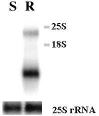
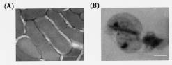
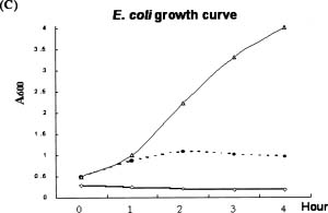
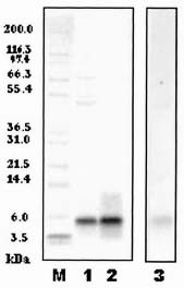
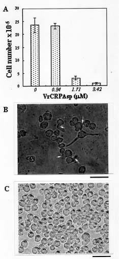
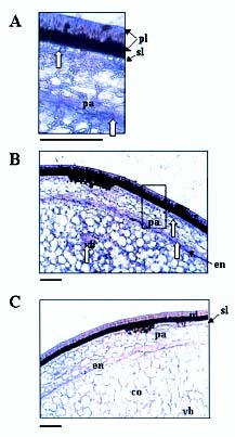
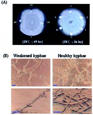
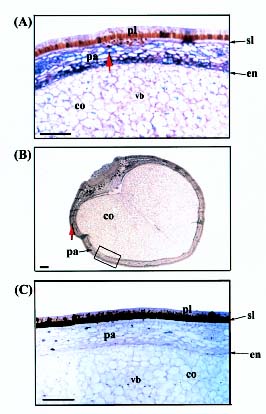
Abstract. A cDNA clone, VrCRP (GenBank accession no. AF326687), encoding an antimicrobial protein was isolated from the differentially expressed cDNAs between a pair of near isogenic lines of mid-matured mung bean seeds. Northern blot analysis showed that VrCRP gene is preferentially expressed in bruchid-resistant developing mung bean seeds. The mature VrCRP is a basic cysteine-rich protein with a predicted molecular mass of 5.9 kDa and a calculated isoelectric point of 8.23. Eight cysteine amino acids present in the primary structure of VrCRP compose four disulfide bridges. Biological analysis showed that VrCRP is toxic to E. coli and completely arrested growth of insect cell (Spodoptera frugiperda, Sf21) at a low concentration of 3.42 µM. VrCRP also inhibited the growth of Rhizoctonia solani. Immunolocalization and in situ hybridization indicated that VrCRP was predominantly present in the parenchyma cells of the seed coat. Based on the biological activities and the protein localization, it is suggested that VrCRP may act as an evolutionary barrier in mung bean seeds that is important in coping with invasions of pathogens and herbivores.

Keywords: Antimicrobial protein; Immunolocalization; In situ hybridization; Mung bean.

Introduction

Plant seeds are usually sown in natural surroundings rich in microorganisms and insects. During evolution, various defense mechanisms developed in plants-such as low water content in the seed and a hard seed coat to provide effective physical defenses against phytopathogens and herbivorous animals. Another array of defense mechanisms includes the production of antimicrobial phytoalexins (Van Etten et al., 1989), pathogenesis-related proteins (Linthorst, 1991; Ponstein et al., 1994), and a large group of cysteine-rich proteins such as lipid transfer protein (Garcia-Olmedo et al., 1995), antimicrobial peptide HvAMP1 (Harrison et al., 1997), Rs-AFP1, Rs-AFP2 (Terras et al., 1992), plant defensins (Broekaert et al., 1995; Terras et al., 1995), hevein (Van Parijs et al., 1991), knottin-type peptides (Cammue et al., 1992; Broekaert et al., 1997), four-cysteine-type peptide (Duvick et al., 1992), 2S albumins (Terras et al., 1992) and thionins (Bohlmann, 1994). The thionins and plant defensins are among the best characterized defensive proteins.

The defensin family is very diverse, and several subfamilies can be recognized on the basis of amino acid sequence homologies (Broekaert et al., 1995), including g-thionin. Plant defensins consist of a triple-stranded an

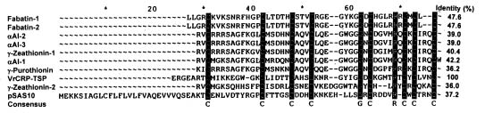
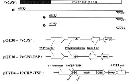
tiparallel b-sheet and a single a-helix lying in parallel with the b-sheet. The Cys-X-X-X-Cys segment of the a-helix is connected by two disulfide bridges to the Cys-X-Cys segment in the third b-strand, forming a cystine-stabilized a-helix motif (Kobayashi et al., 1991). We have isolated a novel cDNA from Vigna radiata, termed VrCRP (Cysteine-rich Protein of Vigna radiata), which encodes a cysteine-rich protein constituting a new member of the plant defensin family. This paper describes the cloning, biological activities, and localization in the seed of this mung bean defensin VrCRP.

Materials and Methods

Plant Varieties

Vigna radiata L. VC1973 and VC6089A were bred in the Asian Vegetable Research and Development Center (AVRDC). Seeds of the former are susceptible to bruchid Callosobruchus chinensis, whereas the latter have been shown to resist bruchid attack. VC6089A (BC6S2) population was bred by crossing a bruchid-resistant wild mung bean, TC1966 (as donor parent) with a breeding line VC1973A (as recurrent parent).

RNA Preparation

Total RNA was extracted from the 15 DAF (days after flowering) developing seeds of VC1973A and VC6089A by hot phenol method (Verwocrd et al., 1989).

*Corresponding author. Tel: 886-2- 2789-9591; Fax: 886-2-2785-8936; E-mail: chingsan@gate.sinica.edu.tw


Botanical Bulletin of Academia Sinica, Vol. 43, 2002

Suppression Subtractive Hybridization (SSH)

SSH was performed between VC6089A cDNA and matched VC1973A cDNA, using the PCR Select cDNA Subtraction kit (Clontech) according to the manufacturer's recommendations. A full-length cDNA, VrCRP was isolated using this method.

Northern Blot Analysis

VrCRP cDNA was amplified with PCR, purified on an agarose gel and used as a probe for Northern blot analysis as follows: 10 µg of total RNAs purified from 15 DAF seeds of VC1973A and VC6089A were electrophoresed in an 1% agarose-0.41 M formaldehyde gel and blotted onto Hybond-H+ membranes (Amersham) with Chomczynski's alkaline downward transfer method (Chomczynski, 1992). The membranes were incubated in 4 mL prehybridization buffer (Church and Gilbert, 1984) containing 7% SDS, 0.25 M sodium phosphate, pH 7.2, 1 mM EDTA, 1% albumin, and 2.5 mg mL-1 denatured salmon sperm single-strand DNA for 5 h at 65°C and then in hybridization buffer containing a random-primed [32P] cDNA probe (1 × 10⁶ cpm mL-1) (Feinberg and Vogelstein, 1984). Hybridization was performed at 65°C for 18 h. The membranes were washed twice with 2 × SSC, 1% SDS and 0.2 × SSC, 1% SDS for 15 min at 65°C. Signals were visualized and analyzed using a PhosphorImager system (Molecular Dynamics). The blots were striped and rehybridized with a probe specific to 25S ribosomal RNA to normalize them for equivalent loading of total RNA.

Construction of Expression Vector

The schematic diagrams for construction of expression vector are shown in Figure 3. The 73 amino acid open reading frame of the VrCRP clone was amplified by PCR with DNA polymerase (Saiki et al., 1988) and three pairs of primers (VrCRF1: 5´-GCGGATCCGA TGACGATGAC AAAATGGAGA GAAAAACTTT CA-3´, VrCRR1: 5´-GGCAAGCTTT CAACAGTTGA CGAGGCAAT-3´; VrCRF2: 5´-GCGGATCCGA TGACGATGAC AAAGAGAGAG GAGAGGCTAG AAC-3´, VrCRR1: 5´-GGCAAGCTTT CAACAGTTGA CGAGGCAAT-3´; VrCRF3: 5´-CATGCCATGG AGAGAGGAGA GGCTAGAAC-3´, VrCRR2: 5´-TCCCCCGGGA CAGTTGACGA GGCAATA-3´), to produce three PCR amplified fragments. VrCRF1, VrCRF2 and VrCRF3 have sense orientation and endonuclease cutting sites (BamH1: VrCRF1, VrCRF2; NcoI: VrCRF3). VrCRR1 and VrCRR2 both have antisense orientation and endonuclease cutting sites (HindIII: VrCRR1; SmaI: VrCRR2). The pQE30 expression vector that has BamH1 and HindIII sites on its multiple cloning sites (MCS) was digested with BamH1 and HindIII. The pTYB4 expression vector was digested with NcoI and SmaI. The PCR amplified fragments were restricted with endonucleases and ligated with T4 DNA ligase (Promega) into the corresponding restriction site of the pQE30 and pTYB4 expression vectors. These constructs were named pQE30-VrCRP, pQE30-VrCRP-TSP and pTYB4-VrCRP-TSP (TSP represents truncated signal peptide), respectively.

Expression and Purification of VrCRP-TSP

The construct (pTYB4-VrCRP-TSP) was transferred to E. coli BL21 (DE3), and the expressed VrCRP-TSP was purified by an intein-mediated purification system with an affinity chitin-binding tag according to the method previously described (Chong et al., 1997). VrCRP-TSP was eluted from the chitin affinity column and further purified with an FPLC gel filtration system using a Superdex peptide HR 10/30 column (10 × 300-310 mm, bed volume 24 mL) (Pharmacia) to remove small amounts of contaminating proteins. The purified VrCRP-TSP was homogeneous as examined by 4-12% gradient NuPAGE (Invitrogen, USA) after silver staining.

Effect of VrCRP-TSP on the Growth of Spodoptera frugiperda Cells

Fall armyworm (Spodoptera frugiperda, Sf21) cells were grown in TNM-FH medium (TNM-FH insect medium, Sigma) containing 8% fetal bovine serum (FBS) at 28°C for 3 days. The cells were counted under microscope and adjusted to 6 × 10⁵ cells mL-1. Appropriate amounts of VrCRP-TSP filtrated through 0.2 µm bacteria filter were added and cultured for 3 days, after which cells were recounted and examined under phase-contrast light microscope.

Antimicrobial Activity Assay

For antifungal activity assay, Rhizoctonia solani was first grown on potato-dextrose agar (PDA) plates at 28°C until the diameter of the fungal colony was around 3-cm. With a sterile cork borer, a mycelial plug was removed from a plate colony and placed mycelium-side down in the center of a 9-cm Petri dish containing PDA. Before protein samples were added, Whatman filter papers with a diameter of 0.5-cm were laid on the wells (0.5-cm diameter and 0.3-cm depth) which punched in the medium 3-cm from the center. The plates were then incubated at 28°C to observe the inhibition zone of the fungal growth lawn around the filter papers.

Western Blot Analysis

Proteins were resolved with 4-12% gradient NuPAGE and trans-blotted to a PVDF (Polyvinylidene fluoride) membrane using capillary transfer (Zeng et al., 1999). The membrane was washed with TBST (10 mM Tris-HCl pH 8.0, 150 mM NaCl, 0.05% tween-20) and equilibrated with TBST containing 1% bovine serum albumin. The blot on the membrane was treated with anti-VrCRP-TSP antiserum for 90 min and washed before being treated with the second antibody (anti-rabbit IgG, AP-linked) for another 90 min. After washing, the membrane was incubated with the coloring reagent 4-nitro blue tertrazolium chloride (NBT) / 5-bromo-4-chloro-3-indolyl-phosphate 4-toluidine salt (BCIP) at 25°C for 4 min. The reaction was terminated by washing the membrane with distilled water for 10 min.

Fixation for Light Microscopy

Hand-sections of whole mung bean seeds (VC6089A) as well as of total preparations of excised embryonic axes


Chen et al. — An antimicrobial protein cDNA from mung bean

were fixed with 3% paraformaldehyde in PBS (phosphate buffer saline, pH 7.4) at 4°C overnight. After dehydration in a graded ethanol series and substitution of ethanol for HISTO-CLEAR II (National Diagnostics, GA, USA) the sections were infiltrated and embedded in Histoplast wax within 5 d at 60°C with a changing of pure Histoplast (Shandon Scientific Ltd., Pittsburgh, USA) every 12 h.

Preparation of Anti-VrCRP-TSP Antiserum

New Zealand white rabbit was used for raising antibodies against the purified VrCRP-TSP. The purified VrCRP-TSP was covalently coupled to keyhole limpet hemocyanin (KLH) using the bifunctional reagent glutaraldehyde and mixed with complete adjuvant (TiterMax Antiserum Preparation Gold). The rabbit was injected with the antigen-KLH-adjuvant mixture three times with a four-week interval between injections. Amounts of VrCRP-TSP used for the injections were 150, 100 and 50 µg for the first, second and third injection, respectively. The rabbit was bled from the carotid artery one month after the final injection. The blood clot was centrifuged for 10 min at 400 g at 4°C. The clear serum was removed, and the complement system was inactivated by incubating at 56°C for 30 min. The serum was then lyophilized and stored at -70°C.

Immunolocalization and in situ Hybridization

The Histoplast-embedded specimens were sectioned with a sledge microtome (Leitz: wetzlar) by means of a C-profile microtome steel knife. The sections were about 7 µm thick. These were floated on distilled water, transferred to Vectabond (VECTOR Lab. SP-1800) coated slides for 0.5 h at 42°C, and then incubated at 37°C overnight. The Histoplast wax was removed from the sections with HISTO-CLEAR, and the sections were rehydrated with a graded ethanol series and PBS. Subsequently, they were blocked with 1% BSA in PBS overnight.

VrCRP was detected using polyclonal antibody raised against recombinant VrCRP-TSP from E. coli. Sites of antibody binding were visualized using a secondary antibody and alkaline-phosphatase conjugates with NBT/BCIP as substrate. The immunostained sections were dehydrated and mounted under coverslips using Entellan (Merck). Additionally, VrCRP mRNA was also detected by an antisense RNA probe of VrCRP. DIG-labeled antisense and sense RNA probes were transcribed from T-vector containing VrCRP insert. After hybridization and washing, the tissue sections were detected by an anti-DIG (alkaline-phosphatase conjugated) antibody with NBT/BCIP as substrate. Control slides containing sections of the same tissue were hybridized with the sense RNA probe.

Results and Discussion

Isolation of VrCRP cDNA from VC6089A

A full-length cDNA, VrCRP, was isolated from the bruchid resistant line VC6089A by suppression subtractive hybridization. Northern blot analysis indicated that

VrCRP was preferentially expressed in bruchid-resistant developing mung bean seeds (Figure 1). This cDNA encodes a protein of 73 amino acids containing 8 cysteine residues and a prediction signal peptide of 22 amino acids. VrCRP possesses a cystine stabilized a-helix motif (CSH motif), as do a number of cysteine-rich proteins (Kobayashi et al., 1991; Froy and Gurevitz, 1998). It also shows amino acid sequence homology to fabatin-1 and fabatin-2 isolated from broad bean (Vicia faba) (47.6% identity) (Zhang and Lewis, 1997), a-amylase inhibitor from sorghum (Sorghum bicolor) (42.6% identity) (Bloch and Richardson, 1991), g-zeathionin-1 (40.4% identity) and g-zeathionin-2 (36.0% identity) from Zea mays (Kushmerick et al., 1998), an antimicrobial protein pSAS10 from cowpea (Vigna unguiculata) (37.2% identity) (Ishibashi et al., 1990), and to g-purothionin from poulard wheat (Triticum turgidum) (36.2% identity) (Colilla et al., 1990) (Figure 2). It is suggested that VrCRP is a new plant defensin based on amino acid sequence homology and conserved amino acid residues in the primary structure.

Expression of VrCRP in E. coli

Expression of VrCRP in E. coli that had been transformed with pQE30 harboring a signal peptide containing full-length VrCRP was unsuccessful. Electro-microscopic observation indicated that E. coli cells maintained a nor

Figure 1. Agarose gel electrophoresis of 10 µg of total RNA purified from 15 DAF seeds of two mung bean varieties VC1973A and VC6089A, then blotted onto Hybond-H+ membrane. Experimental details are given in Materials and Methods. VrCRP cDNA was used as probe. S and R stand for VC1973A and VC6089A mung bean, respectively. The blot was striped and rehybridized with a probe specific to 25S ribosomal RNA to normalize it for equivalent loading of total RNA. 25S and 18S stand for 25S ribosomal RNA and 18S ribosomal RNA, respectively.


Botanical Bulletin of Academia Sinica, Vol. 43, 2002

Figure 2. The alignment of the peptides used for determining the amino acid sequences of 10 plant defensins. Dashes indicate gaps introduced to maximize homology by multiple alignment program. Dark columns indicate all residues identical or similar. The following sequences were derived from protein sequencing: g1-P (Collila et al., 1990); aAI-1 ,2, 3 (Bloch and Richardson, 1991; Nitti et al., 1995); g-1Z (Kushmerick et al., 1998); fabatin-1 and fabatin-2 (Zhang and Lewis, 1997). The following sequences were deduced from cDNA clones: pSAS (Ishibashi et al., 1990) and VrCRP. The VrCRP sequence was aligned with sequences of related peptides, which was performed by using the Genetic Computer Group (GCG) sequence analysis software package.

mal shape before b-D-thiogalactopyranoside (IPTG) induction (Figure 4A) but exploded after IPTG induction (Figure 4B). On the other hand, E. coli cells harboring signal peptide truncated VrCRP (abbreviated as VrCRP-TSP) grew normally at an early stage, but their growth rate slowed down after 1 h IPTG induction and stopped after 2 h (Figure 4C). The results indicated that VrCRP is toxic to E. coli. The response of E. coli cells to VrCRP may involve a membrane alteration leading to depolarization and a channel-mediated influx of ions (Evans et al., 1989; Hughes et al., 2000) or to the membrane becoming an ion channel blocker (Kushmerick et al., 1998). We therefore used another system, IMPACT (Chong et al., 1997), to express 22-amino-acid signal peptide truncated VrCRP. VrCRP was expressed as a VrCRP-TSP/chitin binding domain (CBD) fusion protein with a molecular mass approximately 10-fold that of VrCRP-TSP. The fusion protein was bound to a chitin affinity column, and VrCRP-TSP, with a molecular mass of 5.9 kDa, was cleaved from the bound fusion protein and eluted from the column with 1,4-dithiothreitol (DTT). The eluate was further purified by FPLC on a Superdex HR 10/30 column to remove small amounts of contaminant proteins (Figure 5).

Effect of VrCRP-TSP on Growth of Spodoptera frugiperda Cells

To test the effect of VrCRP-TSP on the growth of insect cells, VrCRP-TSP was added to the fall armyworm (Spodoptera frugiperda, Sf21) cell culture and incubated

Signal peptide (22 a.a.)

Figure 3. Schematic diagrams for construction of expression vectors.

Æ

Figure 4. The toxic effect of full-length VrCRP and signal peptide truncated VrCRP (VrCRP-TSP) on E. coli after IPTG induction. (A, B) Electron microscopic observation of E. coli: Normal cell growth (A); disrupted cells harboring pQE30-VrCRP after IPTG induction (B). Bars = 500 nm. (C) E. coli harboring pQE30-VrCRP (¯—¯), pQE30-VrCRP-TSP (l—l) and without VrCRP (r—r) were shaked at 37°C to reach A600= 0.3-0.5. IPTG was added to these cultures at a final concentration of 0.3 mM, at 0 h. Growth of the cultures was followed by measuring A600 up to 4 h.


Chen et al. — An antimicrobial protein cDNA from mung bean

Rhizoctonia solani is a wide-host-range pathogenic fungus, infecting both dicotyledonous and monocotyledonous crops such as mung bean, soybean, azuki bean, cotton, tobacco, potato, maize, and rice. Thus, VrCRP may help prevent or reduce yield losses in these crops if an effective VrCRP-based bioinsecticide or transgenic crops

Figure 5. Gradient NuPAGE (4-12%) of VrCRP-TSP expressed in E. coli using IMPACT system. The crude extract was applied to a chitin column, and the column was washed. VrCRP-TSP was eluted from the column with a cleavage buffer. VrCRP-TSP was further purified by FPLC on a Superdex Peptide HR 10/30 column. Lane M: protein markers; lane 1: 10 µg eluate (VrCRP-TSP from the chitin column); lane 2: 10 µg VrCRP-TSP purified by FPCL; lane 3: Western blotting assay by anti-VrCRP antiserum and visualized by an alkaline-phosphatase conjugated secondary antibody with NBT/BCIP as substrate.

for 3 days at 28°C. The growth of Sf21 cells was completely arrested under 3.42 µM of the purified VrCRP-TSP (Figure 6A). Ruptured Sf21 cells were observed under electron-microscopy (Figure 6B). The half amount lethal concentration (LC50) of VrCRP-TSP to Sf21 cells was measured as 1.7 µM.

Antifungal Activity

To explore the biological activity of VrCRP, a standard antifungal assay was followed using R. solani as test strain (Schumbaum et al., 1986). The antifungal activity of VrCRP-TSP protein was clearly shown in Figure 7A. Inhibitory activity was observed at a VrCRP-TSP dose as low as 6 µg. Hyphae near the inhibitory halo caused by VrCRP-TSP were subjected to microscopic examination for their morphological change. Hyphae at the center of the plate exhibited a healthy state while those at the border of the inhibition halo showed extensive septum formation and thinning of the unhealthy hyphae. Enlarged tips were also seen on the unhealthy hyphae, showing a marked thinning and lysis (Figure 7B).

Figure 6. Toxicity of VrCRP-TSP to Spodoptera frugiperda (Sf 21) cell. The cell number was adjusted to about 6 × 10⁵ on the first day. Three different levels of VrCRP-TSP were added as indicated to the Sf 21 cell cultures, and cell numbers were counted after 3 days (A). Sf 21 cells without (C) and with (B) VrCRP-TSP (3.42 µM) treatment were observed by phase-contrast light microscope after 3 days. Bars = 80 µm. Arrow indicates rupture of Sf21 cells.


Botanical Bulletin of Academia Sinica, Vol. 43, 2002

can be developed. This soil-borne fungus attacks the roots during mung bean germination, causing seedling damping-off. Mung bean cultivar is also susceptible to Erysiphe polygoni (mungbean powdery mildew), Cercospora canescens (mungbean leaf spot), Thanatephorus cucumeri (mungbean leaf blight), Uromyces vignae (mungbean rust), Xanthomonas campestris pv. phaseoli (mungbean bacterial spot), and Fusarium oxysporium f. sp. tracheiphilum (mungbean wilt). Whether or not VrCRP is also resistant against these pathogens awaits further investigation.

Spatial Distribution of VrCRP mRNA and Protein in the Seed Coat of Mung Bean (VC6089A)

To localize VrCRP transcripts to specific tissues and cells within the seeds, in situ hybridization was performed with DIG-labeled antisense VrCRP RNA on DAF 14 seeds of VC6089A (Figure 8). The results indicated that VrCRP transcripts accumulated in-in order of decreasing amount-endosperm cells, parenchyma cells, in the vascular bundle, and in the subepidermal layer (Figure 8A, B), but there were no detectable VrCRP transcripts in the control seeds that were probed with DIG-labeled sense VrCRP RNA (Figure 8C). VrCRP protein, however, was found to be predominantly present in parenchyma cells of the seed coat

Figure 8. Localization of VrCRP RNA in seeds of VC6089A by in situ hybridization. Cross sections of 14-DAF seeds were hybridized to VrCRP specific DIG-labeled sense (C) and antisense (A, B) RNA probes. Examples for positive VrCRP signals in cells or tissues are indicated by vertical arrows. (A) shows a higher magnification of a rectangular area in (B). pl, palisade layer; sl, subepidermal layer; pa, parenchyma; en, endosperm; co, cotyledon; vb, vascular bundle. Size bars = 50 µm.

(Figure 9A, B). None was detectable in the seed's other cells. A number of defense proteins have recently been found in the seed coats or testa. Borisjuk et al. (1995) reported the presence of legumins and vicilins in the seed coats of broad bean (Vicia faba). Recent reports have raised the possibility that vicilins play a defensive, antifungal role in germinating seeds (Chung et al., 1997; Marcus et al., 1999). Our results indicate that VrCRP might play an important role as one of the defense components in mung bean seeds protecting the seeds from pathogen infection. The results also indicated that localization of VrCRP protein was not exactly the same as VrCRP transcripts. The former appeared predominantly in parenchyma cells while the latter also accumulated in endosperm

Figure 7. Antifungal activity of VrCRP-TSP against Rhizoctonia solani. (A) Numbers indicate the concentrations (in micrograms) of proteins applied to the discs and tested for antifungal activity. The control buffer consisted 20 mM Tris, 50 mM NaCl, 0.1 mM EDTA, pH 8.0; (B) microscopic analysis of the antifungal effect. Comparison of R. solani healthy hyphae growing at the middle of the Petri dish and weakened hyphae growing at the border of the inhibition halo. Bars = 20 µm.


Chen et al. — An antimicrobial protein cDNA from mung bean

and Ms. Huei-Mei Chen at AVRDC and Dr. Yu-Chan Chao at the Institute of Molecular Biology, Academia Sinica for their valuable comments and stimulatory discussions. This work was supported by the National Science Council of the Republic of China (Grant No. NSC 87-2311-B-001-006-B13, NSC 88-2317-B-001-003, NSC 89-2317-B-001-001).

Literature Cited

Bloch, C. Jr. and M. Richardson. 1991. A new family of small (5 kDa) protein inhibitors of insect a-amylases from seeds of sorghum (Sorghum bicolor (L.) Moench) have sequence homologies with wheat g-purothionins. FEBS Lett. 279: 101-104.

Bohlmann, H. 1994. The role of thionins in plant protection. Critical Rev. Plant Sci. 13: 1-16.

Borisjuk, L., H. Weber, R. Panitz, R. Manteuffel, and U. Wobus. 1995. Embryogenesis of Vicia faba L.: histodifferentiation in relation to starch and storage protein synthesis. J. Plant Physiol. 147: 203-218.

Broekaert, W.F., F.R.G. Terras, B.P.A. Cammue, and R.W. Osborn. 1995. Plant defensins: Novel antimicrobial peptides as components of the host defense system. Plant Physiol. 108: 1353-1358.

Broekaert, W.F., B.P.A. Cammue, M.F.C. De Bolle, K. Thevissen, G.W. De Samblanx, and R.W. Osborn. 1997. Antimicrobial peptides from plants. Critical Rev. Plant Sci. 16: 297-323.

Cammue, B.P.A., M.F.C. De Bolle, F.R.G. Terras, P. Proost, J. Van Damme, S.B. Rees, J. Vanderleyden, and W.F. Broekaert. 1992. Isolation and characterization of a novel class of plant antimicrobial peptides from Mirabilis jalapa L. seeds. J. Biol. Chem. 267: 2228-2233.

Chomczynski, P. 1992. One-hour downward alkaline capillary transfer for blotting of DNA and RNA. Anal. Biochem. 201: 134-139.

Chong, S., F.B. Mersha, D.G. Comb, M.E. Scott, D. Landry, L.M. Vence, F.B. Perler, J. Benner, R.B. Kucera, C.A. Hirvonen, J.J. Pelletier, H. Paulus, and M.Q. Xu. 1997. Single-column purification of free recombinant proteins using a self-cleavable affinity tag derived from a protein splicing element. Gene 192: 271-281.

Chung, R.P.T., G.M. Neumann, and G.M. Polya. 1997. Purification and characterization of basic proteins with in vitro antifungal activity from seeds of cotton, Gossypium hirsutum. Plant Sci. 127: 1-16.

Church, G.M. and W. Gilbert. 1984. Genomic sequencing. Proc. Natl. Acad. Sci. USA 81: 1991-1995.

Colilla, F.J., A. Rocher, and E. Mendez. 1990. g-Purothionins: amino acid sequence of two polypeptides of a new family of thionins from wheat endosperm. FEBS Lett. 270: 191-194

Duvick, J.P., T. Rood, A.G. Rao, and D.R. Marshak. 1992. Purification and characterization of a novel antimicrobial peptide from maize (Zea mays L.) kernels. J. Biol. Chem. 267: 18814-18820.

Evans, J., Y.D. Wang, K.P. Shaw, and L.P. Vernon. 1989. Cellular responses to Pyrularia thionin are mediated by Ca2+ influx and phospholipase A2 activation and are inhibited by thionin tyrosine iodination. Proc. Natl. Acad. Sci. USA 86: 5849-5853.

Figure 9. Immunolocalization of VrCRP protein in seed coat of VC6089A. Cross sections of 14-DAF seeds were immunolabeled with anti-VrCRP antibody (A, B) or without anti-VrCRP antibody (C). Then distribution of specifically bound primary antibody was detected using a second antibody conjugated with alkaline phosphatase. Vertical arrows indicate cells with strongly positive VrCRP signal. (A) shows a higher magnification of an area in (B). Size bars = 50 µm. pl, palisade layer; sl, subepidermal layer; pa, parenchyma; en, endosperm; co, cotyledon; vb, vascular bundle.

cells, the vascular bundle, and the subepidermal layer, with the largest amounts in endosperm cells. The question immediately arises as to how VrCRP protein is transported from the site of its synthesis to its physiologically and evolutionally determined site. Studies on the structure, function, and regulation of the leader sequence and promoter of VrCRP gene may provide insight into the mechanism underlying biosynthesis and trafficking of VrCRP during development of mung bean seeds.

Acknowledgments. We thank the Asian Regional Center of the Asian Vegetable Research and Development Center (AVRDC) for providing us with a pair of near isogenic lines, Vigna radiata L. VC1973A and VC6089A; Dr. Chien-An Liu


Botanical Bulletin of Academia Sinica, Vol. 43, 2002

Feinberg, A.P. and B. Vogelstein. 1984. A technique for radio-labeling DNA restriction endonuclease fragments to high specific activity. Addendum. Anal. Biochem. 137: 266-267.

Froy, O. and M. Gurevitz. 1998. Membrane potential modulators: a thread of scarlet from plants to humans. FASEB J. 12: 1793-1796.

Garcia-Olmedo, F., A. Molina, A. Segura, and M. Moreno. 1995. The defensive role of nonspecific lipid-transfer proteins in plants. Trends Microbiol. 3: 72-74.

Harrison, S.J., J.P. Marcus, K.C. Goulter, J.L. Green, D.J. Maclean, and J.M. Manners. 1997. An antimicrobial peptide from the Australian native Hardenbergia violacea provides the first functionally characterized member of a subfamily of plant defensins. Aust. J. Plant Physiol. 24: 571-578.

Hughes, P., E. Dennis, M. Whitecross, D. Llewellyn, and P. Gage. 2000. The cytotoxic plant protein, beta-purothionin, forms ion channels in lipid membranes. J. Biol. Chem. 275: 823-827.

Ishibashi, N., D. Yamauchi, and T. Minamikawa. 1990. Stored mRNA in cotyledons of Vigna unguiculata seeds: nucleotide sequence of cloned cDNA for a stored mRNA and induction of its synthesis by precocious germination. Plant Mol. Biol. 15: 59-64.

Kobayashi, Y., H. Takashima, H. Tamaoki, Y. Kyogoku, P. Lambert, H. Kuroda, N. Chino, T.X. Watanabe, T. Kimura, S. Sakakibara, and L. Moroder. 1991. The cystine-stabilized a-helix : a common structural motif of ion-channel blocking neurotoxic peptides. Biopolymer 31: 1213-1220.

Kushmerick, C., M. de Souza Castro, J. Santos Cruz, C. Jr. Bloch, and P.S. Beirao. 1998. Functional and structural features of g-zeathionins, a new class of sodium channel blockers. FEBS Lett. 440: 302-306.

Linthorst, H.J.M. 1991. Pathogenesis-related proteins of plants. Crit. Rev. Plant Sci. 10: 123-150.

Marcus, J.P., J.L. Green, K.C. Goulter, and J.M. Manners. 1999. A family of antimicrobial peptides is produced by processing of a 7S globulin protein in Macadamia integrifolia

kernels. Plant J. 19: 699-710.

Ponstein, A.S., S.A. Bres-Vloemans, M.B. Sela-Buurlage, P.J.M. van den Elzen, L.S. Melchers, and B.J.C. Cornelissen. 1994. A novel pathogen- and wound-inducible tobacco (Nicotiana tabacum) protein with antifungal activity. Plant Physiol. 104: 109-118.

Saiki, R.K., D.H. Gelfand, S. Stoffel, S.J. Scharf, R. Higuchi, G.T. Horn, K.B. Mullis, and H.A. Erlich. 1988. Primer-directed enzymatic amplification of DNA with a thermostable DNA polymerase. Science 239: 487-491.

Schumbaum, A., F. Mauch, U. Vogeli, and T. Boller. 1896. Plant chitinases are potent inhibitors of fungal growth. Nature 324: 365-367.

Terras, F.R.G., K. Eggermont, V. Kovaleva, N.V. Raikhel, R.W. Osborn, A. Kester, S.B. Rees, S. Torrekens, F. Van Leuven, J. Vanderleyden, B.P.A. Cammue, and W.F. Broekaert. 1995. Small cysteine-rich antifungal proteins from radish: The role in host defense. Plant Cell 7: 573-588.

Terras, F.R.G., H.M.E. Schoofs, M.F.C. de Bolle, F. van L, S.B. Rees, J. Vanderleyden, B.P.A. Cammue, and E.F. Broekaert. 1992. Analysis of two novel classes of plant antifungal proteins from radish (Raphanus sativus L.) seeds. J. Biol. Chem. 267: 15301-15309.

Van Etten, H.D., D.E. Mattews, and P.S. Mattews. 1989. Phytoalexin detoxification: Importance for pathogenicity and practical implications. Annu. Rev. Phytopath. 27: 143-164.

Van Parijs, J., W.F. Broekaert, I.J. Goldstein, and W.J. Peumans. 1991. Hevein: an antifungal protein from rubber-tree (Hevea brasiliensis) latex. Planta 183: 258-264.

Verwocrd, T.C., B.M.M. Dekker, and A. Hockema. 1989. A small-scale procedure for the rapid isolation of plant RNAs. Nucleic Acids Res. 17: 2362.

Zeng, L., R. Tate, and L.D. Smith. 1999. Capillary transfer as an efficient method of transferring proteins from SDS-PAGE gels to membranes. BioTechniques 26: 426-430.

Zhang, Y. and K. Lewis. 1997. Fabatins: new antimicrobial plant peptides. FEMS Microbiol Lett. 149: 59-64.


Chen et al. — An antimicrobial protein cDNA from mung bean