Bot. Bull. Acad. Sin. (2002) 43: 261-269

Lin et al. — Construction of infectious transcripts of ZYMV

Construction of in vitro and in vivo infectious transcripts of a Taiwan strain of Zucchini yellow mosaic virus

Shih-Shun Lin1, Roger F. Hou1, and Shyi-Dong Yeh2,*

1Graduate Institute of Agricultural Biotechnology, and 2Department of Plant Pathology, National Chung Hsing University, Taichung, Taiwan 402, Republic of China

(Received September 5, 2001; Accepted May 13, 2002)

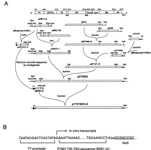
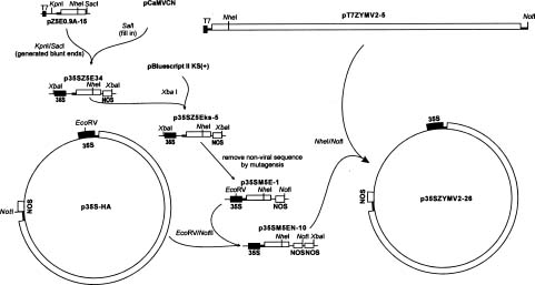
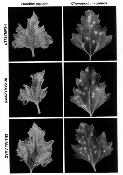
Abstract. The full-length cDNA of a Taiwan strain of Zucchini yellow mosaic virus (ZYMV TW-TN3) was constructed from five overlapping cDNA clones downstream from the bacteriophage T7 promoter in plasmid pT7ZYMV2-5. The plasmid was able to generate an in vitro transcript corresponding to TW-TN3 (9591 nt) with one extra guanosine residue at the 5´ terminus and a poly(A)95 tract at the 3´ end. In addition, pT7ZYMV2-5 was used for the construction of p35SZYMV2-26 that contained the full-length cDNA of TW-TN3 with a Cauliflower mosaic virus (CaMV) 35S promoter and a nopaline synthase (nos) terminator. The capped in vitro transcript generated from pT7ZYMV2-5 and the purified DNA of p35SZYMV2-26 were introduced into zucchini squash plants by mechanical inoculation and particle bombardment, respectively. Both in vitro and in vivo transcripts induced systemic symptoms on zucchini squash 4 to 6 days after inoculation. In addition, both transcripts also induced local lesions on plants of Chenopodium quinoa by mechanical inoculation. The results of infectivity assay, symptomatology, and serologically specific electron microscopy indicated that the in vitro and in vivo TW-TN3 transcripts derived from pT7ZYMV2-5 and p35SZYMV2-26, respectively, are infectious. The ability to generate biologically functional transcripts from the constructed cDNA clones is a significant step for molecular analyses of TW-TN3.

Keywords: In vitro; In vivo; Infectious transcripts; ZYMV TW-TN3.

Introduction

Zucchini yellow mosaic virus (ZYMV) is a member of the genus Potyvirus, the largest group among plant viruses, and is the major limiting factor for production of cucurbits worldwide. The disease is characterized by pronounced reduction in plant growth, yellow mosaic and distortion of leaves, and malformation of fruit (Lisa et al., 1981; Provvidenti et al., 1984). In Taiwan the disease caused by ZYMV has been considered one of the major limiting factors for the production of cucurbits (Lin et al., 1998; Lin et al., 2000). ZYMV particles are flexuous rods, 750 nm in length, and contain a monopartite genome consisting of a positive-sense ssRNA with a 5´ genome-linked protein (VPg) (Siaw et al., 1985; Riechmann et al., 1989; Murphy et al., 1990) and a 3´ poly (A) tract (Hari et al., 1979).

Infectious transcripts generated from cDNA clones of plant RNA viruses have been described for members of most plant virus groups (Boyer and Haenni, 1994). Among potyviruses, transcripts have been synthesized in vitro from full-length cDNA clones with bacterial phage promoters and proved to be infectious for Plum pox virus (PPV) (Riechmann et al., 1990), Zucchini yellow mosaic virus (ZYMV) (Gal-On et al., 1991), Tobacco etch virus (TEV) (Dolja et al., 1992), Pea seed-borne mosaic virus (PSbMV) (Johansen et al., 1996), Peanut stripe virus (PStV) (Flasinski

et al., 1996), Potato virus A (PVA) (Puurand et al., 1996), Tobacco vein mottling virus (TVMV) (Domier et al., 1989; Nicolas et al., 1996), Potato virus Y (PVY) (Jakab et al., 1997), Turnip mosaic virus (TuMV) (Sanchez et al., 1998) and Papaya ringspot virus (PRSV) (Chiang and Yeh, 1997). In vivo infectious transcripts, which are driven by a Cauliflower mosaic virus (CaMV) 35S promoter that can be recognized by host RNA polymerase, have also been reported for PPV (Maiss et al., 1992), ZYMV (Gal-On et al., 1995), Potato virus Y (PVY) (Fakhfakh et al., 1996), PSbMV (Johansen, 1996), Clover yellow vein virus (ClYVV) (Takahashi et al., 1997), PVY (Jakab et al., 1997), TuMV (Sanchez et al., 1998), Lettuce mosaic virus (LMV) (Yang et al., 1998), Plum pox virus (PPV) (Lopez-Moya and Garcia, 2000), TuMV (Sanchez et al., 1998) and PRSV (Chiang and Yeh, 1997). In these cases, the plasmids can be directly introduced to host plants to induce infection.

The complete nucleotide sequence of a severe Taiwan strain ZYMV TW-TN3 has previously been determined by our laboratory (Lin et al., 2001). Comparison of the coat protein (CP) gene and the P1 gene of TW-TN3 with those of other reported ZYMV isolates classified all sequenced ZYMV isolates in four genotypes, among which TW-TN3 and most of ZYMV isolates from Taiwan were placed in genotype I (Lin et al., 2000). The in vitro and in vivo infectious clones of ZYMV have been reported for an Israel isolate (IL) (Gal-On et al., 1991; Gal-On et al., 1995) that was placed in genotype II with closer relationships to US isolates (Lin et al., 2000).

*Corresponding author. Tel: 886-4-2287-7021; Fax: 886-4-2287-7585; E-mail: sdyeh@nchu.edu.tw


Botanical Bulletin of Academia Sinica, Vol. 43, 2002

In this study, the full-length cDNA clone of the severe Taiwan strain ZYMV TW-TN3 was constructed either downstream from the bacteriophage T7 promoter for generating in vitro infectious transcript or downstream from the CaMV 35S promoter for directly using plasmid to generate in vivo infectious transcript. The biological activities were analyzed on systemic host zucchini squash (Cucurbita pepo L. var. Zucchini) and the local lesion host Chenopodium quinoa Willd. by mechanical inoculation or particle bombardment. The ability to generate biologically functional RNA transcripts from those full-length cDNA clones provides a powerful tool to study the gene functions, replication, and pathogenicity of genotype I strains of ZYMV.

Materials and Methods

Construction of the Full-Length cDNA with a T7 Promoter

ZYMV TW-TN3, a severe strain originated from Taiwan (Lin et al., 1998), was propagated in plants of Cucurbita pepo L. var. Zucchini. Virus purification, viral RNA extraction, and cDNA cloning followed the methods previously described (Lin et al., 2001). The strategy for the construction of an in vitro infectious transcript derived from a full-length cDNA clone to TW-TN3 RNA is summarized in Figure 1A. Five overlapping cDNA clones that covered the complete genomic TW-TN3 RNA, including

Figure 1. Construction of the in vitro infectious full-length cDNA of a Taiwan strain Zucchini yellow mosaic virus (ZYMV TW-TN3) in pT7ZYMV2-5. (A), the full-length cDNA clone was constructed downstream from a T7 promoter by ligating suitable restriction fragments from five overlapping cDNA clones corresponding to different genomic regions of TW-TN3. Nonviral sequence upstream from the 5´ end of TW-TN3 sequence was removed by site-directed mutagenesis. The final construct containing a complete cDNA copy of TW-TN3 genome was designated pT7ZYMV2-5. The cDNA of the coding region is indicated by open bars. The 5´ and 3´ non-coding regions of the virus are indicated by thick black lines. The plasmid vectors are indicated by thin lines. The relevant restriction sites for construction are shown; (B), predicted site of initiation and direction of in vitro transcription initiated by T7 RNA polymerase (arrowed). The junction between the T7 promoter and the 5´ end of ZYMV sequence (9591 nt) contained an extra guanosine residue, and the 3´ end of TW-TN3 sequence was followed by a poly(A)95 tract with a NotI site.


Lin et al. — Construction of infectious transcripts of ZYMV

Figure 2. Construction of in vivo infectious clones of ZYMV with a 35S promoter and a nos terminator. The 65 nonviral nucleotides located between the transcription initiation site of the 35S promoter and the first nucleotide of the TW-TN3 sequence in p35SZ5Eks-5 were removed by site-directed mutagensis. The final construct was designated p35SZYMV2-26 and contained a complete cDNA copy of the TW-TN3 genome. The coding region of the cDNA is indicated by open boxes. The 5´ and 3´ non-coding regions of the cDNA are indicated by thick black lines. The plasmid vectors are indicated by thin lines. In addition, the relevant restriction sites for construction are shown.

pZ5E1.8-4, pZM2.7-20, pZF80, pZF31, and pZCP36, were used for construction of the full-length ZYMV genomic cDNA clone by ligating overlapping fragments that shared identical restriction sites. Plasmid pZM2.7-20 was digested with XbaI and ligated with XbaI digested fragment (1.5 kb) released from pZ5E1.8-4 to generate pZ5E27-1. The plasmid pZF31 was digested with StuI and NotI and ligated with StuI-NotI digested fragment (2.4 kb) released from pZF80 to generate pZF31/80-12. Plasmid pZF31/80-12 was digested with MluI and NotI and ligated with MluI-NotI digested fragment (0.85 kb) released from pZCP36 to generate pZF147-15. Plasmid pZ147-15 was digested with ApaI and SalI and ligated with ApaI-SalI fragment (3.7 kb) released from pZ5E27-1 to generate full-length in pZTW20.

Plasmid pBluescript II (SK+) was digested with EcoRI and ligated to the EcoRI fragment (900 bp) released from pZ5E1.8-4 to generate pZ5E0.9A-15. The polylinker sequence (75 nt) located between the T7 promoter and the 5´ terminus of the TW-TN3 genomic sequence in pZ5E0.9A-15 was removed using the site-directed mutagenesis GeneEditor system of Promega (Madison, Wisconsin). Escherichia coli strain XL-1 was used for the cloning of all plasmids and the production of single-stranded DNA via helper phage VCSM13 (Stratagene, La Jolla, California) for the mutagenesis reaction. The oligonucleotide 5´-CGACTCACTATAGAAAATTAAAACAAATCAC-3´ contained 13 nucleotides of the T7 promoter plus an additional guanosine residue and the last 18 nucleotides of the 5´ end of TW-TN3 sequence (underlined) derived from the previous report (Lin et al., 2001). The resulting plasmid pMZ5E-18 was sequenced to verify the deletion. Plasmid pMZ5E-18 was digested with NheI and NotI and ligated with NheI-NotI digested

fragment (8942 bp) released from pZTW20 to generate pT7ZYMV2-5 that contained the T7 promoter and an additional guanosine residue with the full-length cDNA to TW-TN3 genomic RNA (Figure 1B).

Construction of the Full-Length cDNA with a CaMV 35S Promoter

A plasmid pCaMVCN containing a CaMV 35S promoter and a nos terminator was purchased from Pharmacia/LKB (South Plainfield, NJ). Escherichia coli strain JM109 was used for cloning the plasmid, and E. coli strain XL-1 was used for site-directed mutagenesis.

The strategy for the construction of a full-length cDNA clone of TW-TN3 with the 35S promoter and the nos terminator is outlined in Figure 2. The KpnI-SacI fragment of pZ5E0.9A-15 (Figure 1) was converted to blunt ends with the Klenow fragment of DNA polymerase I, and ligated with pCaMVCN that was digested with SalI and filled-in by the Klenow fragment. The resulting plasmid p35SZ5E34 contained 65 extra nucleotides between the 35S promoter and the 5´ end of ZYMV sequence. In order to generate single-stranded DNA for site-directed mutagenesis, p35SZ5E34 was further digested with XbaI and subcloned into XbaI digested pBluescript II (KS+) (Stratagene) to create p35SZ5Eks-5. The 65 nonviral nucleotides located between the transcription initiation site of the 35S promoter and the first nucleotide of TW-TN3 sequence in p35SZ5Eks-5 were then removed by site-directed mutagenesis. The oligonucleotide 5´-GTGATTTGTTTT AATTTTCCTCTCCAAATGAAATGAA -3´, containing the first 18 nucleotides of TW-TN3 RNA (underlined) (Lin et al., 2001) followed by the last 15 nucleotides of the 35S


Botanical Bulletin of Academia Sinica, Vol. 43, 2002

promoter, was used for site-directed mutagenesis. The resulting plasmid p35SM5E-1 was sequenced to verify the deletion.

The EcoRV-NotI fragment of p35M5E-1 was ligated with the same enzyme-digested p35S-HA (Chiang and Yeh, 1997) to generate p35SM5EN-10 to obtain the nos terminator downstream from the NotI site. Plasmid p35SM5EN-10 was then digested with NheI-NotI and ligated to the same enzyme digested fragment (8942 bp) that was excised from the cDNA clone of pT7ZYMV2-5 to produce p35SZYMV2-26, which contained a construct of the full-length TW-TN3 cDNA with a 35S promoter and a nos terminator (Figure 2).

Synthesis of in vitro Transcripts

In vitro transcription of pT7ZYMV2-5 (30 µg in 200 µl final volume) was carried out with the mCAP mRNA capping kit (Stratagene), T7 RNA polymerase (1.2 unit per µl) (Stratagene), RNase inhibitor (Promega, Madison, WI) (0.4 unit per µl), and 500 mM m7GpppG (Stratagene). The mixture was first incubated at 37ºC for 1 h; GTP was then supplemented to 250 mM, and the incubation was further proceeded for 1 h. Size and concentrations of the transcripts were estimated by comparison with a known quantity of viral RNA.

Infectivity Assay

Systemic host Cucurbita pepo L. var. Zucchini at the cotyledon stage and local lesion host Chenopodium quinoa Willd. with four fully expanded leaves were used for bioassay of the cDNA constructs. Transcription mixtures of 20 µl (final quantity of 2-3 µg RNA) were mechanically applied onto a leaf of each carborundum-dusted plant with a sterilized glass spatula. Healthy seedlings were mock-inoculated with 20 µl transcription buffer or with 0.5 µg of ZYMV native RNA per plant as controls.

ZYMV full-length cDNA clone p35ZYMV2-26 was purified by alkaline lysis minipreparation (Sambrook and Russell, 2001). Aliquots of 20 µl, containing 1 µg DNA in sterilized water, were mechanically applied onto a carborundum-dusted leaf of C. quinoa with a sterilized glass spatula. Plants were also inoculated by microprojectile bombardment with a particle delivery system (Bio-Rad, Biolistic PDS-1000/He, Hercules, California) following the procedure described in the user's manual. Briefly, aliquots of 10 µl (containing 0.8 µg DNA) of the DNA-coated gold microcarriers (1.6 µm) were transferred onto the center of the macrocarrier and dried in low humidity under vibration-free conditions. Four zucchini squashes at the cotyledon stage were clustered by rubber bands and placed on a petri dish with the leaves facing the macrocarrier. The petri dish was placed on the holder in the third position down from the microcarrier launch assembly. The vacuum was maintained at 20-25 inches of mercury, and then the microcarriers were delivered into plants at 650 psi. All inoculated plants were kept in a temperature-controlled (23-28ºC) greenhouse for observation of symptom development.

Confirmation of Infection

Inoculated plants were assayed by indirect enzyme-linked immunosorbent assay (ELISA) using ZYMV antiserum (Lin et al., 1998) to detect the presence of ZYMV at 8 days post inoculation (dpi). Results were recorded by measurement of the absorbancy at 405 nm by an ELISA reader (ELx800, Bio-Tek Instruments, Inc., Winooski, Vermont).

Total proteins extracted from leaf tissues of infected plants were separated by SDS polyacrylamide gel (12.5%) electrophoresis, transferred to nitrocellulose membranes (Bio-Rad), and immunostained with the polyclonal antiserum to ZYMV (Lin et al., 1998) as described by Sambrook and Russell (2001).

Virions present in infected squash were also analyzed by immunogold labeling and electron microscopy. The gold labeling of virus particles followed the method of Van Lent and Verduin (1985). Formvar/ carbon-coated grids

Figure 3. Symptoms on zucchini squash induced either by microprojectile bombardment with p35SZYMV2-26 or by mechanical inoculation with the transcript synthesized from pT7ZYMV2-5. In addition, both transcripts also induced local lesions on plants of Chenopodium quinoa by mechanical inoculation. The photographs were taken at 14 days after inoculation.


Lin et al. — Construction of infectious transcripts of ZYMV

were allowed to float on drops of crude sap from leaf tissue of infected plant extracted in Tris-EDTA buffer (10 mM Tris-HCl, 10 mM EDTA, pH 7.4, containing 100 µg/ml bacitracin). Grids were incubated with phosphate-buffered saline (PBS: 0.137 M NaCl, 1.5 mM KH2PO4, 8.1 mM Na2HPO4, 2.7 mM KCl, 3.1 mM NaN3, pH 7.4) containing 1% (w/v) BSA for 10 min and transferred to drops of primary antisera (1:200 in PBS buffer containing 1% BSA) for 30 min. The grids were then washed with 30 drops of PBS and 10 drops of distilled water. Grids were incubated with gold conjugated protein A (Zymed Laboratories, South San Francisco, CA) with 20 nm gold (1:50 in PBS buffer containing 1% BSA) for 30 min, washed with 30 drops of PBS and 10 drops of distilled water, and stained with 2% uranyl acetate. Results of immunodecoration were examined with a JEOL 200CX electron microscope and photographed with Kodak 4489 EM film.

Results

Full-Length cDNA Clones with a T7 Promoter or a 35S Promoter

The clone pT7ZYMV2-5 contained the full length cDNA sequence of ZYMV TW-TN3 (9591 nt) downstream from the T7 promoter, with one extra G in between the promoter and the 5´ end of the ZYMV sequence and a poly (A) tail of 95 nucleotides at the 3´ end followed by a NotI site for run-off transcription (Figure 1B). This full-length cDNA clone was further used for the construction of the full-length cDNA clone with a CaMV 35S promoter (Figure 2).

The plasmid p35SZYMV2-26 contained a construct in the arrangement of the 35S promoter, the complete ZYMV cDNA, a 95 poly (A) tract with 10 additional nucleotides including a NotI site, and a nos polyadenylation signal as a terminator (Figure 2).

Infectivity Assay of in vivo and in vitro Infectious Clones

The biological activities of both infectious clones were analyzed on the systemic host zucchini squash and the local lesion host C. quinoa. Capped in vitro transcript generated from template pT7ZYMV2-5 infected squash plants (4 out of 6 inoculated plants) after they were mechanically inoculated. In addition, the in vitro transcripts also infected plants of C. quinoa (4 out of 4 inoculated plants) after they were mechanically inoculated. In vitro transcripts of pT7ZYMV2-5 were able to induce typical severe symptoms on squash plants 4 to 6 days after inoculation and local lesions on C. quinoa plants 5 to 7 days after inoculation, respectively, similar to those induced by ZYMV TW-TN3 (Figure 3).

The plasmid p35SZYMV2-26 did not infect any of the squash plants (0 out of a total of 12 inoculated plants from three independent experiments) mechanically inoculated. However, 100% infection (16 out of a total of 16 inoculated plants from four independent experiments) was obtained when the plants were particle-bombarded with

Figure 4. Western blot analysis of zucchini squash plants infected with ZYMV infectious clones, using the polyclonal antibody to ZYMV (Lin et al., 1998). Extracts (5 µl) from equal amounts of leaf tissue (0.05 g /500 µl) collected at 7 days after inoculation (0.5 cm diameter of disk from three different leaves ground in 500 µl extraction buffer) were loaded in each lane. Lane 1, leaf extract from a plant infected with ZYMV TW-TN3; Lanes 2 and 3, leaf extracts from plants infected with in vitro transcript of pT7ZYMV2-5 by mechanical inoculation and infected by the plasmid of p35SZYMV2-26 by particle bombardment, respectively; Lane 4, an uninfected plant as control; M, protein markers.

p35SZYMV2-26. Also, an infection rate of 100% was obtained after the plasmid was mechanically introduced onto C. quinoa (20 out of a total of 20 inoculated plants in four independent experiments). Inoculated squash plants showed severe symptoms of yellow mosaic 4 to 6 days after inoculation, similar to those on plants mechanically inoculated with TW-TN3 (Figure 3). Appearance of the symptoms induced by p35SZYMV2-26 was not significantly delayed when compared with those induced by the virus. Local lesions also developed on plants of C. quinoa 5 to 7 days after mechanical inoculation with p35SZYMV2-26. The lesions showed characteristics typical to those induced by ZYMV TW-TN3 (Figure 3).

Squash plants showing symptoms of viral infection after inoculation with either p35SZYMV2-26 or in vitro transcript of pT7ZYMV2-5 were used as sources of inocula to inoculate new sets of plants. In all cases, 100% infection rates were obtained (6 plants per inoculation tested, four independent experiments).

Confirmation of Infection

All inoculated plants were analyzed by ELISA with the antiserum to ZYMV to confirm infection. Squash plants that developed severe symptoms strongly reacted with the


Botanical Bulletin of Academia Sinica, Vol. 43, 2002

local lesion host C. quinoa. In addition, ELISA, western blotting, and electron microscopy confirmed that the artificial viruses created from both in vitro and in vivo transcripts were similar to the native ZYMV.

Typical symptoms of ZYMV infection appeared on plants of zucchini squash and C. quinoa inoculated with infectious in vitro and in vivo transcripts of ZYMV at about the same time as those induced by native RNA or virus particles of ZYMV. These results are similar to the infectivity assay of in vitro and in vivo infectious transcripts of PRSV (Chiang and Yeh, 1997). However, our results are different from those induced by the other six infectious potyviral transcripts, which have a slight lag of 1 to 13 days for symptom development following mechanical inoculation (Domier et al., 1989; Riechmann et al., 1990; Gal-On et al., 1991; Dolja et al., 1992; Jakab et al., 1997; Sanchez et al., 1998; Yang et al., 1998).

The infectivity of a number of viral 35S-cDNA clones by manual inoculation onto plants has been reported, including Pea early browning virus (PEBV, Tobravirus) (MacFarlane et al., 1992); Tobacco mosaic virus L strain (ToMV-L, Tobamovirus) (Weber et al., 1992); Plum poxvirus (PPV, Potyviridae) (Maiss et al., 1992); Cowpea mosaic virus (CPMV, Comoviridae) (Dessens and Lomonossoff, 1993); Alfalfa mosaic virus (AlMV, Bromoviridae) (Neeleman et al., 1993); Cucumber mosaic virus (CMV, Bromoviridae) (Ding et al., 1995); ZYMV (Potyviridae) (Gal-On et al., 1995); and PRSV (Potyviridae) (Chiang and Yeh, 1997). However, low infectivity following the mechanical inoculation of plants using intact plasmid was noticed (Neeleman et al., 1993; Ding et al., 1995;

antiserum, while the uninfected control or plants inoculated with Papaya ringspot virus (PRSV) showed negative reactions. By immunoblotting analysis, a 33 kDa protein corresponding to the CP of ZYMV was detected in the plants infected by the in vitro transcripts derived from pT7ZYMV2-5 or in vivo transcripts derived from p35ZYMV2-26. The quantity of the CP in plants infected with both of them was similar to that infected with wild-type ZYMV (Figure 4).

To confirm that the development of symptoms in zucchini squash plants was induced by p35SZYMV2-26, the crude sap from leaf tissues of infected squash plants was examined by electron microscopy. Numerous filamentous virus particles of 750 nm in length were observed in the samples from infected plants (Figure 5A). When the crude sap was further analyzed by immunogold labeling, the gold particles were specifically located along the entire length of the virions decorated by ZYMV CP antiserum (Figure 5B). Similarly, filamentous particles decorated by gold particles were also found in the samples infected with capped in vitro transcripts derived from pT7ZYMV2-5.

Discussion

In this investigation, full-length cDNA clones of ZYMV TW-TN3 were constructed downstream from a bacteriophage T7 RNA polymerase promoter or a CaMV 35S promoter. The transcripts produced either by in vitro transcription driven by the T7 promoter or by in vivo transcription derived from the 35S promoter were proven infectious when introduced to the systemic host zucchini squash and the

Figure 5. Particles of ZYMV observed in the crude sap of systemically infected leaves of zucchini squash plants inoculated with the p35SZYMV2-26. (A), filamentous particles about 750 nm in length were observed; (B), serologically specific electron microscopy with immunogold labeling on virions prepared from a zucchini squash infected with p35SZYMV2-26. Arrows indicate that ZYMV virions were labeled by 20 nm gold particles after virions were decorated by the antiserum to ZYMV CP (Lin et al., 1998).


Lin et al. — Construction of infectious transcripts of ZYMV

Gal-On et al., 1995). In this study, the plasmid p35SZYMV2-26 failed to establish infection by mechanical inoculation on the four tested squash plants. We repeated the manual inoculation several times on more than 20 squash plants (data not shown), but still no infectivity was noticed. However, particle bombardment of zucchini squash plants resulted in successful infection. Our results agree with those of Dagless et al. (1997), who showed that the construct p35SU1R of TMV was unable to infect some TMV host plants by mechanical inoculation but infected all TMV host plants by particle bombardment. In addition, Gal-On et al. showed that particle bombardment increased the infectivity of pKS35SZYMVNOS from 19% by manual inoculation to 100% (Gal-On et al., 1995). Through particle bombardment, Yamashita et al. (1991) showed a strong positive correlation (>90%) between transient gene expression and the direct delivery of a gold particle into the cell nucleus. The unsuccessful delivery of p35ZYMV2-26 into plant nuclei may be responsible for the lack of infection by mechanical inoculation on zucchini squash plants.

On the other hand, mechanical inoculation of p35SZYMV2-26 on C. quinoa resulted in 100% infection. This result is similar to the results of Weber et al. (1992) that only 1 µg of 35S-TMV-L cDNA was highly infectious when introduced into C. quinoa. The bioassay on C. quinoa does not need any expensive in vitro transcription kits or instruments such as the biolistic delivery system to introduce infectious plasmids, and many infectious clones can be assayed at the same time with only a few C. quinoa plants. Moreover, over 15 local lesions were caused by 1 µg of p35ZYMV2-26 plasmid on C. quinoa plants at 5-6 dpi. Thus, we consider bioassay on plants of C. quinoa a simple, reliable, and economical method for testing the infectivity of in vivo infectious cDNA clone of ZYMV. Under the microscope, there were no trichomes on the epidermis of C. quinoa plants; however, a lot are found on the epidermis of cucurbit plants. The trichomes on the leaf epidermis may prevent the entry of p35SZYMV2-26 into cell nuclei to initiate replication.

The infectious clones of p35SZYMV2-26 and pT7ZYMV2-5 in E. coli were stored at -80ºC for long- term preservation. After one month, when these bacteria were activated from -80ºC and plated at 37ºC, problems of low plasmid yield and low colony numbers of p35SZYMV2-26 were noticed. However, this phenomenon did not occur with pT7ZYMV2-5 in E. coli. Instability of full-length cDNA clones in bacteria have been reported for several animal and plant viruses, possibly due to the potential toxicity and instability of viral sequences in bacteria (Boyer and Haenni, 1994). In some particular cases of potyviruses, the construction of a full-length clone of PVY in E. coli has been unsuccessful to date (Fakhfakh et al., 1996). Instability of the full-length clones of PStV and TuMV in E. coli strain DH5a have also been reported (Flasinski et al., 1996; Sanchez et al., 1998). The reasons for the instability have been attributed to potential expression of toxic product (PVY) (Fakhfakh et al., 1996) and to the intact P3 gene or its protein product (PSbMV)(Johansen, 1996). These notions are supported by the fact

that insertion of introns into the genome of PStMV and PPV strongly attenuates the instability problems (Johansen, 1996; Lopez-Moya and Garcia, 2000). The instability detected in full-length clones of TuMV has been overcome by growing solid and liquid cultures containing 100 µg/ml ampicillin at 30ºC (Sanchez et al., 1998). In our laboratory, a large amount of original infectious clones p35SZYMV2-26 were stockpiled at -80ºC and used to transform E. coli whenever needed.

TW-TN3 is classified in genotype I (Lin et al., 2000), and this report represents the first ZYMV infectious clone in Eastern Asia. Our ability to synthesize biologically active TW-TN3 from full-length cDNA template is a significant step forward in molecular studies of the functions of TW-TN3 genes and the roles of specific sequences of TW-TN3 RNA in encapsidation, pathogenicity, transmission, replication, and translation. Currently, TW-TN3 infectious clone is being modified by mutagenesis to obtain mild strains for cross protection for control of ZYMV in Taiwan or Eastern Asia. This ZYMV-based system that permits both efficient systemic spread and high insertion capacity is suitable for the development of a viral vector for expressing foreign proteins in cucurbits. In addition, the NIa proteinase of ZYMV that recognizes sequence-specific cleavage sites can be designed along with foreign proteins for their processing as free form in infected tissues.

Acknowledgments. This study was supported in part by NSC grants (87-2312-B005-005 and 89-2312-B-005-001) of the Republic of China on Taiwan.

Literature Cited

Boyer, J.C. and A.L. Haenni. 1994. Infectious transcripts and cDNA clones of RNA viruses. Virology 198: 415-426.

Chiang, C.H. and S.D. Yeh. 1997. Infectivity assays of in vitro and in vivo transcripts of papaya ringspot potyvirus. Bot. Bull. Acad. Sin. 38: 153-163.

Dagless, E.M., M.H. Shintaku, R.S. Nelson, and G.D. Foster. 1997. A CaMV 35S promoter driven cDNA clone of tobacco mosaic virus can infect host plant tissue despite being uninfectious when manually inoculated onto leaves. Arch. Virol. 142: 183-191.

Dessens, J.T. and G.P. Lomonossoff. 1993. Cauliflower mosaic virus 35S promoter-controlled DNA copies of Cowpea mosaic virus RNAs are infectious on plants. J. Gen. Virol. 74: 889-892.

Ding, S.W., J.P. Rathjen, W.X. Li, R. Swanson, H. Healy, and R.H. Symons. 1995. Efficient infection from cDNA clones of cucumber mosaic cucumovirus RNAs in a new plasmid vector. J. Gen. Virol. 76: 459-464.

Dolja, V.V., H.J. McBride, and J.C. Carrington. 1992. Tagging of plant potyvirus replication and movement by insertion of beta-glucuronidase into the viral polyprotein. Proc. Natl. Acad. Sci. USA 89: 10208-10212.

Domier, L.L., K.M. Franklin, A.G. Hunt, R.E. Rhoads, and J.G. Shaw. 1989. Infectious in vitro transcripts from cloned cDNA of a potyvirus, Tobacco vein mottling virus. Proc.


Botanical Bulletin of Academia Sinica, Vol. 43, 2002

Natl. Acad. Sci. USA 86: 3509-3513.

Fakhfakh, H., F. Vilaine, M. Makni, and C. Robaglia. 1996. Cell-free cloning and biolistic inoculation of an infectious cDNA of Potato virus Y. J. Gen. Virol. 77: 519-523.

Flasinski, S., U.B. Gunasinghe, R.A. Gonzales, and B.G. Cassidy. 1996. The cDNA sequence and infectious transcripts of Peanut stripe virus. Gene 171: 299-300.

Gal-On, A., Y. Antignus, A. Rosner, and B. Raccah. 1991. Infectious in-vitro RNA transcripts derived from cloned cDNA of the cucurbit potyvirus Zucchini yellow mosaic virus. J. Gen. Virol. 72: 2639-2644.

Gal-On, A., E. Meiri, H. Huet, W.J. Hua, B. Raccah, and V. Gaba. 1995. Particle bombardment drastically increases the infectivity of cloned DNA of zucchini yellow mosaic potyvirus. J. Gen. Virol. 76: 3223-3227.

Hari, V., A. Siegel, C. Rozek, and W.E. Timberlake. 1979. The RNA of Tobacco etch virus contains poly(A). Virology 30: 568-571.

Jakab, G., E. Droz, G. Brigneti, D. Baulcombe, and P. Malnoe. 1997. Infectious in vivo and in vitro transcripts from a full-length cDNA clone of PVY-N605, a Swiss necrotic isolate of potato virus Y. J. Gen. Viro. 78: 3141-3145.

Johansen, I.E. 1996. Intron insertion facilitates amplification of cloned virus cDNA in Escherichia coli while biological activity is reestablished after transcription in vivo. Proc. Natl. Acad. Sci. USA 93: 12400-12405.

Johansen, I.E., W.G. Dougherty, K.E. Keller, D. Wang, and R.O. Hampton. 1996. Multiple viral determinants affect seed transmission of pea seedborne mosaic virus in Pisum sativum. J. Gen. Virol. 77: 3149-3154.

Lin, S.S., R.F. Hou, C.H. Huang, and S.D. Yeh. 1998. Characterization of Zucchini yellow mosaic virus (ZYMV) isolates collected from Taiwan by host reactions, serology, and RT-PCR. Plant Prot. Bull. 40: 163-176.

Lin, S.S., R.F. Hou, and S.D. Yeh. 2000. Heteroduplex mobility and sequence analyses for assessment of variability of Zucchini yellow mosaic virus. Phytopathology 90: 228-235.

Lin, S.S., R.F. Hou, and S.D. Yeh. 2001. Complete genome sequence and genetic organization of a Taiwan isolate of Zucchini yellow mosaic virus. Bot. Bull. Acad. Sin. 42: 243-250.

Lisa, V., G. Boccardo, G. D'Agostino, G. Dellavalle, and M. D'Aquilio. 1981. Characterization of a potyvirus that causes zucchini yellow mosaic. Phytopathology 71: 667-672.

Lopez-Moya, J.J. and J.A. Garcia. 2000. Construction of a stable and highly infectious intron-containing cDNA clone of plum pox potyvirus and its use to infect plants by particle bombardment. Virus Res. 68: 99-107.

MacFarlane, S.A., D. Gilmer, and J.W. Davies. 1992. Efficient inoculation with CaMV 35S promoter-driven DNA clones of the tobravirus PEBV. Virology 187: 829-831.

Maiss, E., U. Timpe, A. Brisske-Rode, D.E. Lesemann, and R. Casper. 1992. Infectious in vivo transcripts of a plum pox potyvirus full-length cDNA clone containing the Cauliflower mosaic virus 35S RNA promoter. J. Gen. Virol. 73:

709-713.

Murphy, J.F., R.E. Rhoads, A.G. Hunt, and J.G. Shaw. 1990. The Vpg of Tobacco etch virus RNA is the 49-kDa proteinase or the amino-terminal 24-kDa part of the proteinase. Virology 178: 285-288.

Neeleman, L., E.A.G. van der Vossen, and J.F. Bol. 1993. Infection of tobacco with Alfalfa mosaic virus cDNAs sheds light on the early function of the coat protein. Virology 196: 883-887.

Nicolas, O., T.P. Pirone, and G.M. Hellmann. 1996. Construction and analysis of infectious transcripts from a resistance-breaking strain of tobacco vein mottling potyvirus. Arch. Virol. 141: 1535-1552.

Provvidenti, R., D. Gonsalves, and H.S. Humaydan. 1984. Occurrence of Zucchini yellow mosaic virus in cucurbits from Connecticut, New-York, Florida, and California. Plant Dis. 68: 443-446.

Puurand, U., J.P.T. Valkonen, K. Makinen, F. Rabenstein, and M. Saarma. 1996. Infectious in vitro transcripts from cloned cDNA of the potato A potyvirus. Virus Res. 40: 135-140.

Riechmann, J.L., S. Lain, and J. A. Garcia. 1989. The genome-linked protein and 5´ end RNA sequence of plum pox potyvirus. J. Gen. Virol. 70: 2785-2790.

Riechmann, J.L., S. Lain, and J.A. Garcia. 1990. Infectious in-vitro transcripts from a plum pox potyvirus complementary DNA clone. Virology 177: 710-716.

Sambrook, J. and D.W. Russell. 2001. Molecular Cloning: A Laboratory Manual, 3nd edn. Cold Springs Harbor Laboratory, New York.

Sanchez, F., D. Martinez-Herrera, I. Aguilar, and F. Ponz. 1998. Infectivity of turnip mosaic potyvirus cDNA clones and transcripts on the systemic host Arabidopsis thaliana and local lesion hosts. Virus Res. 55: 207-219.

Siaw, M.F.E., M. Shahabuddin, S. Ballard, J.G. Shaw, and R.E. Rhoads. 1985. Identification of a protein covalently linked to the 5´ terminus of Tobacco vein mottling virus RNA. Virology 142: 134-143.

Takahashi, Y., T. Takahashi, and I. Uyeda. 1997. A cDNA clone to clover yellow vein potyvirus genome is highly infectious. Virus Genes 14: 235-243.

Van Lent, J.W.M. and B.J.M. Verduin. 1985. Specific gold-labelling of antibodies bound to plant viruses in mixed suspensions. Neth. J. Pl. Path. 91: 205-213.

Weber, H., P. Haeckel, and A.J.P. Pfitzner. 1992. A cDNA clone of Tomato mosaic virus is infectious in plants. J. Virol. 66: 3909-3912.

Yamashita, T., A. Lida, and H. Morikawa. 1991. Evidence that more than 90 percent of beta glucuronidase-expressing cells after particle bombardment directly receive the foreign gene in their nucleus. Plant Physiol. 97: 829-831.

Yang, S.J., F. Revers, S. Souche, H. Lot, O. Le Gall, T. Candresse, and J. Dunez. 1998. Construction of full-length cDNA clones of lettuce mosaic virus (LMV) and the effects of intron-insertion on their viability in Escherichia coli and on their infectivity to plants. Arch. Virol. 143: 2443-2451.


Lin et al. — Construction of infectious transcripts of ZYMV