Bot. Bull. Acad. Sin. (2003) 44: 19-24

Lo et al. — Actin-like protein filaments inside mitochondria

The presence of actin-like protein filaments in higher plant mitochondria

Yih-Shan Lo, Yuh-Tai Wang, Wann-Neng Jane, Lin-June Hsiao, Long-Fang O. Chen, and Hwa Dai*

Institute of Botany, Academia Sinica, Taipei, Taiwan 11529, Republic of China

(Received May 2, 2002; Accepted July 25, 2002)

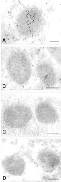
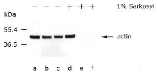
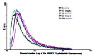
Abstract. In this report we demonstrate that a 42 kDa actin-like protein filament may exist in mung bean seedling mitochondria. The results of a fine structure study, immunogold localization, immunoblot, fluorescent microscopy, and flow cytometric analyses all point to the existence of this actin-like protein inside mung bean mitochondria.

Keywords: Actin-like protein; Plant mitochondria.

Introduction

The cytoskeleton in eucaryotic cells has been well studied. It determines the cell shape and directs the movement of various entities in the cytoplasm. The transport action of mitochondria from one place to another in cytoplasm, controlled by the actin cytoskeleton, has also been well characterized (Kuznetsov et al., 1992; Simon et al., 1995). However, very little is known about how mitochondria keep their shape or how newly synthesized mitochondrial gene-encoded proteins are transported to their final destination for function. Most studies on mitochondrial biogenesis in the past two decades have focused on the import pathway of nucleus gene-encoded proteins into mitochondria (Baker and Schatz, 1991; Pfanner et al., 1992; Fujiki and Verner, 1993).

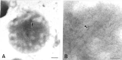
The existence of actin-related protein inside the chloroplast and mitochondria has been suggested before (McCurdy and Williamson, 1987; Murgia et al., 1995). That the F-actin is probably present in organelle dividing rings was suggested by Kuroiwa et al. (1995). Miyagishima et al. (2001) proved that a 5 nm novel filament was involved in the plastid-dividing ring. The tubulin homologous protein-FtsZ, an essential cell division protein in bacteria, was well studied (Bi and Lutkenhaus, 1991; Erickson, 1995, 1997). Osteryoung and Vierling (1995) demonstrated that cpFtsZ, a homologue of tubulin protein, can be imported into the chloroplast of Arabidopsis. Mitochondrial FtsZ in a Chromophyte Alga was also reported recently (Beech et al., 2000). Most importantly, an actin-like protein (MreB) was found in prokaryotes (Jones et al., 2001; Van den Ent et al., 2001). In this report, we found about 5 nm of filamentous structures inside mitochondria. Immunoblot, immunolocalization, fluorescent analyses, and flow

cytometry all showed that 42 kDa actin-like protein filaments are present in mung bean seedling mitochondria.

Materials and Methods

Mitochondria, Mitoplast Isolation and EM Examination

Mitochondria were isolated from four-day-old etiolated mung bean seedlings (TN-5) with M-1 buffer as described previously (Leaver et al., 1983; Dai et al., 1991; Cheng and Dai, 2000). They were then treated with digitonin following the methods of Kfzlfwski and Zagorski (1998). Mitochondria were dissolved in medium B (0.6 M sorbitol, 0.2% BSA, 10 mM Tris-maleate, pH 7.0) at a protein concentration of 40 mg/ml. The digitonin concentration for mitochondria treatment was 0.12 mg/ml followed by a pre-optimized analysis. Mitoplasts were further purified with a 40-65% sucrose gradient buffered with 10 mM Tris-maleate (pH 7.0) by centrifuging at 100,000 g at 4°C for 90 min. Whole-mount EM analysis on mitoplast was carried out by fixing the sample with 1% paraformaldehyde followed by 1% uranyl acetate (pH 4.5) staining for 1-2 min.

Immunolocalization Analysis of Mitochondria in Situ

The four-day-old mung bean tissue was fixed in 10 mM pipes (pH 7.2), containing 2.5% paraformaldehyde and 1.25% glutaraldehyde at 4°C overnight. Dehydration was done after five washings in the same buffer. Infiltration and polymerization were carried out in Lowicryl K4M resin (Roth et al., 1981). Thin tissue sections were labeled by sequential incubations with monoclonal anti-actin antibody (Boehringer Mannheim, C4 clone) followed by reaction with a secondary antibody, goat-anti-mouse antibody conjugated with 20 nm gold. The maize mitochondria specific monoclonal antibody, PM028 (against a yet unidentified maize mitochondrial protein with no cross

*Corresponding author. Fax: 886-2-2782-7954; E-mail: bodaihwa@gate.sinica.edu.tw


Botanical Bulletin of Academia Sinica, Vol. 44, 2003

reactivity toward mungbean mitochondria; purchased from GTMA, Lincoln, NE) and a anti- COXIII (mitochondrial marker protein) were used as a control in the immunolocalization study.

Trypsin/Proteinase K Treatment on Mitochondria

Mitochondria, 50 µg/ml in buffer as previously described (Glick et al., 1992; 0.6 M sorbital, 50 mM K+-HEPES [pH 7.0], 50 mM KCl, 10 mM MgCl2, 2.5 mM EDTA, 2 mM KH2PO4, 2 mM ATP, 1 mg/ml fatty acid-free bovine serum albumin), were treated with various concentrations of trypsin (0.5 mg, 1.0 mg, 2.5 mg, 10 mg/ml) at room temperature for 30 min and then stopped by 2 mM PMSF. Proteinase K (0, 5, 10 µg/ml) treatment was carried out in import buffer used by Chaumont et al. (1990): 0.25 M Mannitol, 20 mM HEPES, pH 7.5, 50 mM KCl, 2 mM MgCl2, 1 mM KH2PO4, 1 mM DTT, 1 mM ATP, 20 µM ADP, 10 mM malate) with and without 1% Sarkosyl.

Immunoblot Analysis on Proteinase K-Treated Mitochondria

Mitochondria with and without proteinase K treatment were analyzed by 12% SDS-PAGE and then transferred to PVDF membrane (Millipore). Protein blots were incubated with anti-actin antibody. Then immunoreactive proteins were detected with goat anti-mouse antibody conjugated with alkaline phosphatase, followed by staining with 2-naphthyl phosphate/fast red RC (Sigma) according to the published procedure (Sambrook et al., 1989).

Fluorescent Analysis on F-Actin in Mitochondria

Mung bean mitochondria were treated with 1mg/ml trypsin as described above. Trypsin treated mitochondria were fixed with 3.7% paraformaldehyde in M-5 buffer, ex

tracted with 0.1% Triton X-100 in M-5 buffer, washed twice with PBS buffer and then stained with 20 units/ml of BODIPY FL phallacidin (Molecular Probes) at room temperature for 30 min in the dark. The unbound dye was removed by centrifugation, and the pelleted mitochondria were washed twice with M-1 buffer by centrifugation. Sample was visualized under a Zeiss Axioplan microscope (EX. 450-490/EM. 520 nm).

Flow Cytometric Analysis of Trypsin-Treated Mitochondria

Mung bean mitochondria were treated with various concentrations of trypsin as described above. Trypsin-treated mitochondria were fixed with 3.7% paraformaldehyde in M-5 buffer, extracted with 0.1% Triton X-100 in M-5 buffer, washed twice with PBS buffer, and then stained with 10 units/ml of BODIPY FL phallacidin at room temperature for 30 min in the dark. The unbound dye was removed by centrifugation, and the pelleted mitochondria were washed twice with M-1 buffer by centrifugation.

Flow Cytometric analysis was carried out on a FACScan (Becton Dickinson, San Jose, CA) equipped with a 488 nm argon laser. Mitochondrial fluorescence was measured for at least 50,000 events per sample. The green fluorescence was measured through a 550 +/- 15 nm band pass filter. Graphics were plotted by means of the Lysys software program (Becton Dickinson).

Results

Fine Structure Study on Mitochondria

The mitoplast obtained from digitonin-treated mitochondria followed by sucrose gradient purification. Whole-mount electron microscopy analysis on polymerized mitoplast after staining with uranyl acetate revealed that a filamentous bundle, each filament about 5 nm in width,

Figure 1. Whole-mount EM micrograph of mitoplast stained with 1% uranyl acetate. Arrowhead in B shows filamentous bundle (each filament about 5 nm in width) inside mitoplast. B is an enlarged portion of A (indicated by arrowhead). Bars equal to 200 nm in A and 100 nm in B.


Lo et al. — Actin-like protein filaments inside mitochondria

exists in mitoplast (Figure 1B, arrowhead). Figure 1B is an enlargement of Figure 1A (enlarged portion indicated by arrowhead).

Immunolocalization Analysis

Immunolocalization analysis against anti-actin antibody demonstrated that anti-actin antibody-reacted protein exists in mitochondria (Figure 2). Some gold particles arranged into a line are shown in Figure 2, A and B. We named this anti-actin antibody-reacted protein in mung bean seedling mitochondria actin-like protein. The ratio of gold particles per mitochondrion in the anti-actin antibody-labeled tissue section to gold particles per mitochondrion in the control section was 28 to 0.6. A total of 250 mitochondria were counted. The immunogold label of anti-COXIII antibody (mitochondrial marker protein) was used as a positive control (Figure 2D).

Immunoblotting Analysis of Proteinase K-Treated Mitochondria

In order to eliminate the cytoskeletons residing outside mitochondria, the mitochondria were treated with different concentrations of Proteinase K, 5 and 10 µg/ml. Figure 3 shows the result of immunoblot analysis on Proteinase K treated mitochondria against anti-actin

Figure 3. Immunoblot analysis on Proteinase K treated mitochondria. Mitochondria (5 µg protein) treated with different concentrations of Proteinase K (lanes a, b and c represents 0, 5 and 10 µg/ml of Proteinase K treatment, respectively) followed by intensive washing with M-1 buffer were analyzed by electrophoresis on a 12.5% SDS-polyacrylamide gel. Lanes d, e and f present the mitochondria treated with 0, 5 and 10 µg/ml Proteinase K, respectively, with 1% Sarkosyl in reaction buffer and then electrophoresis carried out as described above. Immunoblotting study against 2000 X diluted anti-actin monoclonal antibody was carried out as described in Materials and Methods.

Figure 2. In situ immunogold localization of anti-actin antibody-reacted protein. Monoclonal anti-actin antibody was used as first antibody followed by reaction with 20 nm-gold conjugated secondary antibody (A and B). C is the control of A and B. Monoclonal antibody PM028 against maize mitochondrial protein was used as first antibody. PM028 showed no cross reaction with mung bean mitochondria by immunoblot analysis. Same result was obtained when using the mouse lysate as control for immunogold labeling on in situ mitochondria. D is the positive control of mitochondria: an antibody of mitochondrial marker enzyme COXIII was used as a first antibody followed by reaction with gold conjugated secondary antibody. Bars equal to 200 nm in A and D and 400 nm in B and C.


Botanical Bulletin of Academia Sinica, Vol. 44, 2003

Figure 4. Fluorescent analysis on F-actin in mitochondria under microscopy. Mitochondria treated with 1mg/ml trypsin were stained with 20 units/ml BODIPY FL phallacidin for 30 min at room temperature. A is the fluorescent image of trypsin-treated mitochondria. B is the corresponding DIC image of A. Enlargement is 1000X for A and B.

antibody. After various concentrations of Proteinase K treatment on mitochondria, a 42 kDa actin-like protein stays constant in immunoblotting assay (Figure 3, lanes a-c). Under the same Proteinase K treatment conditions, except for the addition of 1% Sarkosyl in reaction buffer, the 42 kDa actin-like protein in mitochondria was completely degredated. (Compare Figure 3, lanes e and f, to corresponding lanes b and c.) This result indicates clearly that a 42 kDa actin-like protein, shown in lanes b-c after Proteinase K treatments, was protected by mitochondrial membrane and hence must reside in mitochondria. After this membrane was dissolved by Sarkosyl, the actin-like protein is all diminished by Proteinase K (Figure 3, compare lanes e and f to lane d). The same results were obtained after various concentrations of trypsin treatment (unpublished data).

Fluorescent Analysis of F-Actin Inside Mitochondria

Figure 4 indicates that after removing protein located outside mitochondria by trypsin, the F-actin specific fluorescent dye BODIPY FL phallacidin may stain F-actin and exhibit a green fluorescent image under microscopy (A). This result indicates that inside mitochondria actin-like proteins exist in a polymeric filamentous form that binds to phallacidin. Figure 4B exhibits a mitochondrial DIC image of 4A.

Flow Cytometric Analysis

Figure 5 shows the fluorescent density of trypsin-treated mitochondria stained with BODIPY FL phallacidin (F-actin specific). For each sample, a fluorescence of 50,000

Figure 5. Fluorescent detection of F-actin inside mitochondria under Flow cytometric analysis. Mitochondria treated with various concentrations of trypsin: 0 mg/ml (red), 0.5 mg/ml (black), 1.0 mg/ml (green), 2.5 mg/ml (blue), and 10 mg/ml (purple) were stained with 10 units/ml BODIPY FL phallacidin followed by intensive washing. Flow cytometric analysis was then carried out on a FACScan (Becton Dickinson) as described in Materials and Methods.


Lo et al. — Actin-like protein filaments inside mitochondria

mitochondria was measured. Mitochondria treated with lower concentrations of trypsin, 0.5 mg/ml (black) and 1.0 mg/ml (green) show slightly lower fluorescent density then the fluorescent density of mitochondria without trypsin treatment (red). In contrast, mitochondria treated with high concentrations of trypsin, 10 mg/ml (purple) show slightly higher fluorescence density compared to control (red).

Discussion

Whole mount electron microscope analysis on mitoplast stained with uranyl acetate revealed clearly that a filamentous bundle (each filament about 5 nm in width) existed in mitoplast (Figure 1). Uranyl acetate stainning in our experimental conditions (see Materials and Methods) may penetrate the membrane and exhibit a positive stained image as shown in Figure 1.

In order to identify the actin-like protein in mitochondria, an immunolocalization analysis on mitochondria was carried out. This analysis against anti-actin antibody indicated that an actin-like protein, which may cross react with a monoclonal anti-actin antibody, exists in mitochondria (Figure 2A, B). The gold particles either arrange in line or scatter over the cytoplasm of mitochondria. No immunogold labeling could be detected in the control study (Figure 2C). Ultrathin sections of mitochondria under electron microscopy also showed that filaments of about 5 nm exist inside mitochondria (unpublished data).

Immunoblot analysis of Proteinase K treated mitochondria showed that a 42 kDa anti-actin antibody-reacted protein was protected by mitochondrial membrane and remained undigested after various concentrations of Proteinase K treatment on mitochondria. After this membrane was dissolved by 1% Sakosyl, this actin-like protein had totally disappeared after Proteinase K treatment under the same enzyme concentrations (Figure 3, compare lanes e and f to b and c, respectively). This result indicates strongly that the 42 kDa actin-like protein is located inside mitochondrial membrane. Figure 4 again exhibits that an F-actin specific fluorescent dye, BODIPY FL phallacidin, may stain F-actin residing inside mitochondria and shows a green fluorescent image under microscopy after mitochondria were treated by trypsin.

Flow Cytometric analysis provides further evidence that phallacidin may bind well to actin-like protein filaments inside mitochondria which were lack of actin outside mitochondria by trypsin or proteinase K (unpublished data) treatment. The slight decrease of fluorescence of mitochondria treated with 0.5 mg/ml and 1.0 mg/ml trypsin may have resulted from the elimination of 42 kDa actin filaments outside mitochondria. Intensive trypsin treatment (5 or 10 mg/ml) on mitochondria may cause some serious disruption to the mitochondrial membrane. It is possible to allow more BODIPY FL phallacidin to bind to actin-like protein filaments inside mitochondria.

Based on the results described above, we suggest that a 42 kDa actin-like protein filament resides inside mitochondria. The existence of actin and other cytoskeletal

filaments in bacteria, chloroplast has been reported before (McCurdy and Williamson, 1987; Bi and Lutkenhaus, 1991; Osteryoung and Vierling, 1995; Erickson, 1995; 1997; Van den Ent et al., 2001). A 60 kDa actin-related protein co-localized with mitochondria was also suggested by Murgia et al. (1995). Various studies on yeast mitochondria have shown that actin may be directly related to mitochondrial morphology, distribution, and biogenesis (Drubin et al., 1993; Lazzarino et al., 1994; Simon et al., 1995; Smith et al., 1995; Yaffe, 1999). Kuroiwa et al. (1995) suggested that F-actin is probably present in organelle dividing rings, and Miyagishima et al. (2001) demonstrated that 5 nm novel filaments constitute the cytosolic ring of the plastid-dividing ring. Our previous study also showed that some filamentous structure appeared in "slow-sedimenting nascent mitochondrion" (Dai et al., 1998). Taken together, this report suggests that a 42 kDa actin-like protein filaments is present inside mung bean mitochondria. We believe that this novel actin-like protein filament may be involved in mitochondrial biogenesis during mitochondrial development in higher plant.

Acknowledgement. This research was supported by research grants from the National Science Council and from Academia Sinica, ROC.

Literature Cited

Baker, K.P. and G. Schatz 1991. Mitochondrial essential for viability mediate protein import into yeast mitochondria. Nature 349: 205-208.

Beech, P.L., T. Nheu , T. Schultz, S. Herbert, T. Lithgow, P.R.Gilson, and G. I. McFadden. 2000. Mitochondrial FtsZ in a Chromophyte Alga. Science 287: 1276-1279.

Bi, E. and J. Lutkenhaus. 1991. FtsZ ring structure associated with division in Escherichia coli. Nature 354: 161-164.

Chaumont, F., V. O'Riordan, and M. Boutry. 1990. Protein transport into mitochondria is conseerved between plant and yeast species. J. Biol. Chem. 265: 16856-16826.

Cheng, Y. and H. Dai. 2000. Sequence and transcript analysis of the orf140-nad3-rps12-atp1 gene cluster in the mitochondrial DNA of mung bean (Vigna radiata L. (Wilzek) cv. Tainan No. 5). Bot. Bull. Acad. Sin. 41: 183-190.

Dai, H., Y.S. Lo, C.Y. Wu, C.L. Tsou, G.H. Hsu, C.G. Chern, M. Ruddat, and K.S. Chiang. 1991. Protein synthesis in isolated mitochondria of rice (Oryza sativa L.) seedlings. Plant Physiol. 96: 319-323.

Dai, H., Y.S. Lo, W.N. Jane, L.W. Lee, and K.S. Chiang. 1998. Population heterogeneity of higher-plant mitochondria in structure and function. Euro. J. Cell. Biol. 75: 198-209.

Drubin, D.G., H.D. Jones, and K.F. Wertman. 1993. Actin structure and function: roles in mitochondrial organization and morphogenesis in budding yeast and identification of the phalloidin-binding site. Mol. Biol. Cell 4: 1227-1294.

Erickson, H.P. 1995. FtsZ, a prokaryotic homolog of tubulin. Cell 80: 367-370.

Erickson, H.P. 1997. FtsZ, a tubulin homologue in prokaryote cell division. Trends Cell Biol. 7: 362-367.

Fujiki, M. and K.J. Verner. 1993. Coupling of cytosolic protein


Botanical Bulletin of Academia Sinica, Vol. 44, 2003

774.

Murgia, I., S.K. Maciver, and P. Morandini. 1995. An actin-related protein from Dictyostelium discoideum is developmentally regulated and associated with mitochondria. FEBS Letters 360: 235-241.

Osteryoung, K.W. and E. Vierling. 1995. Conserved cell and organelle division. Nature 376: 473-474.

Pfanner, N., J. Rassow, I.J. Van der Klei, and W. Neupert. 1992. A dynamic model of the mitochondrial protein import machinery. Cell 68: 999-1002.

Roth, J., M. Bendayan, E. Carlemalm, W. Villiger, and M.J. Baravito. 1981. Enhancement of structural preservation and immunocytochemical staining in low temperature embedded pancreatic tissue. Histochem. Cytochem. 29: 663-671.

Sambrook, J., E.F. Fritsch, and T. Maniatis. 1989. In: Molecular Cloning. A laboratory manual, second edition, Cold Spring Harbor laboratory Press. Cold Spring Harbor, New York. USA, pp. 9.31-9.62 and pp. 18.60-18.75.

Simon, V.R., T.C. Swayne, and L.A. Pon. 1995. Actin-dependent mitochondrial motility in mitotic yeast and cell-free systems: identification of a motor activity on the mitochondrial surface. J. Cell Biol. 130: 345-354.

Smith, M.G., V.R. Simon, H. O`Sullivan, and L.A. Pon. 1995. Organelle-Cytoskeletal interactions: Actin mutations inhibit meiosis-dependent mitochondrial rearrangement in the budding yeast Saccharomyces cerevisiae. Mol. Biol. Cell 6: 1381-1369.

Ven den Ent, F., L.A. Amos, and J. Lowe. 2001. Prokaryotic origin of the actin cytoskeleton. Nature 413: 39-44.

Yaffe, M.P. 1999. The machinery of mitochondrial inheritance and behavior. Science 283: 1493-1497.

synthesis and mitochondrial protein import in yeast : evidence for cotranslational import in vivo. J. Biol. Chem. 268: 1914-1920.

Glick, B.S., A. Brandt, K. Cunningham, S. Müller, R.L. Hallberg, and G. Schatz. 1992. Cytochromes c1 and b2 are sorted to the intermembrane space of yeast mitochondria by a stop-transfer mechanism. Cell 69: 809-822.

Jones, L.J.F., R. Carballido-Lopez, and J. Errington. 2001. Control of cell shape in bacteria: helical, actin-like filaments in Bacillus subtilis. Cell 104: 913-922.

Kfzlfwski, M. and W. Zagorski. 1988. Stable preparation of yeast mitochondria and mitoplasts synthesizing specific polypeptides. Anal. Biochem. 172: 382-391.

Kuroiwa, T., K. Suzuki, R. Itou, K. Toda, T.C. O'Keefe, and H. Kuroiwa. 1995. Mitochondria-dividing ring :ultrastructural basis for the mechanism of mitochondrial division in Cyanidioschyzon merloae. Protoplasma 186: 12-23.

Kuznetsov, S.A., B.M. Langford, and D.G. Weiss 1992. Actin-dependent organelle movement in squid axoplasm. Nature 356: 722-725.

Lazzarino, D.A., I. Boldogh, M.G. Smith, J. Rosand, and L.A. Pon. 1994. Yeast mitochondria contain ATP-sensitive, reversible actin-binding activity. Mol. Biol. Cell 5: 807-818.

Leaver, C.J., E. Hack, and B.G. Forde. 1983. Protein synthesis by isolated plant mitochondria. Methods Enzymol. 97: 476-484.

McCurdy, D.W. and R.E. Williamson 1987. An actin-related protein inside pea chloroplasts. J. Cell Sci. 87: 449-456.

Miyagishima, S., M. Takahara, and T. Kuroiwa. 2001. Novel filaments 5 nm in diameter constitute the cytosolic ring of the plastid division apparatus. Plant Cell Physiol. 42: 768-