Bot. Bull. Acad. Sin. (2003) 44: 25-30

Huang et al. — Developmental phase reversal of Sequoia sempervirens

Association of mitochondrial plasmids with rejuvenation of the coastal redwood, Sequoia sempervirens (D. Don) Endl.

Li-Chun Huang1, Teh-Yuan Chow1, Tsung-Che Tseng1, Ching-I Kuo1, Su-Mei Liu1, Moon-Geok Ngoh1, Toshio Murashige1, and Hao-Jen Huang2,*

1Institute of Botany, Academia Sinica, Nankang, Taipei, Taiwan

2Department of Biology, National Cheng-Kung University, Tainan, Taiwan

(Received May 9, 2002; Accepted August 16, 2002)

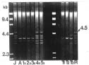
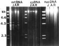
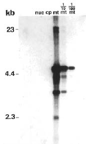
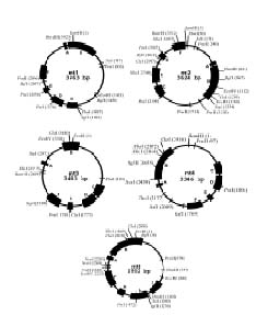
Abstract. Repeated grafting of shoot apices from mature Sequoia sempervirens (D. Don) Endl. trees onto young seedling shoots in vitro causes emergence of rejuvenated, or phase-reversed, scion shoots. The phase reversal is indicated by a restored rooting competence and renewed vigor of roots and shoots. We earlier reported of restriction fragment length polymorphism between juvenile or rejuvenated and adult shoot mtDNA (mitochondrial DNA). This investigation of undigested mtDNA disclosed that S. sempervirens shoots contained at least six small mtDNA molecules, four of which were uniquely associated with juvenile and rejuvenated shoots. The small molecules remained observable in continuously subcultured shoots. Results of cloning and sequencing indicated they are circular in form; we thus, called them plasmids. Significant sequence homology, 38 to 56%, was found among the molecules, indicating highly conserved regions and possibly common origin. No similar phase-associated relationships were observed for unrestricted cpDNA (chloroplast DNA) or nucDNA (nuclear DNA). The plasmids hybridized with the larger, electrophoretically less mobile mtDNA, but not with cpDNA or nucDNA, suggesting their origin in the master circle mtDNA. The distinctness of these plasmids has remained unchanged after more than 20 years of subculturing.

Keywords: Mitochondrial plasmids; Rejuvenation; Sequoia sempervirens.

Introduction

Development of the sexually reproductive mature adult plant normally begins with a strictly vegetative juvenile phase. The maturation, or phase change, is completed within weeks among annuals, but can span several years among perennials. In trees, the process is frequently accompanied by ancillary morphological and physiological traits, most commonly a loss of competence for adventitious rooting and of overall vigor. Since a plant's developmental phase is determined in its shoot apical meristems, reversing the phase of the meristems should result in emergence of rejuvenated shoots. Indeed, new growths with reversed phases have been obtainable variously, including through gibberellin (Rogler and Hackett, 1975) or cytokinin (Bouriquet et al., 1985; Franclet et al., 1987) applications to plants; through continuous subculturing of shoots, especially in cytokinin-containing media (Fouret et al., 1885; Lyrene, 1981; Walker, 1986; Brand and Lineberger, 1992); and through repeatedly grafting shoot apices from mature trees onto juvenile rootstocks in vivo (Doorenbos, 1954; Muzik and Cruzado, 1958) or in vitro (Monteuuis, 1986; Pliego Alfaro and Murashige, 1987; Huang et al., 1992a, 1992b; Ewald and Kretzschmar, 1996; Revilla et al., 1996).

Our earlier investigation of S. sempervirens disclosed mtDNA restriction fragment length polymorphism between juvenile and adult shoots (Huang et al., 1995). Juvenile and rejuvenated shoots contained 4.0 and 3.6 kb BamHI-restricted fragments that were absent in adult shoots. We now report on finding distinctive mitochondrial plasmids in juvenile and rejuvenated shoots.

Materials and Methods

Tissue Culture

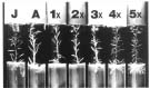
Continuously cultured Sequioa sempervirens (D. Don) Endl. shoots from freshly germinated seedlings served as the juvenile rootstocks, and those from mature trees provided the adult shoot meristems. The continuous cultures were initiated from terminals of juvenile and adult shoots and from scion-growth remnants following regrafting of shoot tips. One- centimeter long terminals were subcultured at 6-wk intervals in nutrient media containing Murashige and Skoog salts (Murashige and Skoog, 1962), 3% sucrose, 0.2% gelrite, and, in µM: 555 i-inositol, 3 thiamine·HCl, 2.4 pyridoxine·HCl, 4.1 nicotinic acid, and 26.6 glycine. Grafting was done by inserting the obliquely cut base of a 1.5-cm long shoot terminal into a longitudinal incision made in a rooted 1-cm tall juvenile stem segment. Regrafting was done at 8-wk intervals. Shoot cultures and grafted plants were maintained at 27°C and under 16-h daily exposure to 22.5 µmol m-2 s-1 cool-white

*Corresponding author. Tel: +886-6-2757575 ext. 65539; Fax: +886-6-2742583; E-mail: haojen@mail.ncku.edu.tw


Botanical Bulletin of Academia Sinica, Vol. 44, 2003

fluorescent light. Rooting bioassays were performed by pre-culturing 2-cm long terminal shoot sections in a medium of 0.2% gelrite, 1.5% sucrose, and 492 µM IBA (indole-3-butyric acid) for 14 days, then culturing in the shoot culture medium.

DNA Extraction, Purification and Electrophoresis

Extraction and purification of mtDNA followed protocols of Smith et al. (1987) and Chase and Pring (1986). Quantities of 100 g of 2- to 4-cm tall etiolated Sequoia shoots, obtained after pre-culturing shoots in darkness (3 wk for juvenile and 6 wk for adult shoots), were employed per extraction. The isolated mtDNA was purified by CsCl density gradient ultra-centrifugation. Gel electophoresis followed the protocol of Sambrook et al. (1989) and employed 0.8% agarose (BioRad ultra-pure DNA grade) containing 0.5 µg ml-1 ethidium bromide and TBE as running buffer. Solutions containing 3 to 5 µg mtDNA were mixed with front indicating dye, then placed in gel wells. Electrophoresis was run overnight at a constant 33 volts and room temperature. Gels were rinsed with deionized water and photographed, using 254 nm UV illumination.

cpDNA was prepared according to the protocol of Smith et al. (1987). Sequoia shoots for cpDNA extraction were grown under 16-h daily illumination (2 to 3 wks for juvenile and 3 to 4 wks for adult shoots). Each extraction employed 100 g of terminal 5-cm segments. nucDNA preparation was based on protocols of Cox and Goldberg (1988) and Watson and Thompson (1986). For each extraction, 25 g of etiolated shoots were employed. Purification and gel electrophoresis of cpDNA and nucDNA followed the same protocols as employed for mtDNA.

Cloning and Southern Hybridization

Cloning of mtDNA molecules was done by excising bands that were visible under a hand-held UV lamp and eluting the DNA via QIAquick Gel Extraction Kit (QIAGEN GmbH, 4072 Hilden, Germany). Separated DNAs were restricted with BamHI or EcoRI. Cloning plasmid pBluescript II was simultaneously restricted with BamHI or EcoRI, and the DNAs were ligated into pBluescript II with T4 ligase (Promega, Madison, WI). Competent E. coli strain JM101 cells were transformed with the ligated cloning plasmids, and ampicillin resistant colonies were selected and cultured in LB broth containing ampicillin. The plasmids were subsequently isolated from bacteria via the Wizard Plus Minipreps or Maxipreps Kit (Promega, Madison, WI). They were then restricted with BamHI or EcoRI, and their mtDNA was separated from cloned plasmid vectors by gel electrophoresis. Gel sections containing mtDNA were excised and eluted with the QIAGEN Qiaquik Gel extraction kit. For probes, 10 ng to 3 µg of eluted mtDNA were rendered luminescent with the DIG kit (Roche Diagnotics GmbH, Roche Molecular Biochemicals, Mannheim).

Southern hybridizations relied on commercial kits. Agarose gels of DNAs were run, photographed with UV, cut to size, and DNA de-purinated with HCl. Unrestricted

nucDNA and cpDNA were added to gel in 13 ng quantities. mtDNA was employed at rates of 13, 1.3 (1/10th) and 0.13 (1/100th) ng per well. The DNAs in washed gels were denatured with NaCl/NaOH, and gels were incubated in Transferring Solution and blotted with nylon membranes. DNAs were transferred to membranes with the Turboblotter System (Schleicher and Schuell, Inc., Keene, NH). Membranes were washed with 5X SSPE, UV crosslinked (UV Stralinker 1800; Stratagene, La Jolla, CA), dried at 65°C, and placed in hybridization bags (Life Technologies, Taipei, TW). Hybridization solution was added, and pre-hybridization was performed. After pre-hybridization the hybridization buffer was removed from the hybridization bag and replaced with fresh hybridization solution, plus denatured probe. The membranes were probed with cloned 4.5 kb mtDNA. Hybridization occurred overnight, and membranes were washed with 0.1 x SSPE and 0.1% SDS and placed in a new plastic bag. They were washed with Buffer 2, treated with anti-dig-AP solution, and washed with Buffer 1 and then with Buffer 3 as supplied by the commercial kit. Finally, CSPD solution was applied, and the membrane exposed to X-ray film.

Nucleotide Sequencing

Cloned mtDNAs were subcloned using the Erase-a-Base system (Promega, Madison, WI) to generate nested deletion sets. Complete sequences of both strands were determined by the dideoxy chain termination method, using Sequenase (Sambrook et al., 1989). The data were analyzed with the GCG software package.

Results and Discussion

Depicted in Figure 1 are sample shoot cultures of a recent series of repeatedly grafted shoot tips from a 60-year-old adult S. sempervirens tree. Rejuvenated shoots resulted after four successive grafts onto juvenile rootstocks. These and the juvenile rootstock shoots rooted

Figure 1. Rejuvenated S. sempervirens shoots produced by repeated grafting of adult shoot apices onto juvenile stem cuttings in vitro. Left to right, shoots arising from J = juvenile seedling, A = ungrafted adult tree shoot apex, and 1× to 5× = 1- to 5-times grafted adult shoot apex. Restored rooting competence and plagiotropic leaves can be seen in 4× and 5×-grafted shoots.


Huang et al. — Developmental phase reversal of Sequoia sempervirens

Figure 2. Unrestricted mtDNA isolated from juvenile (J), adult (A, T1, T2, and T3) and grafted adult S. sempervirens shoots. 1× to 5× = 1 to 5 successive grafts of adult shoot apices onto rooted juvenile stems. Lanes 1 and 9 are molecular size markers, in kb, of HindIII digested lambda phage DNA. Adult shoot cultures were established from shoot apices of A = 60-year-old tree in 1976; those of adult trees identified by T1, T2 and T3 were initiated in 1994. R = shoots from an earlier 5 times grafted adult.

Figure 3. Phase change in relation to DNA of other organelles. Molecular size markers are in lanes just left of each set of organelle DNAs. J = juvenile, A = adult, and R = rejuvenated shoots. The additional lanes to the immediate left of J under mtDNA as well as cpDNA are DNA of juvenile shoots obtained from green shoots for mtDNA and employed in amounts larger than prescribed for cpDNA. Standard quantities of DNA employed per well were 5 µg for mtDNA, 10 µg for cpDNA and 2 µg for nucDNA.

profusely and branched more, their leaves were softer and tended to be plagiotropic, and their stems elongated faster and were thinner than those of ungrafted adult or grafted but un-rejuvenated adult shoots. They also produced less callus. These traits are very stable in vitro. Juvenile and rejuvenated shoots continue to display juvenile morphology, organogenic competence, and greater vigor after more than 20 years of subculturing; adult shoots remain poorly rooting and slow growing.

The developmental phases were very closely associated with small mtDNA molecules. Gel electrophoresis of unrestricted mtDNA clearly revealed six molecules ranging in size from 3.2 to 4.5 kb (Figure 2). A large molecule, presumably the master circle, migrated very slowly; its size was about 23 kb. Four of the small molecules, 3.2, 3.9, 4.0 and 4.5 kb, were found only in juvenile (J) shoots. Two molecules, 3.4 and 3.7 kb, were common to both phases. When juvenility was restored by repeated grafting (4x and 5x), all six small mtDNA molecules became evident in the rejuvenated shoots. Conversely, the still unrejuvenated shoots after 1 to 3 successive grafts continued to lack the four juvenility-associated molecules. Reproducibility of the association between juvenility and mtDNA molecules is exemplified by R (Figure 2), or shoots from a previously 5x-grafted adult shoot tip, where all six mtDNA molecules are now observable. The reappearance of mtDNA molecules in rejuvenated shoots might suggest their translocation through the graft union, from juvenile rootstock to adult shoot meristem, and replication in the adult meristem. However, the currently more accepted explanation is that a phytohormone was transferred and caused the resumption of juvenile phase mtDNA synthesis in adult meristem

Figure 4. Southern hybridization of juvenile mtDNA with juvenile nucDNA (nuc), cpDNA (cp) and mtDNA (mt). In this instance, a juvenile specific 4.5 kb mtDNA molecule was used as probe. The mtDNA was also electrophoresed at 1/10th and 1/100th the prescribed quantity.


Botanical Bulletin of Academia Sinica, Vol. 44, 2003

cells. The stability of the rejuvenated states and their continued corelationship with specific mtDNA molecules in subcultures implicate an involvement of mtDNA in developmental phase change.

Absence of the juvenility-associated mtDNA molecules was not unique to the 60-year-old S. sempervirens tree, the shoot culture of which was established in 1976. Two additional mature trees, T1 and T2, also lacked these molecules (Figure 2). Shoots of a third additional tree, T3, contained two juvenility-associated molecules, 3.2 and 4.0 kb, a characteristic coincident with their readily rooting and faster elongating behavior. Shoot cultures of T1-3 were initiated in 1994.

No relationship was observed between phase change and small molecules of cpDNA or nucDNA. As apparent

in Figure 3, juvenile (J) and rejuvenated (R) shoots displayed the distinctive mtDNA molecules. In contrast, electrophoresis of the unrestricted nucDNA revealed mainly a diffuse, intensely stained region of the large molecule; the unrestricted cpDNA was composed of a single band of large molecule. For unknown reasons, the mtDNA molecules from light-grown, or green, shoots stained more intensely than those of etiolated shoots.

No Southern hybridization occurred between cpDNA or nucDNA with small plasmid mtDNA, suggesting that the plasmid mtDNA molecules were not derived from cpDNA or nucDNA (Figure 4). Probes prepared from plasmid mt1 DNA molecules hybridized readily with the large mtDNA molecule, and to varying degrees with other small mtDNA molecules. This indicates that the mtDNA mol

Figure 5. Restriction map of five circular mtDNA molecules. High homology regions of mtDNA sequence are represented in the boxes (A to F).


Huang et al. — Developmental phase reversal of Sequoia sempervirens

ecules shared a common origin, most probably in the large mtDNA molecule.

Five of the six small mtDNA molecules were sequenced. Lack of an effective restriction enzyme precluded sequencing of one molecule, mt5 DNA plasmid. The sequenced molecules showed homologies of 38 to 56%. The relatively high homology indicated highly conserved sequences and reinforced a common origin. In Figure 5 are shown a restriction map and high homology regions of five plasmid mtDNA molecules. A search of DNA records indicated that the mtDNA molecules contained polypeptide encoding regions, although not of known proteins.

When cloning the molecules, restriction with BamHI or EcoRI resulted in polynucleotides with increased electrophoretic mobility. The increased mobility is therefore interpreted as indicating circularity of the original isolates. In order to prove the cloned mtDNA molecules were circular forms, pairs of specific primers to each cloning site were designed. Sequences around the flanking regions of each cloning site were obtained by PCR amplification with the unrestricted mtDNA serving as templates. All five sequenced plasmid mtDNA were found to be circular forms. Using this process a 112 bp of BamHI fragment within the BamHI cloning site was recovered from the original mt2 DNA molecule shown in Figure 5. The circular structure and their apparently autonomous replication enabled classification of the small mtDNA molecules as plasmids.

The role of nucDNA in the phase change of plants is undeniable, since the vast majority of genes for development resides in the nucleus. On that basis, Poethig (1990) and Ronemus et al. (1996) have made a strong case for nucDNA methylation as underlying plant maturation. Our HPLC analysis also revealed slightly more methylated cytosine in adult S. sempervirens nucDNA (8.57 ± 0.13%) than in juvenile nucDNA (7.30 ± 0.49%), but we observed no differences in methylation of either mtDNA or cpDNA. Methylation of DNA of the three organelles is being further evaluated by Southern hybridization. Meanwhile, we propose that mtDNA also plays a major role in regulating developmental phase change in plants. Esser et al. (1980) earlier reported that senescence of the fungus, Podospora anserine, was related to changes occurring in mitochondrial plasmids. We have established an association between mtDNA and phase change of S. sempervirens; there now remain the tasks of demonstrating a causal relationship and elucidating the identity of the substance(s) transmitted from juvenile rootstock to adult meristem, as well as the underlying mechanism for mtDNA control of phase change.

Acknowledgements. The investigation was supported by Grant NSC 88-2311-B-001-093 from the National Science Council and Grant 85AST-1.1-FAD-49(16) from the Council of Agriculture, Taiwan, as well as research funds from the Institute of Botany, Academia Sinica. We thank Mr. Y.-H. Liu of the National Taiwan Institute of Technology for the photography, and Bau-Lian Huang, Mei-Ru Ju, Li-Tian Cheng, Su-Pi Lin, and Mei-Fong Tsai for their many hours of assistance with culturing tissues and isolating DNA.

Literature Cited

Bouriquet, R., M. Tsogas, and A. Blaselle. 1985. Essais de rajeunissement de l'epicea par les cytokinines. Ann. AFOCEL 1984: 173-185.

Brand, M.H. and R.D. Lineberger. 1992. In vitro rejuvenation of Betula (Betulaceae): morphological evaluation. Am. J. Bot. 79: 618-625.

Chase, C.D. and D.R. Pring. 1986. Properties of the linear N1 and N2 plasmid-like DNAs from mitochondria of cytoplasmic male-sterile Sorghum bicolor. Plant Mol. Biol. 6: 53-64.

Cox, K.H. and R.B. Goldberg. 1988. Analysis of plant gene expression. In C.H. Shaw (ed.), Plant Molecular Biology: A Practical approach. IRK Press, Oxford, pp. 1-35.

Doorenbos, J. 1954. "Rejuvenation" of Hedera helix in graft combinations. Ned. Akad. Wetensch. Amsterdam, Proc. 57: 99-102.

Esser, K., P. Tudzynski, U. Stahl, and U. Kuck. 1980. A model to explain senescence in the filamentous fungus Podospora anserine by the action of plasmid like DNA. Molec. Gen. Genet. 178: 213-216.

Ewald, D. and U. Kretzschmar. 1996. The influence of micrografting in vitro on tissue culture behavior and vegetative propagation of old European larch trees. Plant Cell Tissue Organ Cult. 4: 249-252.

Fouret, Y., Y. Arnaud, and C. Larrieu. 1985. Rajeunissement in vitro du Sequoia sempervirens. Ann. AFOCEL 1984: 111-137.

Franclet, A., M. Boulay, F. Bekkaoui, Y. Fouret, B. Verschoore Martoouzet, and N. Walker. 1987. Rejuvenation. In J.M. Bonga and D.J. Durzan (eds.), Cell and Tissue Culture in Forestry. Martinus Nijhoff Publishers, Vol. 2, pp. 232-248.

Huang, L.C., C.K. Hsiao, S.H. Lee, B.L. Huang, and T. Murashige. 1992a. Restoration of vigor and rooting competence in stem tissues of mature citrus by repeated grafting of their shoot apices onto freshly germinated seedlings in vitro. In Vitro Cell. Dev. Biol. 28: 30-32.

Huang, L.C., S. Lius, B.L. Huang, T. Murashige, E.F.M. Mahdi, and R. Van Gundy. 1992b. Rejuvenation of Sequoia sempervirens by repeated grafting of shoot tips onto juvenile rootstocks in vitro. Plant Physiol. 98: 166-173.

Huang, L.C., L.Y. Lin, C.M. Chen, L.J.Chen, B.L. Huang, and T. Murashige. 1995. Phase reversal in Sequoia sempervirens in relation to mtDNA. Physiol. Plant. 94: 379-383.

Lyrene, P.M. 1981. Juvenility and production of fast-rooting cuttings from blueberry shoot cultures. J. Am. Soc. Hort. Sci. 106: 396-398.

Monteuuis, O. 1986. Microgreffage de points vegetatifs de Sequoiadendron giganteum Bucholz. Seculaires sur de jeunes emis cultives in vitro. C.R. Acad. Sci. Paris 302: 223-225.

Murashige, T. and F. Skoog. 1962. A revised medium for rapid growth and bioassays with tobacco tissue culture. Physiol. Plant. 15: 473-497.

Muzik, T.J. and H.J. Cruzado. 1958. Transmission of juvenile rooting ability from seedlings to adults of Hevea brasiliensis. Nature 181: 1288.

Pliego Alfaro, F. and T. Murashige. 1987. Possible rejuvenation of adult avocado by graftage onto juvenile rootstocks in vitro. HortSci. 22: 1321-1324.


Botanical Bulletin of Academia Sinica, Vol. 44, 2003

Poethig, R.S. 1990. Phase change and the regulation of shoot morphogenesis in plants. Science 250: 923-930.

Revilla, M.A., J. Pacheco, A. Casares, and R. Rodriguez. 1996. In vitro reinvigoration of mature olive trees (Olea europaea L.) through micrografting. In Vitro Cell. Dev. Biol. Plant 32: 257-261.

Rogler, C.E. and W.P. Hackett. 1975. Phase change in Hedera helix: induction of the mature to juvenile phase change by GA. Physiol. Plant. 34: 141-147.

Ronemus, M.J., M. Galbiat, C. Ticknor, J. Chen, and S.L. Dellaporta. 1996. Demethylation-induced developmental pleiotropy in Arabidopsis. Science 273: 654-657.

Sambrook, J., E.F. Fritsch, and T. Maniatis. 1989. Molecular

Cloning. A Laboratory Manual. Cold Spring Harbor Laboratory, Cold Spring Harbor. 2nd ed, Vol. 1-3.

Smith, R.L., M.K.U. Chowdhury, and D.R. Pring. 1987. Mitochondrial DNA rearrangements in Pennisetum associated with reversion from cytoplasmic male sterility to fertility. Plant Mol. Biol. 9: 277-286.

Walker, N. 1986. Sequoia sempervirens. Réjuvenilisation et culture de méristčmes en cascade. Ann. Rech. Sylv. 1985 AFOCEL, pp. 25-48.

Watson, J.C. and W.F. Thompson. 1986. Purification and restriction endonuclease analysis of plant nuclear DNA. In Methods of Enzymology, Academic Press, San Diego, pp. 57-75.