Bot. Bull. Acad. Sin. (2003) 44: 141-150

Tsai and Wang — Rice protein kinases phosphorylating sucrose synthase

Identification of rice manganese-dependent protein kinases that phosphorylate sucrose synthase at multiple serine residues

Zheng-Chia Tsai and Ai-Yu Wang*

Department of Agricultural Chemistry, National Taiwan University, No. 1, Section 4, Roosevelt Road, Taipei, Taiwan 106

(Received October 2, 2002; Accepted January 16, 2003)

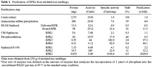
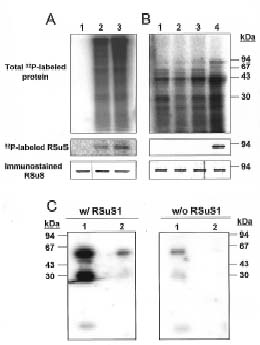
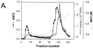
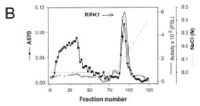
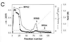
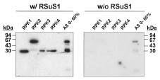
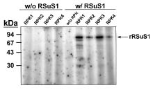
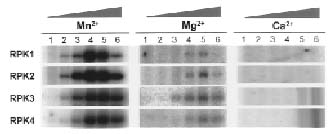
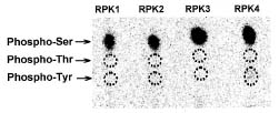
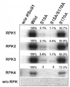
Abstract. Sucrose synthase from the etiolated seedlings of rice (Oryza sativa L. cv. Tainung 67) was phosphorylated both in vivo and in vitro. Four protein kinases that phosphorylated recombinant rice sucrose synthase 1 (RSuS1) in a Mn2+-dependent manner were partially purified and characterized from etiolated rice seedlings. These four kinases, designated as RPK1, RPK2, RPK3 and RPK4, are monomeric enzymes with apparent molecular masses of 34 kDa, 57 kDa, 30 kDa, and 30 kDa, respectively. Phosphoamino acid analysis of the 32P-labeled phosphorylated recombinant RSuS1 indicated that it was phosphorylated at serine residues by these four RPKs. RP-HPLC/ESI-MS analysis of the tryptic peptides of phosphorylated RSuS suggested that the serine residues in the tryptic peptides 13-LHSVR-17 and 168-HLSSK-172 were the target residues for phosphorylation. For confirmation of this finding, mutant recombinant RSuS1, S15A, S170A and S15A/S170A, were purified and subjected to phosphorylation by the four partially purified kinases. The results showed that both Ser15 and Ser170 residues were target residues for RPK1, PRK2 and PRK3 and Ser15 was the major phosphorylation site in RSuS1. Phosphorylation of RSuS1 may not occur exclusively at these two sites since weak phosphorylation of the double mutant protein S15A/S170A was also observed. Phosphorylation of the mutant S15A and S15A/S170A by RPK4 was undetectable, indicating that Ser15 was the only target residue for this kinase.

Keywords: Manganese-dependent protein kinases; Rice; Sucrose synthase.

Introduction

Sucrose synthase (UDPG: D-fructose 2-glucosyl transferase, SuS) was first described by Cardini et al. (1955) and has been characterized in various plant species. The enzyme catalyzes the conversion of sucrose and UDP into fructose and UDPG. Although the reaction is readily reversible, it is thought that SuS functions primarily in the direction of sucrose degradation to provide sugar nucleotides for complex saccharides synthesis (Chourey and Nelson, 1976; Amor et al., 1995; Déjardin et al., 1997; Chourey et al., 1998).

In most plants, SuS is encoded by two or three genes that are spatially and temporally regulated and are differentially modulated by the sugar level, anaerobiosis (Zeng et al., 1998; Wang et al., 1999; Winter and Huber, 2000 and references therein; Barratt et al., 2001; Carlson et al., 2002), and osmotic stress (Déjardin et al., 1999). In potato, SnRK1 (SNF1-related protein kinase) activity is required for normal SuS gene expression (Purcell et al., 1998). On the protein level, the enzyme activity in sucrose synthesis and sucrose cleavage was reported to be differentially affected by divalent metal ions (Delmer, 1972; Tsai, 1974; Huang and Wang, 1998), protein factors (Pontis and Salerno, 1982),

and the redox state of the enzyme (Pontis et al., 1981). In addition, SuS is post-translationally regulated by reversible phosphorylation. Phosphorylation of SuS in vivo and in vitro by endogenous or exogenous protein kinases in a Ca2+-dependent manner has been demonstrated in several plants such as maize (Huber et al., 1996; Winter et al., 1997; Subbaiah and Sachs, 2001), soybean nodule (Zhang and Chollet, 1997; Zhang et al., 1999), tomato (Anguenot et al., 1999), mung bean (Nakai et al., 1998), cotton (Haigler et al., 2001), and rice (Asano et al., 2002). Recently, Chikano et al. (2001) reported that an Arabidopsis SnRK3 protein, AtSR2, expressed in E. coli efficiently phosphorylated SuS in the presence of manganese ions. However, the significance of SuS phosphorylation in vivo is not clear. Phosphorylation/dephosphorylation of the enzyme has been suggested to play a role in regulating its activity (Zhang and Chollet, 1997; Nakai et al., 1998; Anguenot et al., 1999; Zhang et al., 1999; Haigler et al., 2001; Tanase et al., 2002), in its distribution between the cytosol, plasma membrane, and actin cytoskeleton (Amor et al., 1995; Winter et al., 1997; Winter and Huber, 2000), and also in the response of cells to environmental and developmental signals (Subbaiah and Sachs, 2001).

In rice, there are three non-allelic RSus genes encoding SuS (Wang et al., 1992; Yu et al., 1992; Huang et al., 1996; Wang et al., 1999). The gene products of RSus1 and RSus2 are ubiquitously present in suspension-cultured

*Corresponding author. Tel: +886-2-23630231 ext. 3251; Fax: +886-2-23660434; E-mail: aywang@ntu.edu.tw


Botanical Bulletin of Academia Sinica, Vol. 44, 2003

cells, etiolated seedlings, and seeds while those of RSus3 are predominantly found in rice seeds (Wang et al., 1999). Regulation of rice SuS (RSuS) by a calmodulin-like domain protein kinase in a Ca2+-dependent manner in immature rice seeds has been reported recently (Asano et al., 2002). In this study, we demonstrated that RSuS in etiolated rice seedlings could also be phosphorylated by endogenous kinases in a Mn2+-dependent manner. By using the recombinant RSuS expressed in E. coli as a substrate for kinase activity assay, we partially purified and characterized four protein kinases that phosphorylated RSuS1 predominantly on Ser15 and Ser170 residues. To the best of our knowledge, this is the first study to demonstrate that SuS can be phosphorylated at multiple serine residues by different Mn2+-dependent protein kinases.

Materials and Methods

Materials

DEAE Sephacel, CM Sepharose Fast Flow, Sephacryl S-100 HR, Protein A Sepharose CL-4B and protein molecular mass markers were from Amersham Pharmacia Biotech. Complete Protease Inhibitor Cocktail Tablets were purchased from Roche Molecular Biochemicals. [g-32P]ATP (6000 Ci·mmol-1), [32P]Pi (8500-9120 Ci·mmol-1) was from NEN Life Science Products, Inc. The QuickChange Site-Directed Mutagenesis Kit was from Stratagene. Okadaic acid was obtained from Calbiochem. PVDF membranes were from Millipore. All other biochemicals were purchased from Sigma Chemical Co.

Seeds of Oryza sativa L. cv. Tainung 67 were germinated and grown at 30°C in complete darkness for 10 days. The harvested etiolated seedlings were frozen in liquid nitrogen and stored at -80°C until used.

Expression and Purification of Recombinant RSuS1

Plasmid pETsus1, which carries the coding region of RSus1 cDNA under the control of T7 promoter, was transformed into E. coli BLR (DE3). Expression and purification of the recombinant RSuS1 to near homogeneity from E. coli was carried out as described previously (Sayion et al., 1999).

Polyacrylamide Gel Electrophoresis and Western Analysis

Proteins were separated by 10% or 7.5% SDS-PAGE (Laemmli, 1970). After electrophoresis, proteins in gels were stained with Coomassie Blue R-250, or transferred onto PVDF membranes. For detecting RSuS proteins, a mixture of a monoclonal antibody recognizing RSuS1 and RSuS3 and a monospecific anti-peptide antibody recognizing RSuS2 (Wang et al., 1999) was used.

In Vivo Phosphorylation Assay

The 10-day-old etiolated rice seedlings were placed in 5 mL of degassed phosphate buffer (10 µM, pH 7.0) con

taining 0.5 mCi of [32P]Pi. The roots were harvested at the indicated times and were immediately frozen in liquid nitrogen. The samples were extracted with buffer M [100 mM Mops, pH 7.6, 2 mM 2-mercaptoethanol and Complete Protease Inhibitor Cocktail (one tablet per 10 mL buffer)] followed by centrifugation at 27,000 g for 10 min at 4°C. The total proteins in the extracts were separated on 10% SDS-polyacrylamide gels, and the radiolabeled proteins were detected by phosphorimaging. RSuS proteins were immunoprecipitated from the extracts with antibodies according to the method of Anderson and Blobel (1983), followed by SDS-PAGE, Western analysis, and phosphorimage analysis.

In Vitro Phosphorylation Assay

To assay phosphorylation of RSuS in etiolated seedlings by endogenous kinases, 10-day-old etiolated rice seedlings were ground into a fine powder under liquid nitrogen and homogenized with an equal volume of buffer M. The homogenate was centrifuged at 27,000 g for 10 min at 4°C and the pellet was discarded. Aliquots of the supernatant were incubated for 30 min at 30°C with 30 µCi [g-32P]ATP and 0.1 µM okadaic acid with or without divalent ions added. RSuS proteins were purified by immunoprecipitation and analyzed by SDS-PAGE.

To determine the RSuS kinase activity in vitro, the enzyme was assayed in a 20 µL reaction mixture containing buffer A (20 mM Mops, pH 7.6, 2 mM 2-mercaptoethanol), 4 µg purified recombinant RSuS1, 5 µCi [g-32P]ATP and 2 mM MnCl2 at 30 °C for 10 min. The reaction was stopped by adding 20 µL 2×SDS-PAGE sample buffer (125 mM Tris, 2 mM EDTA, 2% SDS, 2-mercaptoethanol, pH 6.8). The samples were analyzed by SDS-PAGE and autoradiography or phosphorimage analysis. To determine the amount of [32P]Pi incorporated, the labeled RSuS bands in gels were excised and counted by a scintillation counter (Beckman LS5000CE). One unit of RSuS kinase activity was defined as 1 pmol of [32P]Pi incorporated into RSuS per min in the standard assay condition.

In-Gel Kinase Assay

The in-gel kinase assay was performed using the method of Hutchcroft et al. (1991) with minor modification. SDS-polyacryamide gels were polymerized with 80 µg purified recombinant RSuS1. For a negative control, a gel without RSuS was used. After electrophoresis, the gels were washed with buffer A six times over a period of 6 h. They were then incubated in buffer A containing 2 mM MnCl2 and 50 µCi [g-32P]-ATP for 60 min at 37°C, followed by Coomassie blue staining and destaining. The destained gels were incubated with Dowex MR-3 resin in H2O for 4 h and then dried and exposed to X-films.

Protein Determination

The protein content of enzyme solutions was determined by the method of Bradford (1976) using bovine serum albumin as the standard.


Tsai and Wang — Rice protein kinases phosphorylating sucrose synthase

Partial Purification of Protein Kinases

All purification steps were carried out at 0 to 4°C. Etiolated rice seedlings were homogenized in 2 volumes (v/w) of extraction buffer (100 mM Mops, pH 7.6, 0.5 mM EDTA, 5 mM 2-mercaptoethanol, 1% polyvinylpolypyplidone). The homogenate was filtered through three layers of cheesecloth and centrifuged at 15,000 g for 10 min. Nucleic acids in the crude extract were precipitated by adding protamine sulfate to 0.2% and removed by centrifugation at 15,000 g for 10 min. Solid ammonium sulfate was added to the centrifugation supernatant to 60% saturation. After centrifugation at 27,200 g for 15 min, the protein pellet was resuspended in buffer A and dialyzed against the same buffer. The enzyme solution was loaded onto a DEAE Sephacel column (2.6 × 20 cm) equilibrated with buffer A. The column was first eluted with buffer A, then with a linear 0-400 mM NaCl gradient in buffer A. Activity was measured in both unbound fractions and NaCl-eluted fractions. The unbound active fractions were pooled and applied onto a CM Sepharose column (1.6 × 15 cm) equilibrated with buffer A. The column was eluted with buffer A followed by a linear gradient (0-600 mM) of NaCl. The column eluate containing kinase activity was concentrated and loaded onto a Sephacryl S-100 column (1.6 × 90 cm). The activity peak eluted with buffer A were concentrated and stored at -20°C. The DEAE-bound fractions containing kinase activity were concentrated, and the buffer was changed to buffer B (20 mM Mops, pH 7.2, 2 mM 2-mercaptoethanol) by ultrafiltration. The enzyme solution was applied onto a phosphocellulose column (1.6 × 20 cm) equilibrated with buffer B. After washing with buffer B, the column was eluted with a linear 0-600 mM NaCl gradient in buffer B. The three peaks with kinase activity (designated RPK2, RPK3 and RPK4) were separately collected and concentrated. RPK2 and RPK3 were further separated on a Sephacryl S-100 column (1.6 × 15 cm) with buffer A as elution buffer. Fractions containing kinase activity were pooled, concentrated, and stored at -20°C.

Phosphoamino Acid Identification

The purified recombinant RSuS1 was phosphorylated in vitro in the presence of 5 µCi [g-32P]ATP and 2 mM MnCl2 by partially purified RPK1, RPK2, RPK3 or RPK4. The 32P-labeled recombinant RSuS1 was separated from kinases by SDS-PAGE and transferred onto a PVDF membrane. The 93-kDa RSuS1 protein band on PVDF was cut out, washed with methanol, and digested with 6 N HCl at 110°C for 1 h. The phosphoamino acids in the acid hydrolysate were analyzed by TLE as described by Jelinek and Weber (1993).

Tryptic Digestion and ESI-MS Analysis

The purified recombinant RSuS1 was phosphorylated in vitro by kinases in the DEAE-bound fractions in the presence of Mn2+ and [g- 32P]-ATP and separated by 10% SDS-PAGE. After electrophoresis, the gel was stained with Commassie blue, and the RSuS1 protein band was excised

from the gel and subjected to tryptic digestion as described by Stone and Williams (1996). The tryptic sample was applied to a reverse-phase HPLC column (LiChrospher WP 300 RP-18, Merck) and eluted with a gradient of 0 to 25% (v/v) acetonitrile in 0.1% (v/v) trifluoroacetic acid. The radioactivity in each fraction was counted. ESI-MS analysis of the fraction containing 32P-label peptides was performed with the VG Platform Electrospray ESI/MS in the Instrumentation Center of National Taiwan University.

Site-Directed Mutagenesis

Plasmid pETsus1 (Sayion et al., 1999) was used as a template for mutagenesis. In vitro mutagenesis was performed using the QuickChange Site-Directed Mutagenesis Kit with the primers 5´-CGCCTCCACGCTGTCAGGGAGC and 5´-GCTCCCTGACAGCGTGGAGGC to change Ser15 to Ala, and the primers 5´-CAGGCATCTGGCTTCGAAGCTCTTCC and 5´-GGAAGAGCTTCGAAGCCAGATGCCTG to change Ser170 to Ala. The changed nucleotides are underlined. Constructs were verified by DNA sequencing. Expression and purification of the mutant recombinant RSuS1 from E. coli was carried out as described above for the wild-type recombinant RSuS1.

Results

Phosphorylation of RSuS in Etiolated Rice Seedlings

To examine whether phosphorylation of SuS occurred in rice, the whole 10-day-old etiolated rice seedling was incubated with [32P]Pi for different time periods. Immunoprecipitation of RSuS from the extracts indicated that RSuS was phosphorylated in vivo (Figure 1A). When soluble extracts of unlabeled etiolated rice seedlings were incubated with [g-32P]-ATP in the presence of various divalent ions, RSuS in the crude extracts was phospholabeled by endogenous protein kinases more intensively in the presence of Mn2+ ions than in the presence of Mg2+ or Ca2+ ions (Figure 1B). The Mn2+-dependent protein kinase activity for phosphorylating RSuS was further identified by in-gel kinase assays using the purified recombinant RSuS1 as a substrate. Two protein bands possessing RSuS-phosphorylation activity were detected in the protein fractions precipitated by 0-60% and 60-100% saturation of ammonium sulfate (Figure 1C).

Partial Purification of the Mn2+-Dependent Kinases Phosphorylating RSuS from Etiolated Rice Seedlings

Purification of the Mn2+-dependent kinases in the 0-60% fraction of ammonium sulfate precipitation was attempted by column chromatography. The purified recombinant RSuS1 was used as a substrate for detecting kinase activity during the purification process. Figure 2A shows the elution profile of DEAE Sephacel ion exchange chromatography. The unbound active peak was loaded onto a CM Sepharose column, and a kinase activity peak,


Botanical Bulletin of Academia Sinica, Vol. 44, 2003

designated RPK1, was eluted with a linear NaCl gradient (Figure 2B). The DEAE-bound fractions containing kinase activity were separated on a phosphocellulose column, three peaks with kinase activity, designated RPK2, RPK3 and RPK4, were obtained (Figure 2C). RPK1, RPK2 and RPK3 were further purified by Sephacryl S-100 gel filtration chromatography (Figure 2D). SDS-PAGE analysis of the four RPKs showed several protein bands in each preparation (data not shown), indicating that they were only partially purified. The purification results are summarized in Table 1. Actual enzyme purity may have been much higher than given in Table 1 because it was not possible to estimate the initial specific activity.

Characterization of RPKs

Figure 3 shows the in-gel kinase assays of these four kinase preparations in the presence of Mn2+ ions. Only a single activity band was found in RPK1, RPK3 and RPK4. The estimated molecular masses were 32 kDa for RPK1 and 34 kDa for both RPK3 and RPK4. The molecular masses of the native enzymes, as determined by Sephacryl S-100 gel filtration chromatography, were 34 kDa, 30 kDa and 30 kDa, for RPK1, RPK3 and RPK4, respectively, indicating that they were monomeric enzymes. The molecule mass of RPK2 determined by Sephacryl S-100 gel filtration chromatography was 57 kDa. However, in addition to a 57-kDa

Figure 1. Phosphorylation of RSuS in etiolated rice seedlings. (A) In vivo phosphorylation of RSuS. The 10-day-old etiolated rice seedlings were incubated with [32P]Pi. Protein extracts from roots harvested at 0, 1 and 2 h (lanes 1 to 3, respectively) were analyzed by SDS-PAGE and phosphorimaging (top panel). RSuS proteins in the extracts were immunoprecipitated with anti-RSuS antibodies and analyzed by SDS-PAGE analysis. After electrophoresis, the gels were subjected to phosphorimage analysis (center panel) or Western analysis using anti-RSuS antibodies (lower panel). (B) In vitro phosphorylation of RSuS by endogenous kinases. Protein extracts from 10-d-old seedlings were incubated with [g-32P]-ATP without divalent ions added (lane 1) or in the presence of Ca2+ (lane 2), Mg2+ (lane 3) or Mn2+ (lane 4). Total phosphorylated proteins and RSuS proteins were analyzed as in (A). (C) Identification of Mn2+-dependent kinase activity by in-gel kinase assay. Proteins from 0-60% (lane 1) and 60-100% (lane 2) fractions of ammonium sulfate precipitation were separated on 15% SDS-polyacrylamide gel polymerized with purified recombinant RSuS1 (left panel) or without RSuS1 (right panel). After electrophoresis, the gels were incubated with [g-32P]-ATP and Mn2+, and then subjected to autoradiography. Molecular weight markers are shown at the margins.


Tsai and Wang — Rice protein kinases phosphorylating sucrose synthase

activity band, a 37-kDa protein band with lower kinase activity was detected in the RPK2 preparation as analyzed by in-gel kinase assay (Figure 3). The latter may be a proteolytic fragment of the former. The in-gel kinase assay in the absence of recombinant RSuS1 (Figure 3, right panel) suggested that these four RPKs may not be able to cata

lyze the autophosphorylation reaction. To confirm this result, in vitro kinase assays were performed by incubating each RPK with [g-32P]-ATP and 2 mM Mn2+ in the presence or absence of purified recombinant RSuS1. The phosphorylated proteins were then analyzed by SDS-PAGE (Figure 4). No phosphorylated protein band was detected

Figure 2. Elution profiles of RSuS kinase activity and protein from each purification step. The enzyme solution resulting from the 0-60% fraction of ammonium sulfate precipitates was loaded onto a DEAE Sephacel column and washed with buffer A followed by elution with a linear 0-400 mM NaCl gradient in buffer A (A). The unbound active fractions were pooled and applied onto a CM Sepharose column and eluted with a linear gradient (0-600 mM) of NaCl. The activity peak was designated as RPK1 (B). The RSuS kinase activity in the DEAE-bound fractions was separated on a phosphocellulose column with a linear 0-600 mM NaCl gradient. The three peaks with kinase activity (designated RPK2, RPK3 and RPK4) were separately collected (C). RPK1, RPK2 and RPK3 were further separated on a Sephacryl S-100 column (1.6 × 15 cm) with buffer A as elution buffer (D). Kinase activity (open circles) was assayed in the presence of Mn2+ using purified recombinant RSuS1 (rRSuS1) as substrate and then analyzed by SDS-PAGE and autoradiography (shown in lower panels). The densities of the 32P-labeled RSuS bands were quantified by an image analyzer. Protein concentrations (closed circles) were determined by the method of Bradford (1976). The dashed lines indicate the NaCl gradients.


Botanical Bulletin of Academia Sinica, Vol. 44, 2003

in any assay in the absence of purified recombinant RSuS1, verifying that none of the four RPKs possessed autophorylation activity. In addition, phosphorylation of RSuS1 was not observed in the absence of RPK (Figure 4, the 5th lane), showing that the purified recombinant RSuS1 did not contain kinase activity from RSuS1 itself or from the E. coli proteins co-purified with the recombinant RSuS1.

To determine the cofactor requirements of these RPKs, kinase activity was examined using various concentrations of Mn2+, Mg2+ or Ca2+ in the in vitro kinase assay. Activity of these enzymes was found to be much higher in the presence of Mn2+ compared with Mg2+ but was undetectable in the presence of Ca2+ ions (Figure 5). Phosphorylation of RSuS by these enzymes could be activated by Mn2+ at concentrations as low as 10 µM. The Mn2+ concentrations required for optimal activity were 2 mM for RPK1 and RPK2, and 10 mM for RPK3 and RPK4. The minimum Mg2+ concentration at which the phosphorylation of RSuS was detectable was 0.1 mM for all four kinases. Optimal Mg2+-activated activity of these enzymes was observed with 10 mM Mg2+.

The activity of the four kinases was inhibited 95% by staurosporine, a broad kinase inhibitor, at a concentration of 50 nM (data not shown). To identify the amino acid residue(s) of RSuS1 targeted by the four RPKs, the purified recombinant RSuS1 was in vitro phosphorylated in the presence of [g-32P]-ATP, Mn2+ and one of the four partially purified RPKs, submitted to SDS-PAGE, and transferred onto a PVDF membrane. The 93-kDa RSuS1 protein band on PVDF was subjected to phosphoamino acid analysis as described in Materials and Methods. As shown in Figure 6, all four RPKs phosphorylated recombinant RSuS1 at serine residue(s).

Phosphorylation Sites on RSuS

In a preliminary experiment, the 32P-labeled recombinant RSuS1 that was phosphorylated in vitro by kinases in the DEAE-bound fraction was isolated from SDS-PAGE, digested with trypsin, and then subjected to off-line RP-HPLC/ESI-MS analysis. The ESI-MS spectrum of the 32P-labeled fraction resolved from RP-HPLC suggested that several tryptic peptides were present in this fraction (data not shown). Peaks at m/z 231.8 and 346.0, 327.0 and 366.2 were predicted to represent the tryptic peptides 13-LHSVR-17 and 168-HLSSK-172, respectively, of the RSuS1 with the addition of one phosphate. Although the result of ESI-MS was inconclusive, phosphorylation of Ser-15 and Ser-170 by the four partially purified RPKs was confirmed using site-directed mutagenized recombinant RSuS1. Ser-15 and/or Ser-170 of the recombinant RSuS1 were mutagenized to Ala and the mutant proteins were expressed and purified from E. coli. Phosphorylation in vitro by RPK1, RPK2 and RPK3 was observed for the S15A, S170A and S15A/S170A mutant proteins, but the extent of phosphorylation was less than that of wild type (Figure 7). The phosphorylation levels of mutant S15A and S170A were 2.1-3.6% and 83.3- 93.9%, respectively, of those of the wild-type RSuS1.

Figure 3. In-gel kinase assay of the partially purified RPKs. The four partially purified kinases that phosphorylated RSuS (RPK1, RPK2, RPK3 and RPK4) and the 0-60% fraction of ammonium sulfate precipitation (AS 0-60%) were separated on SDS-polyacrylamide gels polymerized with purified recombinant RSuS1 (left panel) or without RSuS1 (right panel). After electrophoresis, the gels were incubated with [g-32P]-ATP in the presence of Mn2+, and then subjected to autoradiography. Molecular weight markers are shown at the margins.

Figure 4. In vitro phosphorylation assay of the partially purified RPKs. Activity of the four partially purified RPKs was determined in vitro in the presence of [g-32P]-ATP and 2 mM Mn2+ with or without purified recombinant RSuS1 added. The samples were then analyzed by SDS-PAGE, and the radiolabeled RSuS was detected by phosphorimaging. Molecular weight markers are shown at the margins.

Figure 5. Effect of Mn2+, Mg2+ and Ca2+ ions on the activity of RPKs. Activity of the four partially purified RPKs was determined in vitro using 4 µg purified recombinant RSuS1 as substrate in the presence of different concentrations (lanes 1 to 6, 0, 0.01, 0.1, 2, 10, and 50 mM, respectively) of Mn2+ (left panel), Mg2+ (central panel) or Ca2+ (right panel) ions. The samples were then analyzed by SDS-PAGE and the radiolabeled RSuS was detected by phosphorimaging.


Tsai and Wang — Rice protein kinases phosphorylating sucrose synthase

Replacement of both Ser15 and Ser170 residues with alanine resulted in a further decrease in phosphorylation level as compared to S15A. These results indicated that both Ser15 and Ser170 residues were target residues for RPK1, RPK2 and RPK3 and Ser15 was the major phosphorylation site on RSuS1. In addition, phosphorylation of RSuS1 by these three RPKs may not occur exclusively at these two sites since the double mutant protein S15A/S170A could also be phosphorylated. Phosphorylation of the mutant S15A and S15A/S170A by RPK4 was not detected, indicating that Ser15 was the only target residue for this kinase.

Discussion

Phosphorylation of SuS by protein kinases in a Ca2+-dependent manner has been reported in several studies (Huber et al., 1996; Winter et al., 1997; Zhang and Chollet, 1997; Zhang et al., 1999; Subbaiah and Sachs, 2001; Asano et al., 2002). Although Chikano et al. (2001) has reported that an Arabidopsis SnRK3 protein, AtSR2, expressed in E. coli could phosphorylate recombinant SuS in the presence of manganese ions, the Mn2+-dependent protein kinase phosphorylating SuS has not been purified and characterized from plants. In this study, we demonstrated that RSuS in etiolated rice seedlings could be phosphorylated by endogenous protein kinases in a Mn2+-dependent manner and we partially purified four different protein kinases catalyzing the phosphorylation of RSuS. These enzymes were not sensitive to Ca2+ but required Mn2+ for their maximal activity, indicating that they do not belong to the calcium-dependent protein kinases (CDPKs). The four RPKs phosphorylated RSuS1 at Ser15, which has been shown to be a conserved residue among plant SuS and the phosphorylation site for CDPKs in vitro in several plant species including maize (Huber et al., 1996), soybean nodule (Zhang and Chollet, 1997; Zhang et al., 1999) and rice (Asano et al., 2002). The second phosphorylation site on RSuS1 for the RPK1, RPK2 and RPK3 was found to be Ser170. Analysis of the sequences around Ser15 and Ser170 revealed that the latter conform to the SnRK1 consensus recognition motif Hyd-(Basic-X)-X-X-Ser-X-X-X-Hyd, where Hyd is a hydrophobic residue, and the order of the amino acids in the parenthesis is not critical (Halford and Hardie, 1998). Sequences of the kinase domains in CDPKs and SnRKs have been shown to cluster together in a phylogenetic analysis (Hardie, 2000). The target serine residues for the three Mn2+-dependent RPKs can be phosphorylated by CDPKs or within the recognition motif for SnRK1, revealing that these enzymes may be closely related to CDPKs and SnRKs. However, this postulation requires further investigation.

Phosphorylation of the double mutant S15A/S170A by RPK1, RPK2 and RPK3 indicated that RSuS1 was phosphorylated at multiple serine residues (Figure 7). Alignment of RSuS1 with other 21 SuS sequences in the Swiss-prot database revealed that there are 17 serine residues highly conserved in SuS sequences from various plants. Among these conserved residues, Ser15, Ser157 and Ser170 were

Figure 6. Phosphoamino acid analysis of the recombinant RSuS1 phosphorylated in vitro by RPKs. In vitro phosphorylation of the purified recombinant RSuS1 was performed in the presence of [g-32P]-ATP, Mn2+ and one of the four partially purified RPKs. The samples were separated by SDS-PAGE and transferred to PVDF membrane. The 93-kDa RSuS1 protein band on PVDF was then excised and hydrolyzed by HCl. The phosphoamino acids in the acid hydrolysate were analyzed by one-dimensional TLE as described (Stone and Williams, 1996).

Figure 7. In vitro phosphorylation of the wild-type and site-directed mutagenized recombinant RSuS1 by the four RPKs. Wild-type (WT) and site-directed mutagenized mutant recombinant RSuS1 ( S15A, S15A/S170A and S170A) were expressed in E. coli and purified to near homogeneity. Each purified recombinant RSuS1 (6 µg) was incubated with [g-32P]-ATP, Mn2+ and one of the four partially purified RPKs. The samples were separated by SDS-PAGE and detected by phosphorimaging. The captured images were quantitatively analyzed using an image analyzer. The numbers on the top of each panel indicate the extent of phosphorylation for each mutant protein relative to the wild-type RSuS1.


Botanical Bulletin of Academia Sinica, Vol. 44, 2003

conditioned by the shrunken-1 mutations in maize. Biochem. Genet. 14: 1041-1055.

Chourey, P.S., E.W. Taliercio, S.J. Carlson, and Y.L. Ruan. 1998. Genetic evidence that the two isozymes of sucrose synthase present in developing maize endosperm are critical, one for cell wall integrity and the other for starch biosynthesis. Mol. Gen. Genet. 259: 88-96.

Déjardin, A., C. Rochat, S. Wuillèm, and J.P. Boutin. 1997. Contribution of sucrose synthase, ADP-glucose pyrophosphorylase and starch synthase to starch synthesis in developing pea seeds. Plant Cell Environ. 20: 1421-1430.

Déjardin, A., L.N. Sokolov, and L.A. Kleczkowski. 1999. Sugar/osmoticum levels modulate differential abscisic acid-independent expression of two stress-responsive sucrose synthase genes in Arabidopsis. Biochem. J. 344: 503-509.

Delmer, D.P. 1972. The regulatory properties of purified Phaseolus aureus sucrose synthetase. Plant Physiol. 50: 469-472.

Haigler, C.H., M. Ivanova-Datcheva, P.S. Hogan, V.V. Salnikov, S. Hwang, K. Martin, and D.P. Delmer. 2001. Carbon partitioning to cellulose synthase. Plant Mol. Biol. 47: 29-51.

Halford, N.G. and D.G. Hardie. 1998. SNF1-related protein kinases: global regulators of carbon metabolism in plants? Plant Mol. Biol. 37: 735-748.

Hardie, D.G. 2000. Plant protein-serine/threonine kinases: classification into subfamilies and overview of function. In M. Kreis, J.C. Walker, and J.A. Callow (eds.), Advances in Botanical Research Incorporating Advances in Plant Pathology, Plant Protein Kinases, vol. 32. Academic Press, pp. 1-44.

Huang, D.Y. and A.Y. Wang. 1998. Purification and characterization of sucrose synthase isozymes from etiolated rice seedlings. Biochem. Mol. Biol. Int. 46: 107-113.

Huang, J.W., J.T. Chen, W.P. Yu, L.F. Shyur, A.Y. Wang, H.Y. Sung, P.D. Lee, and J.C. Su. 1996. Complete structures of three rice sucrose synthase isogenes and differential regulation of their expressions. Biosci. Biotech. Biochem. 60: 233-239.

Huber, S.C., J.L. Huber, P.C. Liao, D.A. Gage, R.W.Jr. McMichael, P.S. Chourey, L.C. Hannah, and K. Koch. 1996. Phosphorylation of serine-15 of maize leaf sucrose synthase. Occurrence in vivo and possible regulatory significance. Plant Physiol. 112: 793-802.

Hutchcroft, J.E., M.Jr. Anostario, M.L. Harrison, and R.L. Geahlen. 1991. Renaturation and assay of protein kinases after electrophoresis in sodium dodecyl sulfate-polyacrylamide gels. Methods Enzymol. 200: 417-423.

Jelinek, T. and M.J. Weber. 1993. Optimization of the resolution of phosphoamino acids by one-dimensional thin-layer electrophoresis. Biotechniques 15: 628-630.

Laemmli, U.K. 1970. Cleavage of structure proteins during the assembly of the head of bacteriophage T4. Nature 227: 680-685.

Nakai, T., T. Konishi, X.Q. Zhang, R. Chollet, N. Tonouchi, T. Tsuchida, F. Yoshinaga, H. Mori, F. Sakai, and T. Hayashi. 1998. An increase in apparent affinity for sucrose of mung bean sucrose synthase is caused by in vitro phosphorylation or directed mutagenesis of Ser11. Plant Cell Physiol. 39: 1337-1341.

Pontis, H.G. and G.L. Salerno. 1982. Inhibition of sucrose synthetase cleavage activity by protein factors. FEBS Lett.

predicted to be the most likely phosphorylation sites in RSuS1 with high NetPhos scores (0,996, 0.972 and 0.808, respectively) as analyzed by the program NetPhos 2.0 for prediction of phosphorylation sites (Blom et al., 1999). Whether Ser157 is another phosphorylation site for the RPKs will be determined using synthetic peptides and site-directed mutagenized RSuS1 as substrates. The physiological significance of phosphorylation of RSuS at various serine residues will also be investigated in the future.

Acknowledgements. We thank Dr. Guor-Rong Her and Shu-Yun Sun for ESI-MS analysis, and Dr. Chien-Chih Yang and Dr. Pei-Yeh Chen for helpful discussions on ESI-MS data. This work was supported by grants from the National Science Council of the Republic of China.

Literature Cited

Amor, Y., C.H. Haigler, S. Johnson, M. Wainscott, and D.P. Delmer. 1995. A membrane-associated form of sucrose synthase and its potential role in synthesis of cellulose and callose in plants. Proc. Natl. Acad. Sci. USA 92: 9353-9357.

Anderson, D.J. and G. Blobel. 1983. Immunoprecipitation of proteins from cell-free translation. Methods Enzymol. 96: 111-120.

Anguenot, R., S. Yelle, and B. Nguyen-Quoc. 1999. Purification of tomato sucrose synthase phosphorylated isoforms by Fe(III)-immobilized metal affinity chromatography. Arch. Biochem. Biophys. 365: 163-169.

Asano, T., N. Kunieda, Y. Omura, H. Ibe, T. Kawasaki, M. Takano, M. Sato, H. Furuhashi, T. Mujin, F. Takaiwa, C.Y. Wu, Y. Tada, T. Satozawa, M. Sakamoto, and H. Shimada. 2002. Rice SPK, a calmodulin-like domain protein kinase, is required for storage product accumulation during seed development: Phosphorylation of sucrose synthase is a possible factor. Plant Cell 14: 619-628.

Barratt, D.H.P., L. Barber, N.J. Kruger, A.M. Smith, T.L. Wang, and C. Martin. 2001. Multiple, distinct isoforms of sucrose synthase in pea. Plant Physiol. 127: 655-664.

Blom, N., S. Gammeltoft, and S. Brunak. 1999. Sequence- and structure-based prediction of eukaryotic protein phosphorylation sites. J. Mol. Biol. 294: 1351-1362.

Bradford, M.M. 1976. A rapid and a sensitive method for the quantitation of microgram quantities of protein utilizing the principle of protein-dye binding. Anal. Biochem. 72: 248-254.

Cardini, C.E., L.F. Leloir, and J. Chiriboga. 1955. The biosynthesis of sucrose. J. Biol. Chem. 214: 149-155.

Carlson, S.J., P.S. Chourey, T. Helentjaris, and R. Datta. 2002. Gene expression studies on developing kernels of maize sucrose synthase (SuSy) mutants show evidence for a third SuSy gene. Plant Mol. Biol. 49: 15-29.

Chikano, H., M. Ogawa, Y. Ikeda, N. Koizumi, T. Kusano, and H. Sano. 2001. Two novel genes encoding SNF-1 related protein kinases from Arabidopsis thaliana: differential accumulation of AtSR1 and AtSR2 transcripts in response to cytokinins and sugars, and phosphorylation of sucrose synthase by AtSR2. Mol. Gen. Genet. 264: 674-681.

Chourey, P.S. and O.E. Nelson. 1976. The enzymatic deficiency


Tsai and Wang — Rice protein kinases phosphorylating sucrose synthase

141: 120-123.

Pontis, H.G., J.R. Babio, and G. Salerno. 1981. Reversible unidirectional inhibition of sucrose synthase activity by disulfides. Proc. Natl. Acad. Sci. USA 78: 6667-6669.

Purcell, P.C., A.M. Smith, and N.G. Halford. 1998. Antisense expression of sucrose non-fermenting-1-related protein kinase sequence in potato results in decreased expression of sucrose synthase in tubers and loss of sucrose-inducibility of sucrose synthase transcripts in leaves. Plant J. 14: 195-202.

Sayion, Y., Y.W. Huang, H.M. Chen, Y.C. Liao, and A.Y. Wang. 1999. Expression and characterization of rice sucrose synthase in Escherichia coli. Food Sci. Agri. Chem. 1: 122-128.

Stone, K.L. and K.R. Williams. 1996. Enzymatic digestion of proteins in solution and in SDS polyacrylamide gels. In J.M. Walker (eds.), The Protein Protocols Handbook, Human Press, ToTowa, New Jersey, pp. 415-425.

Subbaiah, C.C. and M.M. Sachs. 2001. Altered patterns of sucrose synthase phosphorylation and localization precede callose induction and root tip death in anoxic maize seedlings. Plant Physiol. 125: 585-594.

Tanase, K., K. Shiratake, H. Mori, and S. Yamaki. 2002. Changes in the phosphorylation state of sucrose synthase during development of Japanese pear fruit. Physiol. Plant. 114: 21-26.

Tsai, C.Y. 1974. Sucrose-UDP glucosyltransferase of Zea mays endosperm. Phytochemistry 13: 885-891.

Wang, A.Y., M.H. Kao, W.H. Yang, Y. Sayion, L.F. Liu, P.D. Lee, and J.C. Su. 1999. Differentially and developmentally

regulated expression of three rice sucrose synthase genes. Plant Cell Physiol. 40: 800-807.

Wang, A.Y., W.P. Yu, R.H. Juang, J.W. Huang, H.Y. Sung, and J.C. Su. 1992. Presence of three rice sucrose synthase genes as revealed by cloning and sequencing of cDNA. Plant Mol. Biol. 18: 1191-1194.

Winter, H. and S.C. Huber. 2000. Regulation of sucrose metabolism in higher plants: localization and regulation of activity of key enzymes. Crit. Rev. Plant Sci. 19: 31-67.

Winter, H., J.L. Huber, and S.C. Huber. 1997. Membrane association of sucrose synthase: changes during the graviresponse and possible control by protein phosphorylation. FEBS Lett. 420: 151-155.

Yu, W.P., A.Y. Wang, R.H. Juang, H.Y. Sung, and J.C. Su. 1992. Isolation and sequences of rice sucrose synthase cDNA and genomic DNA. Plant Mol. Biol. 18: 139-142.

Zeng, Y., Y. Wu, W.T. Avigne, and K.E. Koch. 1998. Differential regulation of sugar-sensitive sucrose synthase by hypoxia and anoxia indicate complementary transcripitional and posttranslational responses. Plant Physiol. 116: 1573-1583.

Zhang, X.Q. and R. Chollet. 1997. Seryl-phosphorylation of soybean nodule sucrose synthase (nodulin-100) by a Ca2+-dependent protein kinase. FEBS Lett. 410: 126-130.

Zhang, X.Q., A.A. Lund, G. Sarath, R.L. Cerny, D.M. Roberts, and R. Chollet. 1999. Soybean nodule sucrose synthase (nodulin-100): further analysis of its phosphorylation using recombinant and authentic root-nodule enzymes. Arch. Biochem. Biophys. 371: 70-82.


Botanical Bulletin of Academia Sinica, Vol. 44, 2003