Bot. Bull. Acad. Sin. (2003) 44: 151-158

Lin et al. — Regulation of circadian protein degradation

Factors affecting the circadian degradation of COP23 in Synechococcus RF-1

Rong-Fong Lin, Kun-Da Tsai, and Tan-Chi Huang*

Institute of Botany, Academia Sinica, Nankang, Taipei, Taiwan 115, Republic of China

(Received October 3, 2002; Accepted February 11, 2003)

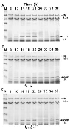
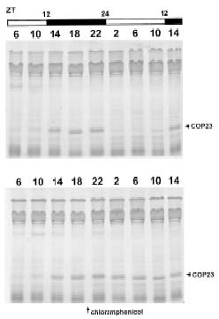
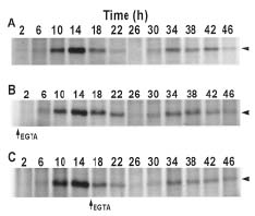
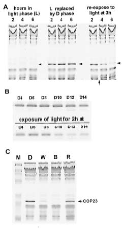
Abstract. The circadian rhythm of COP23, a protein located in the cell membrane of Synechococcus RF-1, was regulated by a circadian synthesis rhythm and a circadian degradation rhythm. The circadian synthesis rhythm was controlled mainly at the transcriptional level (Chen et al., 1996). For the circadian degradation rhythm, some factors were responsible for COP23 degradation. The addition of chloramphenicol to the cultures prevented COP23 degradation, so new protein(s) synthesis before the decline of COP23 must be essential for COP23 degradation. When EGTA was added to the Synechococcus RF-1 cultures before COP23 degradation, the decline of COP23 was prevented. Thus, extracellular Ca2+ was required for the circadian degradation of COP23. However, the addition of EGTA did not affect the synthesis rhythm of COP23. Light was also needed for COP23 degradation. Comparing the light spectra, blue light is more effective for COP23 degradation than white or red light. Phosphorylation could not be detected in vivo or in vitro before COP23 degradation. Thus, COP23 degradation seemed not to be attributed to its phosphorylation.

Keywords: Calcium requirement; Circadian degradation; Circadian rhythm; Light effect; Output pathway; Synechococcus RF-1.

Introduction

Circadian rhythms are a widespread phenomenon regulated by biological clocks. They are responsible for the daily rhythmicity found in a wide variety of organisms, including mammals, insects, higher plants, fungi, algae, and cyanobacteria. The circadian system is generally composed of three basic divisions: (a) a circadian oscillator; (b) input pathways of signal transduction for clock setting; and (c) output pathways of signal transduction for the temporal regulation of specific biological processes. Since a great deal of studies have focused on understanding the nature of the "oscillator," remarkable progress has been made regarding its components and regulation (Young, 1998; Dunlap, 1999; Johnson and Golden, 1999; Lowrey et al., 2000). Thus, the molecular mechanisms of "clock oscillators" have been widely studied in humans (Tei et al., 1997; Sun et al., 1997), mice (Antoch et al, 1997; King et al., 1997), Drosophila (Sehgal et al., 1995; Rosbash et al., 1996; Young et al., 1996), Neurospora (Dunlap, 1996; Crosthwaite et al., 1997), and cyanobacteria (Ishiura et al., 1998; Johnson and Golden, 1999).

In unveiling the circadian clock, it is important to identify how environmental cues such as light or temperature entrain the oscillator (input pathway) and how the information from the clock is transduced to regulate rhythmic cellular processes (output pathway) in addition to the regulation mechanism of the oscillator itself. Several research

works have studied input and output pathways (Huang and Grobbelaar, 1995; Liu et al., 1996; Tsinoremas et al., 1996; Anderson and Kay, 1997; Katayama et al., 1999; Kreps et al., 2000). Some of them were investigated at a biochemical level. For example, the effect of different light conditions on the circadian expression of the gene encoding chlorophyll a/b binding protein was studied for the input pathway (Somers et al., 1998; Bognar et al., 1999). In the control mechanism of the output pathway, the rhythm of CO2 metabolism in Bryophyllum (Wilkins, 1992) and that of bioluminescence or nitrate reductase in Gonyaulax (Morse et al., 1989; Morse et al., 1990; Ramalho et al., 1995) were examined. In addition, a molecular mechanism that links the feedback loop of the central circadian oscillator in the suprachiasmatic nucleus in mice to the regulation of a clock-controlled gene has been discovered (Jin et al., 1999).

In our previous studies, a 23 kDa circadian oscillating protein, COP23, located in the cell membrane of Synechococcus RF-1 was cloned and characterized (Chen et al., 1996). A search of the GenBank uncovered no homologous genes. Detection of COP23 in three other cyanobacteria Synechococcus PCC7942, Synechocystis PCC6803, and Cyanothece ATCC51142 by Southern hybridization using COP23 gene as a probe indicated that hybridization bands could only be detected in the genomic DNA of Cyanothece ATCC51142. Both Synechococcus RF-1 and Cyanothece ATCC51142 are nitrogen-fixing unicellular cyanobacteria. Thus, COP23 might exist exclusively in unicellular cyanobacteria of nitrogen fixation.

The exposure of Synechococcus RF-1 to a diurnal light/dark regimen induced not only a circadian synthesis

*Corresponding author. Fax: 886-2-2782-7954; E-mail:tanchi@gate.sinica.edu.tw


Botanical Bulletin of Academia Sinica, Vol. 44, 2002

rhythm, but also a circadian degradation rhythm of COP23. It is evident that COP23 was regulated rhythmically by an "oscillator" (see "Results"). With an active translational rate and a high protein content, COP23 is an advantageous system for studying the circadian output pathway in Synechococcus RF-1. In this paper, factors involved in the regulation of COP23 degradation are investigated.

Materials and Methods

Organism and Cultivation

The axenic culture of N2-fixing unicellular cyanobacterium, Synechococcus RF-1 (PCC 8801), was isolated from a rice field. Cells were cultivated without aeration or shaking in Erlenmeyer flasks containing nitrate-free BG-11 medium (Stanier et al., 1971) supplemented with 10 mM HEPES (N-[2-hydroxyethyl]piperazine-N´-[3-propanesulfonic acid]) buffer, pH 8.0. The cultures were incubated at 28°C under light (about 35 µmol·m-2·s-1) from white fluorescent tubes. They were grown either in continuous light (L/L) or in a 12 h light/12 h dark (L/D) regimen, depending on the requirement of the experiments. Cell concentration was estimated from measurements of optical density made with a photoelectric colorimeter (Klett-Summerson, USA) fitted with a No. 42 blue filter. Culture in log phase with a cell concentration of 3 ×10⁷ cells/ml was used for [35S]methionine labeling or for protein extraction.

Preparation of Protein Extracts by Boiling in Buffer

A 10 ml sample of culture was withdrawn at appropriate times. Cells were collected by centrifugation at 9,000 rpm (Sigma 3MK, Germany) for 5 min. The pelleted cells were washed twice with distilled water by vortexing at maximal speed for 1 min (Vortex Genie 2, USA). The washed cells were collected by centrifugation, resuspended in 0.1 ml of SDS extraction buffer (62.5 mM Tris, pH 6.8; 2% SDS, 10% glycerol), and then transferred to a 1.5 ml screw-top vial. The suspension was held in a boiling water bath for 5 min and immediately cooled in an ice bath for 3 min. The extracted proteins were collected after centrifugation at 13,000 rpm (Heraeus Biofuge fresco, Germany) for 5 min.

Protein Labeling with [35S]methionine

A 1 ml sample of culture was withdrawn at appropriate times. Cells were collected by centrifugation at 12,000 rpm (Kubota KM15200, Japan) for 1 min and then resuspended in 0.2 ml of fresh medium containing 1.85 MBq of [35S]methionine (specific activity 37 TBq·mmol-1). The cell suspension was incubated under the same conditions of light and temperature as the parent cultures. A 1 ml aliquot of 1% casamino acid (acid-hydrolyzed casein purchased from Difco, USA) was added to the cell suspension after 30 min of incubation and then centrifuged. The pelleted cells were resuspended in 0.1 ml of SDS extraction buffer, boiled for 5 min, cooled on ice, and then collected by centrifugation.

Detection of Protein Phosphorylation in Vivo and in Vitro

Eight ml of the cultures entrained by L/D (12/12) cycles were taken for in vivo phosphorylation. Nitrate-free BG11 medium of the cultures were replaced with the same medium without phosphate 2 h before labeling with [32P]orthophosphate. During labeling, cells were collected by centrifugation at 9,000 rpm (Sigma 3 MK, Germany) for 5 min and then resuspended in 0.4 ml of nitrate-free BG11 without phosphate. To the suspensions were added 1.85 MBq of [32P]orthophosphate and followed with incubation as in the parental culture for 30 min. The reaction was stopped by incubating at 60°C for 10 min and then centrifuging at 12,000 rpm (Kubota KM15200, Japan) for 1 min. The pelleted cells were cooled on ice and washed twice with nitrate-free BG11 without phosphate. The cells were collected by centrifugation, and the proteins were extracted with 0.1 ml of SDS extraction buffer by boiling for 5 min.

For in vitro phosphorylation, 13 ml of L/D-entrained Synechococcus RF-1 cultures were collected by centrifugation at 9,000 rpm (Sigma 3MK, Germany) for 5 min and washed with 1 ml of kinase buffer (25 mM Tris, pH 7.6; 2 mM MnCl2, 10 mM MgCl2, 0.1 mM CaCl2). The pelleted cells were resuspended in 0.1 ml of kinase buffer in the presence of 1 mM PMSF (phenylmethyl sulfunyl fluoride). The suspension was added to 0.1 g of sea-sand (0.1-0.3 mm diameter, Merck) and then broken by vibrating (5,000 rpm) three times for 50 sec with a Mini-Beadbeater (Biospec., USA). The homogenized cells were cooled on ice, then the sea-sand and unbroken cells were removed by centrifugation at 2,000 rpm (Kubota KM15200, Japan) for 3 min. The supernatant was collected, and 3.7 MBq of [g-32P]ATP was added. The reaction mixture was incubated at 28°C for 30 min, stopped by adding 100 µl of SDS extraction buffer, and boiled for 5 min. The supernatant was collected by centrifugation at 12,000 rpm (Kubota KM15200, Japan) for 5 min, and then analyzed by 12% SDS-PAGE and autoradiography.

SDS-PAGE and COP23 Identification

The SDS-PAGE was conducted with the Mini-Protean II dual-slab cell apparatus (BioRad, USA). A 12% polyacrylamide gel was used for electrophoresis. The protein bands were detected by staining the gel with Coomassie Blue. For the [35S]methionine-labeled sample, the gel was vacuum-dried at 80°C after electrophoresis, and then a Kodak X-ray film was exposed to it at room temperature. As reported in the previous paper (28), the electrophoretic mobility and amino acid sequence of COP23 have been determined. In this study, partial N-terminal sequence analysis or Western analysis was used, if needed, for the identification of COP23 in the gel.

Results

New Protein Synthesis was Needed Before the Initiation of Rapid COP23 Degradation


Lin et al. — Regulation of circadian protein degradation

As described in our previous papers, when the Synechococcus RF-1 cultures were transferred to continuous light after 12 h L/12 h D entrainment, the membrane protein COP23 exhibited a circadian rhythm in its mRNA level, synthesis rate, and protein content (Chen et al., 1996). As shown in the upper panel of Figure 1, the protein content of COP23 declined greatly during light phase after L/D entrainment. The decline of COP23 in the light phase must have been due to protein degradation rather than protein modification because Western analysis detected no shifted band(s) (data not shown). If chloramphenicol was added to the RF-1 cultures before COP23 decrease (lower panel of Figure 1), the degradation was inhibited. Thus, new protein synthesis must be involved in the sensitivity of promoting COP23 to proteolysis, either the de novo synthesis of a new protease, or essential for protease activity.

Extracellular Ca2+ was Required for COP23 Degradation

As indicated in Figure 1, the protein content of COP23 declined during the light phase and increased in the dark phase in L/D-entrained Synechococcus RF-1 cultures. When the L/D-entrained RF-1 cultures were transferred to continuous illumination (L/L), the rhythm of COP23 persisted (Figure 2A). If EGTA was added to the cultures before the onset of degradation, the decline of COP23 in the

Figure 2. Effects of EGTA addition and Ca2+ replenishment on the circadian rhythm of COP23 content in Synechococcus RF-1. A, The Synechococcus RF-1 cultures were entrained by diurnal L/D cycles and then transferred to continuous light. B, To the cultures as in A were added 2 mM EGTA (final concentration) at 16 h (arrow) under L/L condition. As compared with A, it showed that COP23 was not degraded after EGTA addition. C, To the EGTA pretreated cultures as in B were added 2.5 mM CaCl2 at 20 h (vertical arrowhead) under L/L condition. COP23 was degraded significantly at 26 h under L/L condition as in A. Proteins were extracted at time intervals with SDS extraction buffer. The relative concentration of protein bands in the SDS-PAGE gel was detected by staining with Coomassie Blue. M refers to the protein marker.

Figure 1. New protein synthesis was required for COP23 degradation. Upper panel, The Synechococcus RF-1 cultures were entrained by diurnal L/D (12/12) regimen. The proteins were extracted with SDS extraction buffer at time intervals of 4 h and then analyzed by 12% SDS-PAGE. Lower panel, the proteins in the same L/D-entrained Synechococcus RF-1 cultures were extracted and analyzed as in the upper panel except that chloramphenicol (100 µg/ml) was added at ZT20 h (D8 h). ZT24 (ZT0) represents light on and ZT12 light off in the L/D cycle.


Botanical Bulletin of Academia Sinica, Vol. 44, 2002

light phase (subjective day) was inhibited, preventing the circadian fluctuation of COP23 content (Figure 2B). However, if Ca2+ was supplemented before COP23 degradation, the rapid degradation resumed at the same time as the cultures without EGTA treatment (Figure 2C).

Although EGTA disturbed the circadian rhythm of COP23 content in the Synechococcus RF-1 cultures (Figure 2B), it did not significantly influence its synthesis rhythm. If EGTA was added to the cultures at different time points, the synthesis rhythm of COP23 was not affected through detection of [35S]methionine incorporation (Figure 3). Based on the data of Chen et al. (1991), amino acid uptake also revealed circadian rhythms. The uptake efficiency of the amino acids including methionine was higher during the light phase than during the dark phase after the Synechococcus RF-1 cultures were entrained by L/D cycles. Thus, the higher synthesis rate of COP23 at dark phase is not due to the uptake rhythm of [35S]methionine (Figure 3). The results indicated that the synthesis of COP23 is not feedback inhibited by accumulation of COP23 itself, but that synthesis of COP23 is controlled by an "oscillator," and the "oscillator" remains running even when extracellular Ca2+ is chelated by EGTA. Nevertheless, the results did not exclude the possibility that calcium is required for clock function. Since EGTA mainly effects on the removal of Ca2+ from the incubation medium (Chen et al., 1988). As a consequence, the concentration of cytosolic Ca2+ may still remain high enough for the "oscillator" to function normally for the duration of the experiment after addition of EGTA.

In the experiments of EGTA-treatment, 97 kDa protein showing no rhythmicity was found to decrease immediately after addition of EGTA to the culture (Figure 2B). It was confirmed to be a Ca2+-binding protein by 45Ca-autoradiography (data not shown). Features of this Ca2+-binding protein will be described in future reports.

Light was Essential for COP23 Degradation

The protein COP23 has been shown to accumulate in the dark phase and decrease during the light phase in the L/D-entrained cultures (proteins at early light phase was shown in the left panel of Figure 4A). However, COP23 was not degraded when the L/D-entrained cultures were kept in darkness during the light phase instead of maintaining the original L/D regime (middle panel of Figure 4A). Degradation of COP23 resumed when the cultures kept in the dark were re-exposed to light (right panel of Figure 4A). The results indicated that light was essential for COP23 degradation.

To examine if light effects on COP23 degradation occurred at specific periods such as during the light phase, the L/D-entrained cultures were transferred to continuous darkness at the beginning of the dark phase and pulse-labeled with [35S]methionine at D0 h (beginning of the dark phase) for 1 h. Samples were taken from the labeled cultures at time intervals initiated at D4 h and then illuminated for 2 h. Then the membrane proteins were extracted and analyzed by SDS-PAGE and autoradiography. It appeared that the newly synthesized COP23 did not decline if the cultures were not illuminated (Figure 4B, upper panel); for the samples exposed to light, decreased levels of COP23 occurred only in those cultures taken after D10 h (Figure 4B, lower panel). The times at which degradation was initiated after illumination were correlated to the light phase of entrainment. Thus, light effects on COP23 degradation occurred at specific light phases rather than at dark phases.

Comparing the effects of different light spectra—including white light (from fluorescent lamps), red light (660 nm), and blue light (425 nm)—revealed that blue light is more effective than red light for COP23 degradation (Figure 4C). Blue light was effective on COP23 degradation when the light intensity was lowered to 20 µmol·m-2·s-1; however, red light had no significant effects even when its intensity was raised to 500 µmol·m-2·s-1. White light with spectra mainly covering the blue light range was also effective, but it had less effect on COP23 degradation than blue light.

Phosphorylation Seemed not to be Coupled with COP23 Degradation

The interconversion of proteins by phosphorylation-dephosphorylation may play important roles in the regulation of protein degradation (Goldberg and John, 1976). Some evidence indicates that the cyclic degradation of the PER protein in Drosophila as well as the FRQ protein in Neurospora (Edery et al., 1994; Dembinska et al., 1997; Liu et al., 2000) may be triggered by phosphorylation. Therefore, it was examined whether COP23, like PER protein, was phosphorylated during the degradation

Figure 3. The circadian synthesis of COP23 in Synechococcus RF-1 was not affected by EGTA addition. A, Cultures without EGTA addition (control experiment). B, EGTA (2 mM) was added at 1 h after the cultures were transferred to constant illumination. C, EGTA (2 mM) was added at 16 h after the cultures were transferred to constant illumination. Analysis was started at 2 h after the diurnal L/D-entrained cultures were transferred to continuous light. Protein samples extracted from [35S]methionine-labeled cultures were analyzed as described in Materials and Methods. The COP23 band is indicated by arrowhead.


Lin et al. — Regulation of circadian protein degradation

periods. According to the experiments of [32P]orthophosphate incorporation in vivo or [g-32P]ATP in vitro, phosphorylation of COP23 band was not detected (data not shown).

Discussion

Several papers have reported that the circadian rhythmicity of some physiological processes are modulated by Ca2+ levels. For example, entrainment or resetting of the rhythmic oscillations such as the conidiation rhythm in Neurospora crassa (Nakashima, 1986), the circadian rhythm of cell division in Euglena gracilis (Tamponnet and Edmunds, 1990), circadian leaflet movement in Robinia pseudoacacia (Gomez and Simon, 1995) and in Cassia fasciculate (Roblin and Fleurat-Lessard, 1984) is affected by intracellular Ca2+ levels. In the organisms with neural systems, Ca2+ is involved in intercellular communication, so it was suggested to play a crucial role in clock entrainment (Geutz and Block, 1994). In plants, Ca2+ is recognized as essential for the signal transduction of phytochrome (Bowler et al., 1994). In the transgenic tobacco and Arabidopsis transformed with Ca2+ -sensitive apoaequorin, the rhythmic luminescence of aequorin, is regulated by cytosolic and chloroplastic Ca2+ levels (Johnson et al., 1995). Thus, Ca2+ might play important roles in the circadian input pathway and then affect the phase-resetting of the clock by light (Neuhaus et al., 1993). However, the function of Ca2+ at the molecular level has not been fully identified, and no evidence proves Ca2+ to be an intrinsic component of the clock "oscillator." Circadian rhythms have been identified in a few prokaryotes (Huang and Grobbelaar, 1995; Johnson and Golden, 1999), and some of them may also be regulated by Ca2+ level. For instance, the circadian nitrogen-fixing activity (Chen et al., 1988) and dark respiration rate (Chen et al., 1989) in Synechococcus RF-1 are both regulated by extracellular Ca2+ levels, as is the circadian degradation of COP23. Therefore, Ca2+ is involved, at least, in the regulation of some circadian output pathways in Synechococcus RF-1 although no papers concerning the influence of intracellular Ca2+ over circadian rhythm in prokaryotes have yet appeared.

The mechanism of light effects on the clock entrainment has been intensely studied, and some reports were concerned with the blue light effects. In Neurospora, the expression of the wc-1 gene is transcriptionally induced by blue light (Ballario et al., 1996; Froehlich et al., 2002). In addition, blue light also triggers the turnover of the TIM protein in Drosophila (Hunter-Ensor et al., 1996; Myers et al., 1996; Zeng et al., 1996; Emery et al., 1998; Stanewsky et al., 1998). In this paper, degradation of COP23 is also regulated by blue light. It is interesting to see that the stabilities of TIM, an intrinsic component of the "oscillator" in Drosophila, and of COP23, the "oscillator"-controlled protein in Synechococcus RF-1, are both significantly affected by blue light. When fruit flies were illuminated at various times in the middle of the night, light caused TIM to disappear prematurely in all cases (Hunter-Ensor et al., 1996). As a consequence, exposure of Drosophila to con

Figure 4. COP23 degradation was light-dependent. A, Light effects on COP23 degradation. Content of COP23 in the diurnal L/D-entrained Synechococcus RF-1 cultures were analyzed at 2, 4, and 6 h, respectively, during light phase (left); the same time points as left panel but the light phase was replaced by dark condition (middle); the cultures after light to dark replacement were re-exposed to light at the time-point (3 h) indicated by an arrow (right). The COP23 band is indicated by arrowhead. B, Effects of light on COP23 degradation at different time points. Upper panel: The L/D-entrained Synechococcus RF-1 cultures were transferred to continuous darkness at the beginning of dark phase. The cultures were pulse-labeled with [35S] methionine at D0 h for 1 h. Samples were taken at 2 h intervals initiated at D4 h. COP23 was assayed by SDS-PAGE and detected by autoradiography. Lower panel: The procedures were the same as upper panel except that the cultures taken at time intervals were exposed to light for 2 h. C, Spectrum effect on COP23 degradation. Content of COP23 was analyzed at 2 h after the light phase of L/D-entrained cultures was replaced by dark condition (D); illuminated by white light from fluorescent lamp (W, 50 µmol·m-2·s-1); by blue light (B, 50 µmol·m-2·s-1); or by red light (R, 50 µmol·m-2·s-1). When the culture was treated with blue or red light, bandpass filter (425BP70 or 660BP70, Omega Optical Inc., VT, USA) was mounted in front of Kodak slide projector to provide light of specified wavelength.


Botanical Bulletin of Academia Sinica, Vol. 44, 2002

stant daylight causes arrhythmicity. However, in Synechococcus RF-1, the rhythm of COP23 persisted under L/L after being entrained by diurnal L/D cycles. As shown in Figure 4B, the light sensitivity of COP23 was effectively limited to the late dark phase as well as the light phase. Therefore, the regulation mechanisms of blue light over both systems could be different. TIM is ubiquitinated in response to light in fruit flies, and so its degradation is suggested to be through a ubiquitin-proteasome mechanism (Naidoo et al., 1999). Nevertheless, it is believed that the ubiquitin-proteasome system is not present in the prokaryotes, and COP23 is a membrane-located protein in the cyanobacterium Synechococcus RF-1, so it is unlikely to be involved in the ubiquitin-proteasome system as in eukaryotes for its degradation. Our preliminary data (not shown) indicated that a light-dependent protein modification system specific to COP23 as well as a rhythmic protease activity occurred in the early light phase are present in the membrane fraction of Synechococcus RF-1. Therefore, it is suggested that the circadian degradation of COP23 is controlled by a circadian protease activity coupled with the light-modulated modification of COP23.

Although the degradation of COP23 was light dependent, it did not mean that COP23 would be degraded if the RF-1 culture was exposed to light. As indicated in Figure 2A, the rhythm of COP23 content persisted under L/L after entrained by diurnal L/D cycles. In our experiments, the light sensitivity of COP23 degradation was effective only when COP23 was programmed to be degraded by the circadian clock (Figure 4B). Therefore, the degradation of COP23 was principally regulated by an "oscillator" and sub-regulated by light.

Conditions in the natural environment, including illumination and temperature, do not always fluctuate uniformly. For the adaptation of an orgamism to variable environments, the "oscillator" offers the capacity for the clock to be reset by daily environmental cues. However, this resetting process takes at least one diurnal cycle. As a consequence, the "oscillator" may not respond as quickly as the daily variation in the environment. Therefore, a circadian output pathway which is principally rather than absolutely regulated by an "oscillator" better allows an organism to adapt to the fluctuation of natural environments, especially the rhythmic proteins which may play major roles in an organism.

Acknowledgements. This work was funded by the Academia Sinica and the National Science Council of the Republic of China.

Literature Cited

Anderson, S.L. and S.A. Kay. 1997. Phototransduction and circadian clock pathways regulating gene transcription in higher plants. Adv. Genet. 35: 1-34.

Antoch, M.P., E.J. Song, A.M. Chang, M.H. Vitaterna, Y. Zhao, L.D. Wilsbacher, A.M. Sangoram, D.P. King, L.H. Pinto, and J.S. Takahashi. 1997. Functional identification of the mouse circadian Clock gene by transgenic BAC rescue. Cell 89: 655-667.

Ballario, P., P. Vittorioso, A. Magrelli, C. Talora, A. Cabibbo, and G. Macino. 1996. White collar-1, a central regulator of blue light responses in Neurospora, is a zinc finger protein. EMBO J. 15: 1650-1657.

Bognar, L.K., A. Hall, E. Adam, S.C. Thain, F. Nagy, and A.J. 1999. Millar, The circadian clock controls the expression pattern of the circadian input photoreceptor, phytochrome B. Proc. Natl. Acad. Sci. USA 96: 14652-14657.

Bowler, G., G. Neuhaus, H. Yamagata, and N.-H. Chua. 1994. Cyclic GMP and calcium mediate phytochrome Phototransduction. Cell 77: 73-81.

Chen, H.-M., C.-Y. Chien, and T.-C. Huang. 1996. Regulation and molecular structure of a circadian oscillating protein located in the cell membrane of the prokaryote Synechococcus RF-1. Planta 199: 520-527.

Chen, T.-H., T.-C. Huang, and T.-J. Chow. 1989. Calcium is required for the increase of dark respiration during diurnal nitrogen fixation by Synechococcus RF-1. Plant Sci. 60: 196-198.

Chen, T.-H., T.-C. Huang, and T.-J. Chow. 1988. Calcium requirement in nitrogen fixation in the cyanobacterium Synechococcus RF-1. Planta 173: 253-256.

Chen, T.-H., T.-L. Chen, L.-M. Hung, and T.-C. Huang. 1991. Circadian rhythm of amino acid uptake by Synechococcus RF-1. Plant Physiol. 97: 55-59.

Crosthwaite, S.K., J.C. Dunlap, and J.J. Loros. 1997. Neurospora wc-1 and wc-2: transcription, photoresponses, and the origins of circadian rhythmicity. Science 276: 763-769.

Dembinska, M.E., R. Stanewsky, J.C. Hall, and M. Rosbash. 1997. Circadian cycling of a period-b-galactosidase fusion protein in Drosophila: evidence for cyclical degradation. J. Biol. Rhyth. 12: 157-172.

Dunlap, J.C. 1996. Genetics and molecular analysis of circadian rhythms. Annu. Rev. Genet. 30: 579-601.

Dunlap, J.C. 1999. Molecular bases for circadian clocks. Cell 96: 271-290.

Edery, I., L.J. Zwiebel, M.E. Dembinska, and M Rosbash. 1994. Temporal phosphorylation of the Drosophila period protein. Proc. Natl. Acad. Sci. USA 91: 2260-2264.

Emery, P., W.V. So, M. Kaneko, J.C. Hall, and M. Rosbash. 1998. CRY, a Drosophila clock and light-regulated cryptochrome, is a major contributor to circadian rhythm resetting and photosensitivity. Cell 95: 669-679.

Froehlich, A.C., Y. Liu, J.J. Loros, and J.C. Dunlap. 2002. White Collar-1, a circadian blue light photoreceptor, binding to the frequency promoter. Science 297: 815-819.

Geutz, M.E. and G.D. Block. 1994. Intracellular free calcium in the entrainment pathway of molluscan circadian pacemakers. Neurosci. Biobehav. Rev. 18: 555-561.

Goldberg, A.L. and A.C.S. John. 1976. Intracellular protein degradation in mammalian and bacterial cells. Annu. Rev. Biochem. 45: 747-803.

Gomez, L.A. and E. Simon. 1995. Circadian rhythm of Robinia pseudoacacia leaflet movement-role of calcium and phytochrome. Photochem. Photobiol. 61: 210-215.

Huang, T.-C. and N. Grobbelaar. 1995. The circadian clock in the prokaryote Synechococcus RF-1. Microbiology 141: 535-540.

Hunter-Ensor, M., A. Ousley, and A. Sehgal. 1996. Regulation of the Drosophila protein TIMELESS suggests a mecha


Lin et al. — Regulation of circadian protein degradation

nism for resetting the circadian clock by light. Cell 84: 677-685.

Ishiura, M., S. Kutsuna, S. Aoki, H. Iwasaki, C.R. Andersson, A. Tanabe, S.S. Golden, C.H. Johnson, and T. Kondo. 1998. Expression of a gene cluster kaiABC as a circadian feedback process in cyanobacteria. Science 281: 1519-1523.

Jin, X., L.P. Shearman, D.R. Weaver, M.J. Zylka, G.J. de Vries, and S.M. Reppert. 1999. A molecular mechanism regulating rhythmic output from the suprachiasmatic circadian clock. Cell 96: 57-68.

Johnson, C.H., M.R. Knight, T. Kondo, P. Masson, J. Sedbrook, A. Haley, and A. Trewavas. 1995. Circadian oscillations of cytosolic and chloroplastic free calcium in plants. Science 269: 1863-1865.

Johnson, C.H. and S.S. Golden. 1999. Circadian programs in cyanobacteria: adaptiveness and mechanism. Annu. Rev. Microbiol. 53: 389-409.

Katayama, M., N.F. Tsinoremas, T. Kondo, and S.S. Golden. 1999. cpmA, a gene involved in an output pathway of the cyanobacterial circadian system. J. Bacteriol. 181: 3516-3524.

King, D.P., Y. Zhao, A.M. Sangoram, L.D. Wilsbacher, M. Tanaka, M.P. Antoch, T.D. Steeves, M.H. Vitaterna, J.M. Kornhauser, P.L. Lowrey, F.W. Turek, and J.S. Takahashi. 1997. Positional cloning of the mouse circadian CLOCK gene. Cell 89: 641-653.

Kreps, J.A., T. Muramatsu, M. Furuya, and S.A. Kay. 2000. Fluorescent differential display identifies circadian clock-regulated genes in Arabidopsis thaliana. J. Biol. Rhyth. 15: 208-217.

Liu, Y., N.F. Tsinoremas, S.S. Golden, T. Kondo, and C.H. Johnson. 1996. Circadian expression of genes involved in the purine biosynthetic pathway of the cyanobacterium Synechococcus sp. strain PCC7942. Mol. Microbiol. 20: 1071-1081.

Liu, Y., J. Loros, and J.C. Dunlap. 2000. Phosphorylation of the Neurospora clock protein FREQUENCY determines its degradation rate and strongly influences the period length of the circadian clock. Proc. Natl. Acad. Sci. USA 97: 234-239.

Lowrey, P.L., K. Shimomura, M.P. Antoch, S. Yamazaki, P.D. Zemenides, M.R. Ralph, M. Menaker, and J.S. Takahashi. 2000. Positional syntenic cloning and functional characterization of the mammalian circadian mutation tau. Science 288: 483-491.

Morse, D., P. Milos, E. Roux, and J.W. Hastings. 1989. Circadian regulation of the synthesis of substrate binding protein in the Gonyaulax bioluminescent system involves translational control. Proc. Natl. Acad. Sci. USA 86: 172-176.

Morse, D.S., L. Fritz, and J.W. Hastings. 1990. What is the clock? Translational regulation of circadian bioluminescence. Trends Biochem. Sci. 15: 262-265.

Myers, M.P., K. Wager-Smith, A. Rothenfluh-Hilfiker, and M.W. Young. 1996. Light- induced degradation of TIMELESS and entrainment of the Drosophila circadian clock. Science 271: 1736-1740.

Naidoo, N., W. Song, M. Hunter-Ensor, and A. Sehgal. 1999. A role for the proteasome in the light response of the timeless clock protein. Science 285: 1737-1741.

Nakashima, H. 1986. Phase-shifting of the conidiation rhythm in Neurospora crassa by calmodulin antagonists. J. Biol. Rhyth. 1: 163-169.

Neuhaus, G., C. Bowler, R. Kern, and N.-H. Chua. 1993. Calcium/calmodulin-dependent and independent phytochrome signal transduction pathways. Cell 73: 937-952.

Ramalho, C.B., J.W. Hastings, and P. Colepicolo. 1995. Circadian oscillation of nitrate reductase activity in Gonyaulax polyedra is due to changes in cellular protein levels. Plant Physiol. 107: 225-231.

Roblin, G. and P.C. Fleurat-Lessard. 1984. A possible mode of calcium involvement in dark- and light-induced leaflet movements in Cassia fasciculata Michx. Plant Cell Physiol. 25: 1495-1499.

Rosbash, M., R. Allada, M. Dembinska, W.O. Guo, M. Le, S. Marrus, Z. Qian, J. Rutila, J. Yaglom, and H. Zeng. 1996. A Drosophila circadian clock. Cold Spring Harbor Symp. Quant. Biol. 61: 265-278.

Sehgal, A., A. Rothenfluh-Hilfiker, M. Hunter-Ensor, Y. Chen, M.P. Myers, and M.W. Young. 1995. Rhythmic expression of timeless: a basis for promoting circadian cycles in period gene autoregulation. Science 270: 808-810.

Somers, D.E., P.F. Devlin, and S.A. Kay. 1998. Phytochromes and cryptochromes in the entrainment of the Arabidopsis circadian clock. Science 282: 1488-1490.

Stanewsky, R., M. Kaneko, P. Emery, B. Beretta, K. Wager-Smith, S.A. Kay, M. Rosbash, and J.C. Hall. 1998. The cryb mutation identifies cryptochrome as a circadian photoreceptor in Drosophila. Cell 95: 681-692.

Stanier, R.Y., R. Kunisawa, M. Mandel, and G. Cohen-Bazire. 1971. Purification and properties of unicellular blue-green algae (order Chroococcales). Bacteriol. Rev. 35: 171-205.

Sun, Z.S., U. Albrecht, O. Zhuchenko, J. Bailey, G. Eichele, and C.C. Lee. 1997. RIGUI, a putative mammalian ortholog of the Drosophila period gene. Cell 90: 1003-1011.

Tamponnet, C. and L.N.Jr. Edmunds. 1990. Entrainment and phase-shifting of the circadian rhythm of cell division by calcium in synchronous cultures of the wild-type Z strain and of the ZC achlorophyllous mutant of Euglena gracilis. Plant Physiol. 93: 425-431.

Tei, H., H. Okamura, Y. Shigeyoshi, C. Fukuhara, R. Ozawa, M. Hirose, and Y. Sakaki. 1997. Circadian oscillation of a mammalian homologue of the Drosophila period gene. Nature 389: 512-516.

Tsinoremas, N.F., M. Ishiura, T. Kondo, C.R. Andersson, K. Tanaka, H. Takahashi, C.H. Johnson, and S.S. Golden. 1996. A sigma factor that modifies the circadian expression of a subset of genes in cyanobacteria. EMBO J. 15: 2488-2495.

Wilkins, M.B. 1992. Circadian rhythms: their origin and control. New Phytol. 121: 347-375.

Young, M.W. 1998. The molecular control of circadian behavioral rhythms and their entrainment in Drosophila. Annu. Rev. Biochem. 67: 135-152.

Young, M.W., K. Wager-Smith, L. Vosshall, L. Saez, and M.P. Myers. 1996. Molecular anatomy of a light-sensitive circadian pacemaker in Drosophila. Cold Spring Harbor Symp. Quant. Biol. 61: 279-284.

Zeng, H., Z. Qian, M.P. Myers, and M. Rosbash. 1996. A light-entrainment mechanism for the Drosophila circadian clock. Nature 380: 129-135.


Botanical Bulletin of Academia Sinica, Vol. 44, 2002