Bot. Bull. Acad. Sin. (2003) 44: 253-256
Chang Tubeufia and Acanthostigma
Tubeufia dactylariae sp. nov. and Acanthostigma scopulum, a new record of Taiwan
H.S. Chang
Institute of Botany, Academia Sinica, Taipei, Taiwan 115, Republic of China
(Received January 28, 2002; Accepted December 2, 2002)
Abstract. A holomorphic fungus connected with a peculiar anamorph resembling the form-genus Dactylaria was discovered and described as a new species of the genus Tubeufia. In addition, a new recorded ascomycete Acanthostigma scopulum was also illustrated and described.
Keywords: Acanthostigma; New record; New species; Taiwan; Tubeufia.
Two interesting ascomycetous fungi were found during our survey on Taiwan freshwater microfungi. One is a new species of Tubeufia, whereas the other, Acanthostigma scopulum, is a new record of Taiwan. They are described and illustrated in this paper.
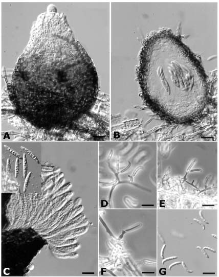
Tubeufia dactylariae H. S. Chang sp. nov. (Figure 1, A-H)
Pseudothecia superficialia, solitaria vel aggregata, globosa vel obovata, brunnea vel atrobrunnea, laevia, carnosa, 100-120 µm diam × 150-200 µm alta. Ostiola leviter papillata. Asci bitunicati, clavati, 80-100 × 9-12 µm. octospori. Ascosporae fusiformes, 32-36 × 6-7 µm, hyalinae vel dilute brunneae, laeves, 7-8 septatae, rectae vel leviter curvatae.
Colonies olivaceous to dark brown on V-8 juice agar, hyphae immersed, smooth. Pseudothecia superficial, solitary to aggregated, clavate to pyriform, brown, smooth, fleshy, with black superficial hyphae growing into the substrate from the base of pseudothecia, 150-200 × 100-120 µm; peridium composed of 4 to 5 layers of pseudoparenchymatous cells. Ostioles slightly papillate. Asci bitunicate, clavate to broadly cylindrical, 8-spored, 80-100 × 9-12 µm. Ascospores fusiform, 32-36 × 6-7 µm, hyaline to pale brown, smooth, 7-8 septate, straight or slightly curved, not constricted at septa.
Dactylaria-like Anamorph. This fungus formed conidial state on V-8 juice agar or on autoclaved corn leaf sections (3 × 2 cm) placed on Sach's medium. Conidiophores micronematous, mononematous, arising laterally from undifferentiated hyphae, stout, simple or branched, hyaline. Conidiogenous cells mono- or polyblastic, hyaline, thin walled. Conidia hyaline, smooth and thin walled, cylindrical, upper part bent and tapered towards the rounded end, 3-4 septate, 18-26 × 4-5 µm.
Specimen examined. TAIWAN. TAIPEI COUNTY: Wulai, on unidentified decayed twig, Jan 21, 1992, H. S. Chang, WL0121-92 (HOLOTYPE, HAST).
Notes. Instead of forming helicosporous conidia as most of the species in the genus Tubeufia (Barr, 1980; Sivanesan, 1983), this fungus forms an undescribed mitosporic species closely similar to species of Dactylaria in its conidiogenous cell and conidiophore forms. However, its conidiophores and conidiogenous cells are also very similar to those of the genus Helicomyces, i.e., formed as short, lateral branches of the repent mycelium, except that the conidia are not helical but cylindrical with a rounded base and a bend towards the tip. The conidial states of the genus Tubeufia, in most cases, belong to the form-genera Helicosporium and Helicoma. However, a Monodictys anamorph has also been reported to be associated with T. amazonensis (Samuels et al., 1979). Taphrophila cornucapreolli, a fungus closely related to Tubeufia, was also demonstrated in culture to be connected with a Mirandina anamorph highly similar to Dactylaria (Scheuer, 1991).
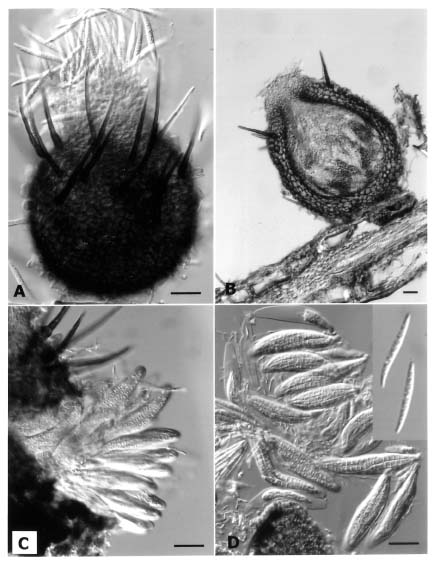
Acanthostigma scopulum (Cooke & Peck) Peck, Bull. New York State Mus. 1: 22. 1887. (Figure 2, A-D)
Sphaeria scopula Cooke & Peck, Ann. Rep. New York State Mus. 32: 51. 1880.
Lasiosphaeria scopula (Cook & Peck) Sacc., Syll. Fung. 9: 852. 1891.
Tubeufia scopula (Cooke & Peck) M. E. Barr, Mycotaxon 12: 1964. 1980.
Pseudothecia solitary, superficial, globose to subglobose, with aseptate or unisepatate setae mostly distributed on upper half of ascomata, 130-180 µm high × 100-120 µm diam. Peridium thick, composed of up to 5 layers of pseudoparechymatous cells. Ostioles papillate. Asci bitunicate, 8-spored, broadly cylindrical, 70-80 × 12-14 µm. Cellular pseudoparaphyses numerous among asci,
E-mail: bododo@ccvax.sinica.edu.tw