Bot. Bull. Acad. Sin. (2003) 44: 275-284

Ming et al. — Liquorice cell tolerance of water stress

Application of external calcium in improving the PEG-induced water stress tolerance in liquorice cells

Ming Li1, Gen-Xuan Wang1,2,*, and Jiou-Sheng Lin1

1State Key Laboratory of Arid Agroecology, Lanzhou University, 730000, Lanzhou, P.R. China

2School of Biology, Zhejiang University, 310027, Hangzhou, P.R. China

(Received October 30, 2002; Accepted May 14, 2003)

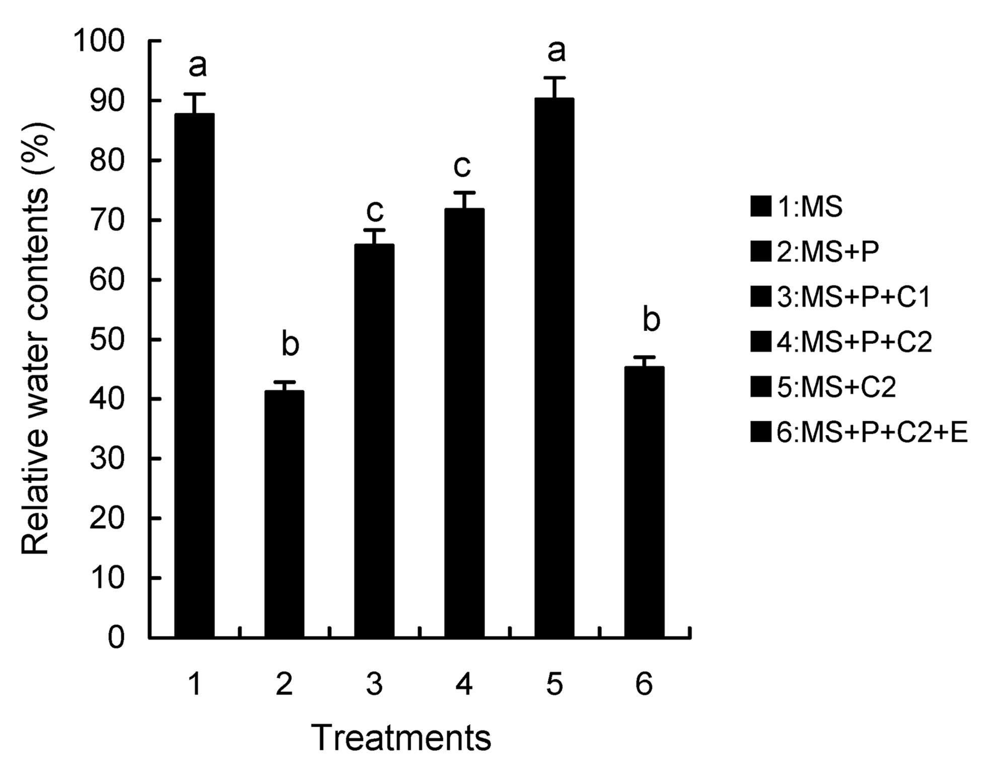
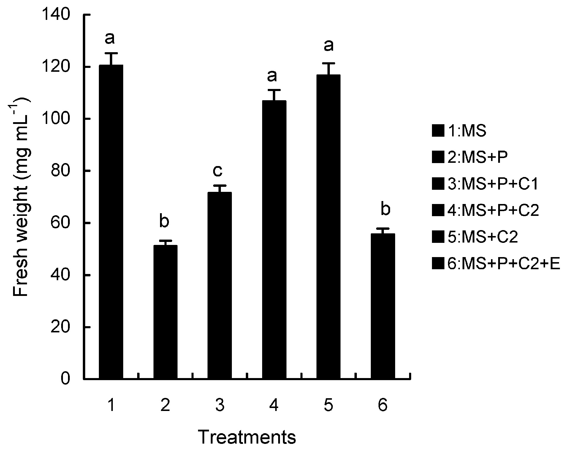
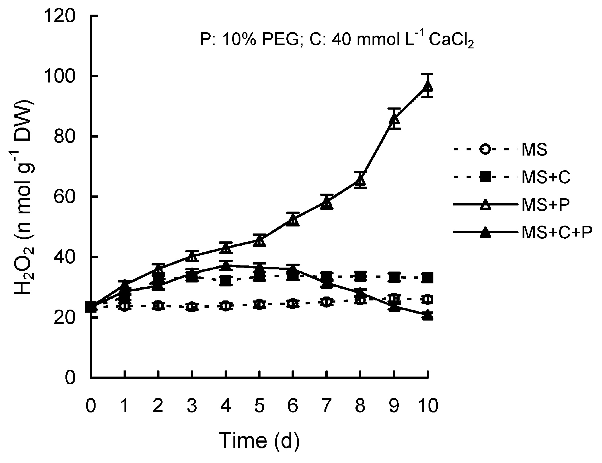
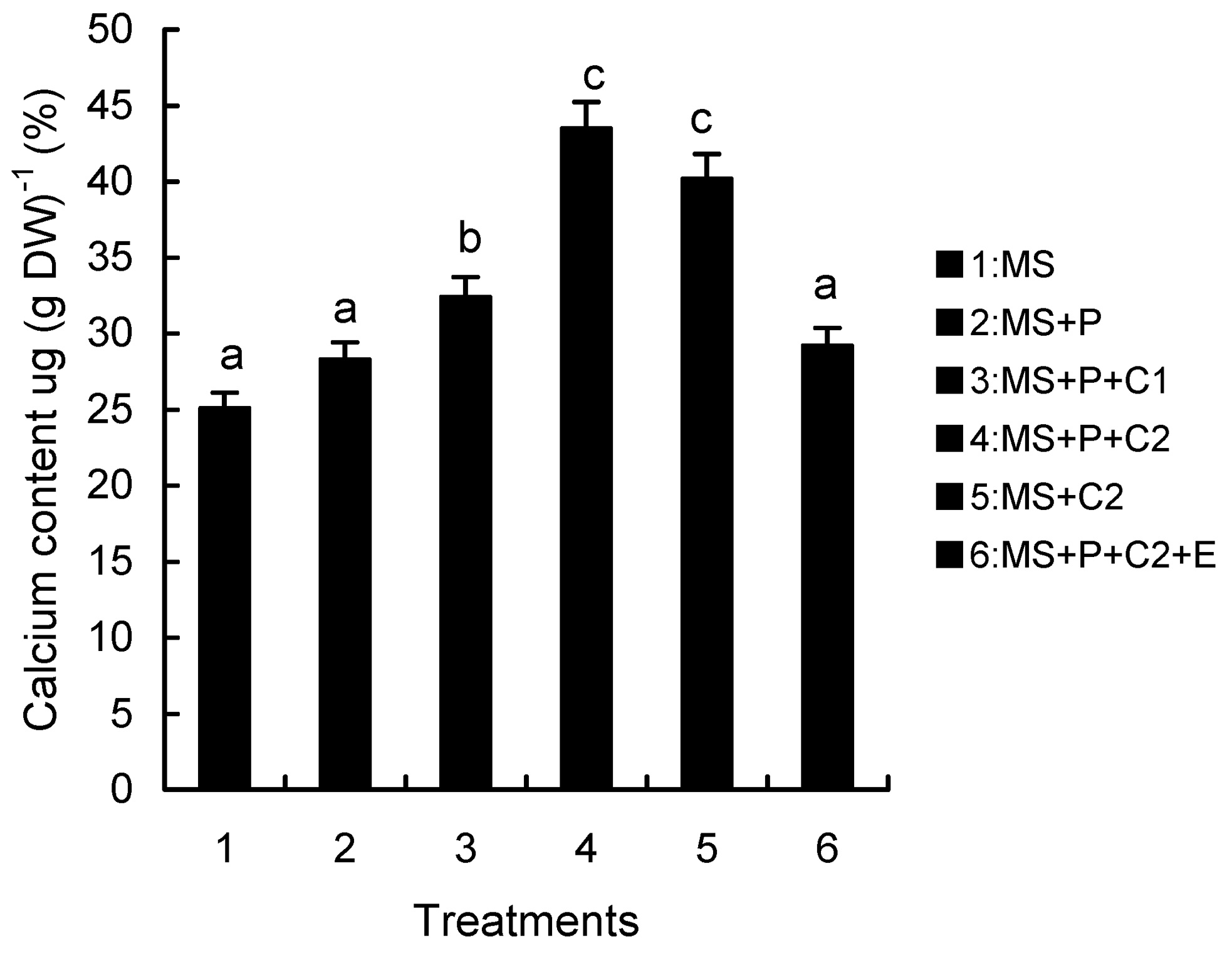
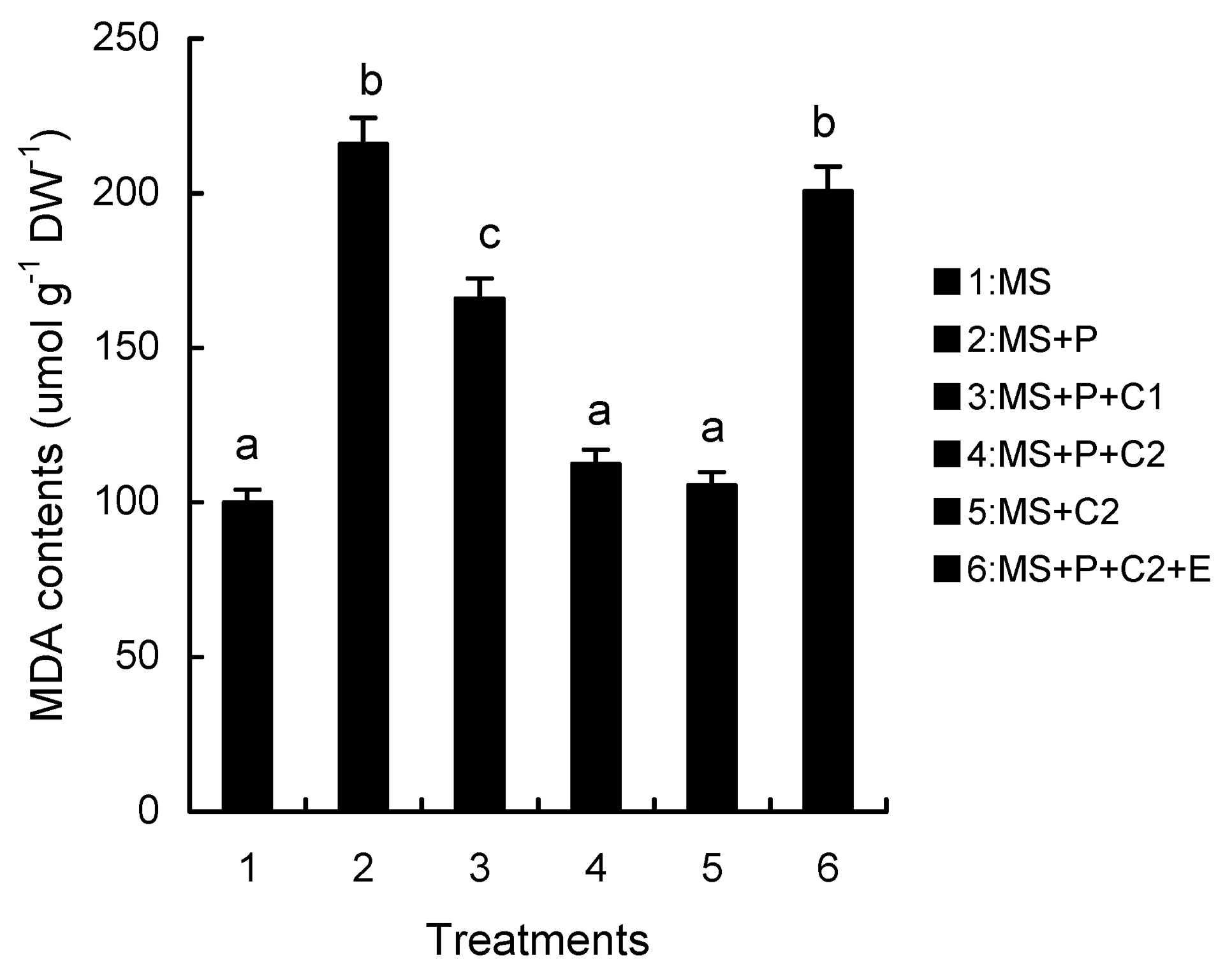
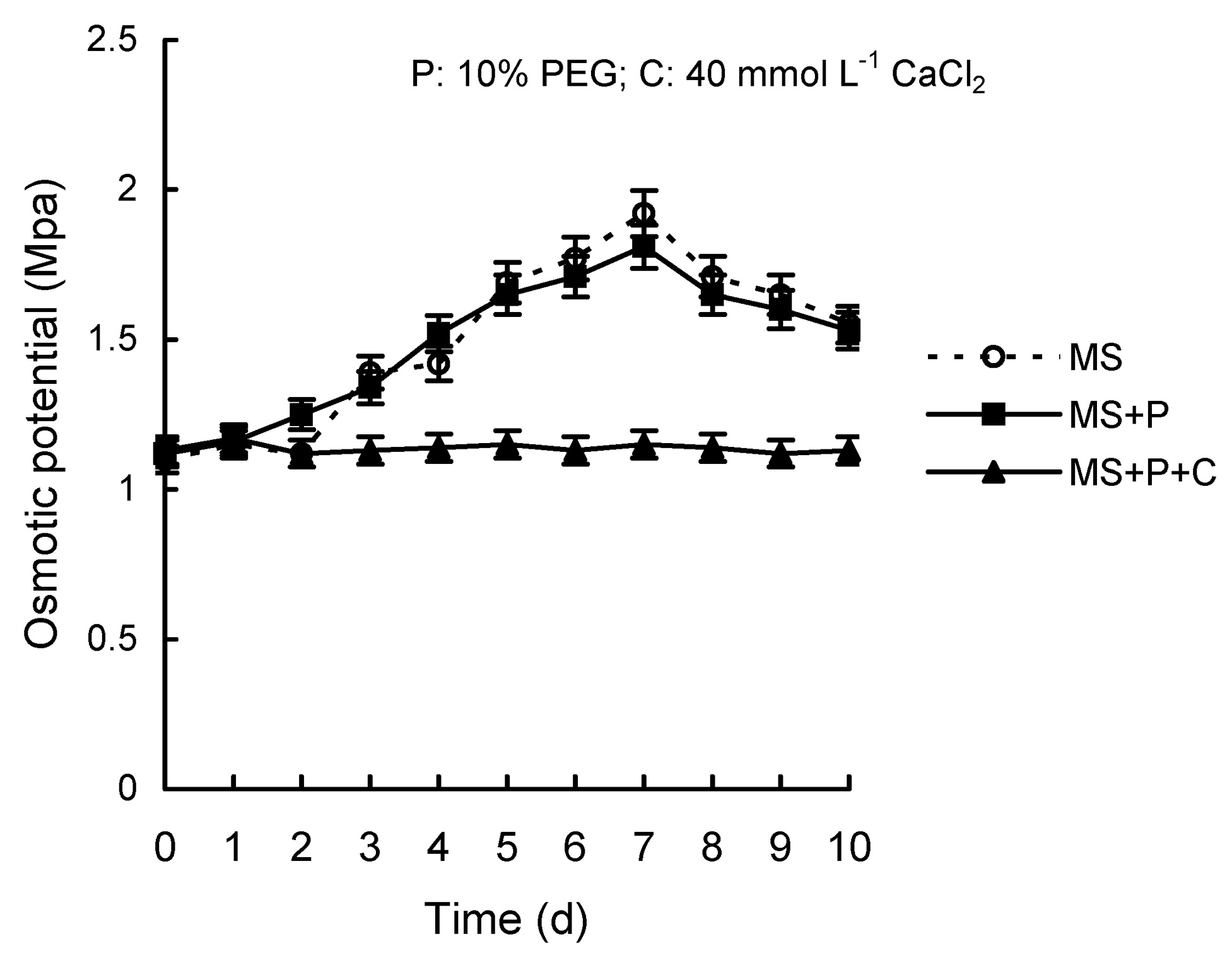
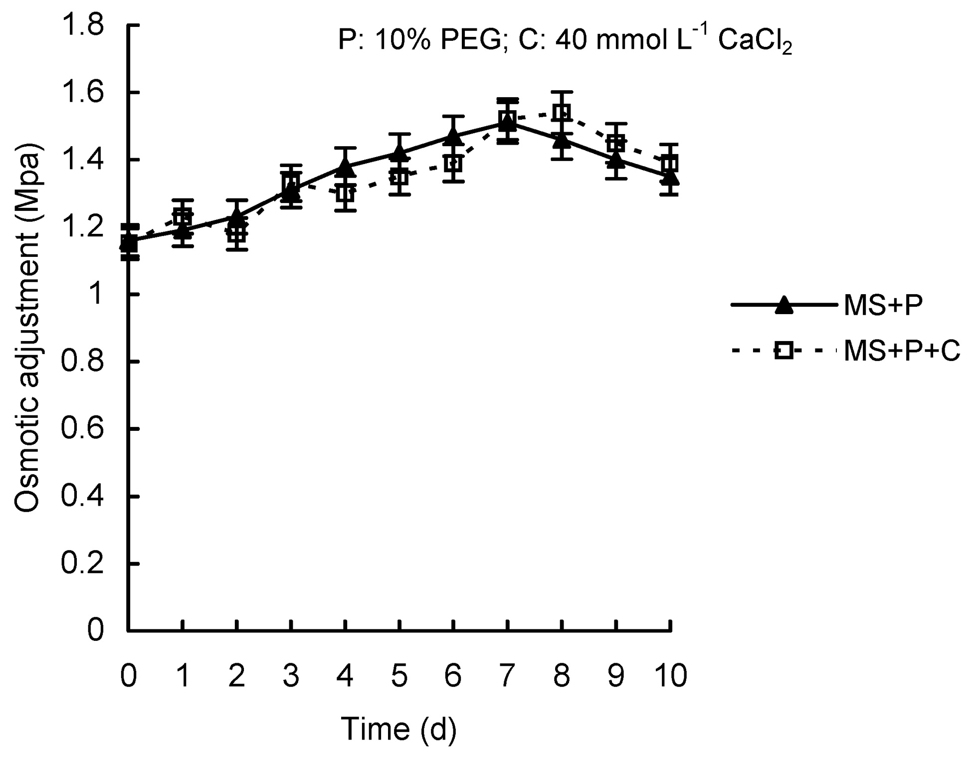
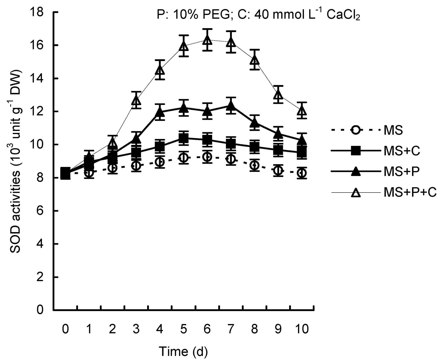
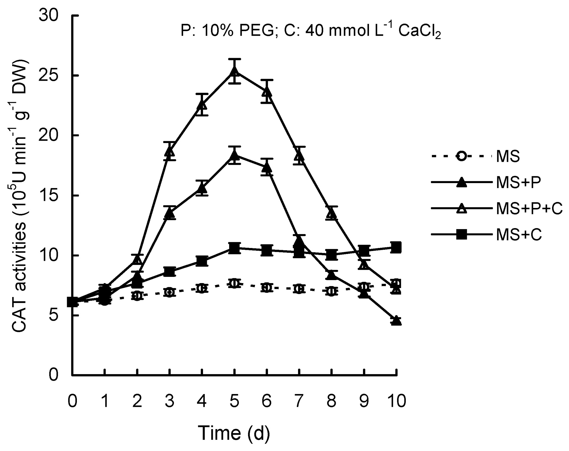
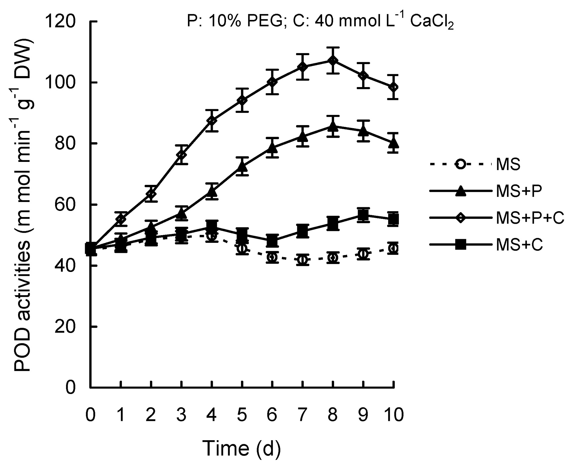
Abstract. Calcium (Ca2+) may be involved in plant tolerance to water stress by regulating antioxidant metabolism. This study was designed to examine whether external Ca2+ treatment would improve drought tolerance in liquorice cells. The results showed that water stressed treatment induced by 10% PEG could reduce significantly the FW and RWC of liquorice cells, but external Ca2+ treatment considerably increased the two factors after 10-days stress. In addition, lesser amounts of MDA and H2O2 accumulated in Ca2+-treated cells than in untreated cells, and the activities of CAT, SOD and POD in Ca2+-treated cells were higher than in untreated cells during the stress period. The measured parameters treated by 40 mmol L-1CaCl2 were higher than those treated by 10 mmol L-1CaCl2. The changes in CAT, SOD and POD activities under stressed conditions were significantly larger than those in non-stressed conditions. Under stressed conditions, the trend of SOD activities was similar to that of CAT, and the activity of CAT was larger than that of SOD. CAT activity changed in relation to H2O2 content. It was indicated that water stress induced oxidative stress in liquorice cells, and application of external calcium (40 mmol L-1) significantly improved water stress tolerance in those cells. In addition, the measured parameters were different between Ca2+-treated cells under stressed and non-stressed conditions, and it is possible that calcium signals were different coming from different stimulations. The investigations also showed that the effect of external Ca2+ on the measured parameters was not due to the regulation of osmotic potential and osmotic adjustment in liquorice cells. The mechanism that allowed extracellular Ca2+ to improve adaptation of liquorice cells to drought was mediated by mitigating oxidative stress.

Keywords: Aatioxidant enzyme; Ca2+; Glycyrrhiza uralensis; Polyethylene glycol; Water stress.

Abbreviations: CAT, catalase; FW, fresh weight; MDA, malondialdehyde; PEG, polyethylene glycol; POD, peroxidase; RWC, relative water content; SOD, superoxide dismutase.

Introduction

Liquorice (Glycyrrhiza uralensis Fisch) is a traditional medicinal plant in China. The species has significant abilities to withstand adverse environmental stresses such as drought, cold, and hot. (Qiou et al., 2000; Wang et al., 2001). In addition, liquorice usually grows in rich-calcium soil in arid or semi-arid areas (Qiou et al., 2000; Zhang et al., 2000; Wang et al., 2001), We are thus interested in the correlation between external calcium and water stress tolerance in liquorice.

Drought is a common and serious problem to plants in arid or semi-arid areas. Plants have developed different morphological, physiological, and biochemical mechanisms to withstand drought stress. Evidence from different lines of research suggests that drought stress can induce oxidative stress in plants (Dhindsa and Matowe, 1981; Mukherjee and Choudhwri, 1981; De Lucad'Oro and Trippi,

1987; Trippi et al., 1989). Oxidative stress—resulting from the generation of AOS, such as superoxide (O2), peroxide hydrogen (H2O2) and hydroxyl radicals (OH·)—is detrimental to plant survival under a water stress environment. To counteract the toxicity of AOS, a highly efficient antioxidative defense system, including both nonenzymic (e.g. ascorbate, carotenoids, a-tocopherol) and enzymic constituents (e.g. SOD [EC1.15.1.1], POD [EC 1.11.1.7], CAT [EC 1.11.1.6]), is present in plant cells and plays an important role in defending plants from AOS damage (Elstner, 1982; Smirnoff, 1993). The system is able to catalyze or participate in the elimination of free oxygen radicals and H2O2 from cells (Schaedle and Bassham, 1977; Asada and Takahashi, 1987; Wang et al., 1989). Under a drought environment, the balance between the generation and elimination of AOS in plants becomes damaged, causing an accumulation of AOS. Thus, the cell membrane system is destroyed, and a series of metabolite responses causes turbulence (Wang, 1988; Chen, 1989). Plants have the capacity of regulation balance between the generation and elimination of AOS within the range of plants tolerancing adverse environments. Many studies have

*Corresponding author. Fax: +86-931-8912893; E-mail: Wanggx@lzu.edu.cn


Botanical Bulletin of Academia Sinica, Vol. 44, 2003

shown that drought causes antioxidative enzyme activity changes in plants (Del Lengo et al., 1993; Iturbe-Ormaetxe et al., 1998; Li and Wang, 2002). The phenomena were the exhibition of regulation balance function between AOS and antioxidative system in plants.

It is well known that calcium functions as a regulator of plant cell metabolism (Kauss, 1987; Pietrobon et al., 1990). In addition, it has been noted that calcium could paticipate in the regulating mechanism in plants adjusting to adverse conditions such as high temperature (Cooke et al., 1986), cold injury (Arora and Palta, 1988), drought stress (Bowler and Fluhr, 2000), and salt stress (Cramer et al., 1985). In several plant cell-elicitor systems, some evidence has been obtained that the activation of defense responses depends on the presence of extracellular Ca2+ (Ebel and Cosio, 1994; Gong et al., 1997). Some authors have proposed that Ca2+ induces increases in antioxidant enzyme activities and decreases in MDA content in a plant's response to a water stress environment (Gao et al., 1999; Shu and Fan, 2000). However, in our view, direct cell-level evidence of the relation between external calcium and water stress tolerance in plants is lacking. Also, certain characteristics of liquorice-make it an appropriate experimental material for researching the mechanisms of external calcium to water stress tolerance in plants.

Cells/tissue culture is useful and convenient tool for investigating cell mechanisms. Much research has used cultured cells as a model system to study the cellular responses under various abiotic stresses involving physiological and biochemical changes (Fallon and Phillips, 1989; Leone et al., 1994). Because cultured cells must adapt to water stress conditions through cellular processes, investigations of such cells could help us to elucidate the cellular mechanisms involved in water stress. Cell lines of liquorice, obtained in our laboratory, are capable of growth in liquid medium containing up to 10% PEG. In this study, a water deficit environment was obtained by adding PEG (mol wt 8000) in culture medium. PEG itself did not contribute significantly to the osmotic adjustment (Handa et al., 1982). The cell wall is impermeable to PEG, and PEG does not degrade appreciably and enter the cells.

In this paper we explore further the possible involvement of external calcium in water-stress physiology using the liquorice cell system, again, the effects of external Ca2+ treatment on H2O2 content, antioxidant enzymes as well as osmotic adjustment under water stress. The parameters such as fresh weight (FW), relative water content (RWC), total calcium content, MDA content, H2O2 content, SOD, CAT, POD activities, and osmotic potential in liquorice cells as well as water potential in the medium were measured.

Our objectives were: (1) to characterize whether external calcium participates in the mechanisms of water stress tolerance in liquorice cells, and (2) to characterize the effects of external calcium on antioxidant enzymes activities and H2O2 content in liquorice cells under water stressed conditions. The study also may provide useful information for better understanding the mechanisms of drought tolerance.

Materials and Methods

Plant Material

Seeds of liquorice were obtained from plants that grow in the desert wilderness of MinQin County in Gansu Province, China. The original plant samples were authenticated by the traditional medicine college of Gansu Traditional Chinese Medicine University. The seeds were immersed in 85% H2SO2 for 30 min in order to facilitate germination, followed by rinsing in sterile distilled water for about 10 min. They were then surface sterilized by immersing in 0.1% HgCl2 for 15 min. To produce sterile seedlings, seeds were placed on the surface of MS medium without auxin and supplemented with sucrose 30 g L-1 and Agar 0.8%. The pH was adjusted to 5.8 before the medium was autoclaved, and incubated at 25°C with 9 h darkness and 15 h light of 30 µE·m-2 s-1.

Callus Cultures

The cotyledons were cut about 3 mm off the seedlings and placed on the surface of MS medium containing 3% sucrose and 0.8% agar. Auxins, 2,4-D 1.2 mg L-1, 6-BA 1 mg L-1 and 500 mg L-1 casein were supplemented in the medium. pH was adjusted to 5.8 before the medium was autoclaved and incubated at 25°C with 9 h darkness and 15 h light of 30 µE·m-2 s-1.

Cell Cultures

The suspension cells were derived from the callus tissue. 1 g fresh weight of callus was put in a 250 ml flask, to which was added 50 ml liquid MS culture medium containing 2,4-D 1 mg L-1 and 3% sucrose (pH 5.8). Cells were cultured at 25°C with shaking at 120 rpm on a gyratory shaker in a photoperiod of 15 h at light intensity of 30 µE·m-2 s-1. Cells were subcultured every week with a 4% inoculum until enough cell material was obtained to start them on the stress medium. All cells were less than 6 months old when they were subjected to osmotic stress.

Water Stress, CaCl2 and Calcium Chelating Agent (EGTA) Treatment

PEG (mol wt 8000) was added to the growth medium at concentrations of 10% (w/v) before adjusting the pH of the medium to 5.8. Distributed into each culture tube was 25 ml of medium. The water potential of each medium after autoclaving was -1 Mpa for MS and -4.7 Mpa for that containing 10% PEG, respectively. The suspension-cultured cell of 1 g was transferred to each culture flask containing 25 ml of medium and different concentrations of CaCl2, or CaCl2 + 50 mmol L-1 of calcium chelating agent (EGTA) was supplemented in the growth medium containing 10% PEG. Each treatment was performed five times.

Measurement of H2O2

H2O2 content was measured according to the method of Manuel et al. (1986). The 1 g cells were homogenized with 0.2 g activated charcoal and 12 ml 5% TCA and cen


Ming et al. — Liquorice cell tolerance of water stress

trifuged at 18,000 g for 10 min. The supernatant was filtered through a Millipore filter (0.45 µm) and used for the assay. A 25 µl aliquot was brought to 2 ml with 100 mM potassium phosphate buffer (pH 8.4), and 1 ml of colorimetric reagent was added. This reagent was made by mixing 1:1 (v/v) 0.6 mM 4-(2-pyridylazo) rescorcinol (disodium salt) and 0.6 mM potassium titanium oxalate. The decrease in A508 against distilled H2O was followed up and took the minimal value. Blanks were obtained by adding buffer to 25 µl TCA until it was 2 ml. Content of H2O2 was determined by differences in A508 content between samples and blanks, using 30% H2O2 (10-200 µmol) as a standard.

Measurement of FW and RWC

The cells, which were water stressed and calcium treated for 10 d, were collected by centrifugation at 500 g for 7 min and blotted thoroughly on filter papers, after which their fresh weight was determined. The cells were placed on a sheet of nylon mesh with 300-µm pores under the axenic condition, and cells were floated on water until weights were constant. The cells were blotted dry, and turgid weights were determined. The dry weight was measured after the cells were dried for at least 24 h in the preweighing aluminum lockets at 85 °C. Relative water content was calculated as follows: RWC (%) = (FW_DW / SW_DW)×100%, where FW is the fresh weight of the cells, DW is their dry weight after 85°C for 24 h, and SW is the turgid weight of cells after soaking in water for 4 h at room temperature (approximately 20°C).

Measurement of Enzyme Activities

The frozen cells of 1.0 g (DW) were homogenized in 1 ml of 50 mM sodium phosphate (pH 7.0) that contained 1% PVPP-40. The homogenate was centrifuged at 15,000 g for 30 min. The supernatant was collected and stored at -80°C for further analyses. The activity of SOD was assayed by monitoring its ability to inhibit the photochemical reduction of NBT. Each 3-ml reaction mixture contained 50 mM sodium phosphate (pH 7.8), 13 mM methionine, 75 µM NBT, 2 µM riboflavin, 100 nM EDTA, and 200 µl of the enzyme extract. The production of blue formazan was followed by monitoring the increase in absorbance at 560 nm (Beauchamp and Fridovich, 1971). The activity of CAT was determined by following the consumption of H2O2 (extinction coefficient 39.4 mmol L-1 cm-1) at 240 nm for 2 min. The reaction mixture contained 2 ml of 100 mM sodium phosphate buffer (pH 6.5) and 100 µl of enzyme extract, and the reaction was initiated by adding 10 µl of 30% H2O2. POD activity was based on the determination of guaiacol oxidation (extinction coefficient 26.6 mmol L-1 cm-1) at 470 nm by H2O2. The reaction mixture contained 100 mM potassium phosphate buffer (pH 6.5), 16 mM guaiacol, and 10 µl of 10% H2O2 in a 3 ml volume. The reaction was initiated by adding plant extract and was followed for 10 min.

Lipid peroxidation was measured as MDA determined by the thiobarbituric acid (TBA) reaction. Cells (0.5 g) were homogenized in 4 ml of 0.1% (w/v) trichloroacetic acid

(TCA). The homogenate was centrifuged at at 15,000 g for 20 min. To 500 µl of the aliquot of the supermatant, 500 µl of 20% TCA containing 0.5% (w/v) TBA and 50 µl BHT (butylated hydroxytoluene) (4% w/v in ethanol) were added. The mixture was heated at 95 °C for 30 min and then quickly cooled on ice. The absorbance was measured at 532 nm. The concentration of MDA was calculated using an extinction coefficient of 155 mM-1cm-1.

Measurement of Water Potential, Osmotic Potential, and Calcium Content

Water potential (Yw) and osmotic potential (Ys) were measured using a Wescor dewpoint Microvoltmeter HR-33T and a Wescor thermocouple hygometer sample chamber C-52. The method used to measure Yw and Ys in cells was as described earlier (Brown et al., 1979). For the calcium determination, 1 g dried cell was ashed and the calcium extracted in 5 ml 2 N HCl, which was diluted to 100 ml with distilled H2O before analysis on a Perkin Elmer 303 Atomic Asorption Spectosynthesis at 422.7 nm. Osmotic adjustment was calculated as the difference in osmotic potential at full turgor between control and stressed cells (Blum and Sullivan, 1986; Blum, 1989).

Results

The Effect of External Ca2+ on FW and RWC in Liquorice Cells Under Water Stress Condition

In response to the 10% PEG acclimation treatment, FW and RWC decreased significantly compared with the same parameters in non-stress medias (MS, MS+40 mmol L-1 CaCl2) by the end of 10 days. Application of external calcium (MS+P+10 mmol L-1 CaCl2, MS+P+40 mmol L-1 CaCl2) could elevate greatly FW and RWC of liquorice cells, and the effects of the two concentrations of CaCl2 was different. For 10 mmol L-1 CaCl2 treatment, FW and RWC increased nearly 0.4 and 0.6 fold, respectively, compared with the same parameters in media without external calcium (MS+P); in the case of 40 mmol L-1 CaCl2 treatment, FW and RWC increased approximately 1.1 and 0.7 fold compared with the same parameters in stress medias without external calcium, respectively. In addition, under stress conditions, 40 mmol L-1 CaCl2 treatment could increase FW almost to the level of non-stress conditions (MS, MS+40 mmol L-1 CaCl2). However, FW and RWC of liquorice cells in the stress media containing external calcium and calcium chelating agent (MS+P+40 mmol L-1 CaCl2+50 mmol L-1 EGTA) were reduced to a level similar to that of media without external calcium stress (MS+P). Moreover, under non-stress conditions, external calcium treatment (MS+40 mmol L-1 CaCl2) had no significant difference on FW and RWC in comparison with the same parameters in the MS media (Figures 1 and 2).

The Effect of External Ca2+ on H2O2 Content in Liquorice Cells Under Water Stress Conditions

The trend of H2O2 content was remarkably different be


Botanical Bulletin of Academia Sinica, Vol. 44, 2003

Figure 1. Relative water content as affected by water stress and CaCl2 treatment in liquorice cells. P: 10% PEG; C1: 10 mmol L-1 CaCl2; C2: 40 mmol L-1 CaCl2; E: 50 mmol L-1 EGTA. Vertical bars represent S.E. Each point represents mean ±S.E. (n=5). Different letters indicate significant differences between treatments within each cell line (p<0.01).

Figure 2. Fresh weight as affected by water stress and CaCl2 treatment in liquorice cells. P: 10% PEG; C1: 10 mmol L-1 CaCl2; C2: 40 mmol L-1 CaCl2; E: 50 mmol L-1 EGTA. Vertical bars represent S.E. Each point represents mean ±S.E. (n=5). Different letters indicate significant differences between treatments within each cell line (p<0.01).

tween conditions containing external calcium and those not containing it during the water stress acclimation treatment (Figure 3). H2O2 content increased significantly in stress media containing external calcium (MS+P+40 mmol L-1 CaCl2), but first increased slowly (0-5 days) then declined (5-10 days) in stress media not containing external calcium (MS+P) with the prolonged cultured duration.

Under non-stress conditions, H2O2 content of liquorice cells remained relatively steady with the prolonged cultured duration. In media containing external calcium (MS+40 mmol L-1 CaCl2), H2O2 content was slightly higher than media without external calcium (MS).

The Effect of External Ca2+ Treatment on Total Calcium Content in Liquorice Cells Under Water Stress Condition

Total Ca2+ content increased approximately 0.3 and 0.7 fold in stress media containing 10 mmol L-1 CaCl2 (MS+P+C1) and 40 mmol L-1 CaCl2 (MS+P+C2) compared with non-external calcium media (MS), respectively. In addition, total Ca2+ content in medium containing 10% PEG (MS+P) was slightly higher than in non-external calcium media (MS). 50 mmol L-1 EGTA treatment could greatly influence total calcium content of liquorice cells. The result showed that total Ca2+ content in media containing calcium chelating agent (MS+P+40 mmol L-1 CaCl2+50 mmol L-1 EGTA) decreased approximately to the level of non-EGTA media (MS+P+40 mmol L-1 CaCl2) (Figure 4).

The Effect of External Ca2+ Treatment on Osmotic Potential and Osmotic Adjustment in Liquorice Cells Under Water Stress Condition

Increased trends (0-7 days) were observed in both osmotic potential and osmotic adjustment in liquorice cells

Figure 3. H2O2 content as affected by water stress and CaCl2 treatment in liquorice cells. Vertical bars represent S.E. Each point represents mean ±S.E. (n=5).

during water stress acclimation duration. External Ca2+ treatment (40 mmol L-1 CaCl2) had no significant effects on the two parameters (Figures 5 and 6).

The Effect of External Ca2+ Treatment on MDA Content in Liquorice Cells Under Water Stress Condition

MDA content increased significantly more under stress conditions (MS+P) than under non-stress conditions (MS). Application of external Ca2+ could reduce MDA content considerably compared with non-external Ca2+ stress media. 10 mmol L-1 CaCl2 and 40 mmol L-1 CaCl2 treatment (MS+P+C1, MS+P+C2) reduced MDA content 0.3 and 0.9 fold, respectively, compared with those of non-external


Ming et al. — Liquorice cell tolerance of water stress

Figure 4. Calcium content as affected by water stress and CaCl2 treatment in liquorice cells. P: 10% PEG; C1: 10 mmol L-1 CaCl2; C2: 40 mmol L-1 CaCl2; E: 50 mmol L-1 EGTA. Vertical bars represent S.E. Each point represents mean ±S.E. (n=5). Different letters indicate significant differences between treatments within each cell line (p<0.01).

Figure 7. MDA content as affected by water stress and CaCl2 treatment in liquorice cells. P: 10% PEG; C1: 10 mmol L-1 CaCl2; C2: 40 mmol L-1 CaCl2; E: 50 mmol L-1 EGTA. Vertical bars represent S.E. Each point represents mean ±S.E. (n=5). Different letters indicate significant differences between treatments within each cell line (p<0.01).

calcium stress treatment (MS+P). In media containing calcium chelating agent (MS+P+40 mmol L-1 CaCl2 +50 mmol L-1 EGTA), MDA content increased remarkably, almost to the level of non-external calcium stressed media (MS+P). However, under non-stress conditions, application of external calcium (MS+40 mmol L-1 CaCl2) made no significant difference to MDA content compared with non-external calcium media (MS) (Figure 7).

The Effect of External Ca2+ Treatment on POD, SOD and CAT Activities in Liquorice Cells Under Water Stress Conditions

The SOD and CAT activity trends in liquorice cells were similar in both stress media containing external calcium (MS+P+40 mmol L-1 CaCl2) and containing no external calcium (MS+P) during water stress acclimation period (Figures 8-10). SOD activities increased during the initial period (0-5 days), then maintained a slowing decline (5-10 days) with prolonged stress duration. As with the change of SOD, CAT activities also showed an increased trend within the 0-5 day period and decreased with the subsequent prolonged duration, but the change was larger than with SOD. The activities of POD increased within the period of 0-8 days, and then slowly declined. However, in media containing external calcium stress (MS+P+40 mmol L-1 CaCl2), SOD, CAT, and POD activities were higher than in media without it (MS+P) during cultured duration. Moreover, under non-stress conditions (MS+40 mmol L-1 CaCl2, MS), the activities of POD, SOD and CAT remained

Figure 5. Osmotic potential as affected by water stress and CaCl2 treatment in liquorice cells. Vertical bars represent S.E. Each point represents mean ±S.E (n=5).

¬

Figure 6. Osmotic adjustment as affected by water stress and CaCl2 treatment in liquorice cells. Vertical bars represent S.E. Each point represents mean ±S.E (n=5).


Botanical Bulletin of Academia Sinica, Vol. 44, 2003

Figure 8. SOD activities as affected by water stress and CaCl2 treatment in liquorice cells. Vertical bars represent S.E. Each point represents mean ±S.E (n=5).

Figure 9. CAT activities as affected by water stress and CaCl2 treatment in liquorice cells. Vertical bars represent S.E. Each point represents mean ±S.E (n=5).

relatively steady compared with the same parameters under stress conditions during the culture period, and POD, SOD and CAT of media containing external calcium were slightly higher than the same parameters of media without external calcium (Figures 8-10). In addition, the change was significantly different between stress media containing external calcium (MS+P+40 mmol L-1 CaCl2) and non-stress media containing external calcium (MS+40 mmol L-1 CaCl2).

Discussion

In this study, a water deficit environment was obtained by adding PEG (mol wt 8000) to cultured media. Although some authors have decreased water potential with low-molecular-weight solutes, this approach does not work well because small molecules may be taken up into cells, particular in long-term experiments, and because they penetrate the cell wall readily, they remove water from the cell. In drying soil, water is lost from the cell wall as well as from the cell, and this condition only can be reproduced using high-molecular-weight solutes. The value representing water potential in the medium containing 10% PEG media was -4.7 Mpa. The induced water stress caused damage to liquorice cells, as manifested by the decrease in FW and RWC as well as the MDA content increase in liquorice cells (Figures 1, 2, 7). MDA content is often used as an indicator of lipid peroxidation resulting from oxidative stress (Smirnoff, 1995), and its accumulation is considered a manifestation of the detriment of AOS in plants.

In recent years, it has been widely noted that calcium plays an important role in the adaptation of plants to adverse environments (Mukherjee and Choudhwri, 1981; Mukherjee and Choudhwri, 1985; Abdel, 1998; Bowler and Fluhr, 2000). It was indicated that Ca2+ had the function of preventing cell membrane injury and leakage as well as stabilizing cell membrane structure under adverse environmen

Figure 10. POD activities as affected by water stress and CaCl2 treatment in liquorice cells. Vertical bars represent S.E. Each point represents mean ±S.E (n=5).

tal conditions (Hepler and Wayne, 1985). Studies on the cell wall deposition in spruce hypocotyls cuttings indicated that Ca2+ was essential and that, in vitro, its absence diminished cell wall deposition and made the wall more suseptible to injury (Eklund and Eliasson, 1990). Yang et al., indicated that higher calcium ion concentration was important to maintaining cell membrane integrity under water stressed conditions and that the character was a function specific to calcium ion (Yang et al., 1993). In this study, application of external calcium resulted in higher FW and RWC content and lower MDA content in liquorice cells compared with the contents in media without external calcium under water stress conditions, and the effects of a highly concentrated CaCl2 treatment (40 mmol L-1 CaCl2) were higher than those of a less concentrated


Ming et al. — Liquorice cell tolerance of water stress

treatment (10 mmol L-1 CaCl2). The addition of calcium ion chelator EGTA was able to greatly reduce the function of external calcium. The data suggested that the more concentrated CaCl2 treatment could significantly mitigate the damage of water stress, and the effect was specific to Ca2+ in liquorice cells.

Our data showed that the activities of SOD, CAT and POD in liquorice cells remained relatively steady under non-water stress conditions compared with those under stress conditions with a prolonged culture period. This suggested that SOD, CAT and POD participate in the regulating mechanism of liquorice cells withstanding a water stress environment and is consistent with the result of our previous investigation (Li and Wang, 2002).

H2O2 is a product of peroxisomal and chloroplastic oxidative reactions (Del Rio et al., 1992) and is itself an active oxygen species (AOS). Water stress increased H2O2 content, and the application of external calcium was able to reduce it in liquorice cells over a period of prolonged stress. Increasing evidence suggests that many environmental stresses cause damage directly or indirectly through the formation of AOS (Elstner, 1982; Smirnoff, 1993). Plants have many potential sources of AOS intermediates (Mittler, 2002). Under normal conditions, the production of AOS in cells is low, but many stresses that disrupt the cellular homeostasis of cell enhance the production of AOS (Polle, 2001). As stated previously, major AOS-scavenging mechanisms of plants include SOD, CAT and POD (Asada and Takahashi, 1987; Bowler et al., 1992). CAT and POD remove H2O2 very efficiently, and SOD scavenges the superoxide anion (Scandalios, 1993).

In this study, the alteration of H2O2 content might be correlated with the change of CAT, SOD, and POD activities under water stress conditions. H2O2 contents were maintained steadily (0-5 days) while CAT and SOD activities were increased (0-5 days); in contrast, H2O2 contents were enhanced (5-10 days) while CAT and SOD activities were decreased (5-10 days). Similar results were reported in other studies involving rice (Dwivedi et al., 1979), jute (Chowdhury and Choudhuri, 1985), and sunflower (Quartacci and Navari-Izzo, 1992). The activities of POD were showed an increasing trend within 0-8 days. An increase of POD activity was also observed in other studies of drought (Dwivedi et al., 1979; Badiani and De Biasi, 1990) and other stress conditions such as salt (Quartacci and Navari-Izzo, 1992). CAT activity seems more susceptible than that of SOD and POD in liquorice cells during water stress acclimation. The principal H2O2-scavenging enzyme in plants is CAT (Willekens et al., 1995). In addition, SOD activity showed a trend similar to that of CAT under stress conditions. SOD is a group of metalloenzymes (Cu/ZnSOD, FeSOD and MnSOD) that catalyze the disproportionation of superoxide free radicals to H2O2 and O2 (Fridovich, 1986). Hydrogen peroxide, produced by SOD in the course of other enzymatic and nonenzymatic reactions, is removed by CAT and POD (Fridovich, 1986; Salin, 1988). Three distinct types of SOD, based on the metal ion in their active sites, have been observed from a wide range of organisms

examined. The differential role of Cu/ZnSOD, FeSOD and MnSOD as well as their subcellular locations in liquorice cells in responding to water stress conditions will be further investigated in subsequent study.

SOD, CAT and POD may act cooperatively, and CAT plays a key role in the mechanism of H2O2-scavenging in liquorice cells. Some authors indicate that the CAT/POD system might act cooperatively to remove H2O2 at a minimal expense of reducing power and at a maximal rate (Hilde et al., 1997). We have proposed the view that an antioxidative enzyme defense system might function in a cooperative way and maintain a steady balance in plants (Li and Wang, 2002).

Calcium is also a primary second messenger in signal transduction and regulates physiological and biochemical processes in the responses of plants to extracellular adverse abiotic environments (Bush, 1995; Shinozaki and Yamaguchi-Shinozki, 1997; Bowler and Fluhr, 2000). The important role of calcium signals in the transduction of environmental change into plant response has been documented for a wide range of stimuli (Bush, 1995; Knight et al, 1996; Takahashi et al., 1997; Kawano et al, 1998). Much of the evidence has shown that external calcium treatment can increase tolerance capacity to adverse environments involving drought (Bowler and Fluhr, 2000), cold (Arora and Palta, 1988), heat (Cooke et al., 1986), and salt stress (Cramer et al., 1985).

The mechanism on SOD, CAT and POD activities regulated by external calcium is still vague. In this investigation, Changes in SOD, CAT and POD activities of liquorice cells were similar in both containing external calcium stress conditions (MS+P+40 mmol L-1 CaCl2) and non-external calcium stress conditions (MS+P), but such activities in media containing external calcium stress were higher than those in the media without external calcium stress. The result indicated that external calcium could elevate the activities of SOD, CAT, and POD in liquorice cells under water stress conditions. In addition, SOD, CAT and POD activities of liquorice cells in media containing external calcium stress (MS+P+40 mmol L-1 CaCl2) were higher than those of non-stress media containing external calcium (MS+40 mmol L-1 CaCl2). The result implies that regulation of external calcium was different between under water stress conditions and non-water stress conditions. We speculate the phenomenon might be related to the role of calcium as a second message. Ca2+ signal, as a multifunctional second messenger, can achieve specificity to the nucleus (Dolmetsch and Lewis, 1997). Ca2+ signal patterns can occur as single transients, repetitive oscillations, or sustained plateaux (Berridge, 1993; Clapham, 1995), and different signaling patterns, such as oscillations and waves, may arise from the selective activation of transcriptional regulators (Dolmetsch and Lewis, 1997). Special calcium signaling coming from different stimuli/conditions should be a mechanism that perceives and transduces different signal's stimuli and causes different physiological responses in plants. In this investigation, external calcium treatment increased total calcium content in liquorice cells.


Botanical Bulletin of Academia Sinica, Vol. 44, 2003

For determining the signal role of Ca2+, intracellular free calcium ion content and subcellular locations of Ca2+ will be measured in our next investigation.

Osmotic adjustment is an important mechanism of a plant's tolerance to a drought environment (Smith et al., 1989; Ludlow et al., 1990). Osmotic adjustment and osmotic potential increased slowly in liquorice cells under prolonged stress, but were not affected significantly by external Ca2+ treatment under water stress conditions. The results indicate that the effect of external Ca2+ on liquorice cells was not due to the regulation of osmotic potential or osmotic adjustment.

In conclusion, external calcium plays an important role in mitigating damage coming from water stress to liquorice cells. The application of external calcium can remarkably increase FW and RWC as well as significantly decrease MDA content in liquorice cells. In addition, the SOD, CAT and POD activities of liquorice cells could be elevated by external calcium treatment under water stressed conditions. In particular, RWC, H2O2 content change, as well as the change of SOD, CAT and POD activitites were significantly various in both conditions containing external calcium stress and non-stress conditions containing external calcium. This phenomena may be related to various calcium signals stimulated by different environmental conditions. The regulation function of external calcium had no relation with osmotic adjustment in liquorice cells. External calcium was able to regulate CAT, SOD and POD activities and affect the generation of H2O2 in liquorice cells. Application of external Ca2+ would protect liquorice cells from the injury of water stress environments by mitigating oxidative stress.

Acknowledgement. The research was supported by National Key Basic Research Special Funds ( G1999011705) and the National Natural Science Foundation of China (30170161).

Literature Cited

Abdel, B.R. 1998. Calcium channels and membrane disorders induced by drought stress in Vicia faba plants supplemented with calcium. Acta. Physiol. Plant. 20: 149-153.

Arora, R. and J.P. Palta. 1988. In vivo perturbation of membrane-associated calcium by freeze-thaw stress in onion bulb cells. Plant Physiol. 87: 622-628.

Asada, K. and M. Takahashi. 1987. Production and scavenging of active oxygen in photosynthesis. In D.J. Kyle, C.B. Osmond, and C.J. Arntzen (eds.), Photoinhibition, Elsevier, Amsterdam, pp. 227-287.

Badiani, M., M.G. De Biasi. M. Colognola, and F. Artemi. 1990. Catalase, peroxidase and superoxide dismutase activities in seedlings submitted to increasing water deficit. Agrochimica 34: 90-102.

Beauchamp, C. and L. Fridovich. 1971. Superoxide dismutase: improved assays and an assay applicable to acrylamide gels. Anal. Biochem. 444: 276-287.

Berridge, M.J. 1993. Inositol trisphosphate and calcium signaling. Nature 361: 315-325.

Blum, A. 1989. Osmotic adjustment and growth of barley geno

types under drought stress. Crop Sci. 29: 230-233.

Blum, A. and C.Y. Sullivan. 1986. The comparative drought resistance of landraces of sorghum and millet from dry and humid regions. Annu. Bot. 57: 838-846.

Bowler, C. and B. Fluhr. 2000. The role of calcium and activated oxygens as signals for controlling cross-tolerance. Trends Plant Sci. 5: 241-243.

Bowler, C., M. Van Montagu, and D. Inze. 1992. Superoxide dismutase and stress tolerance. Annu. Rev. Plant Physiol. Plant Mol. Biol. 43: 83-116.

Brown, D.C.W., D.W.M. Leung, and T.A. Thorpe. 1979. Osmotic requirement for shoot formation in tobacco callus. Physiol. Plant. 46: 36-41.

Bush, D.S. 1995. Calcium regulation in plant cells and its role in signaling. Annu. Rev. Plant Physiol. Plant Mol. Biol. 46: 95-122.

Chen, S.Y. 1989. Lipid overoxidation and the adverse environment stress in plants. Chin. Bull. Bot. 6: 211-217.

Chowdhury, S.R. and M.A. Choudhuri. 1985. Hydrogen peroxide metabolism as an index of water stress tolerance in jute. Physiol. Plant. 65: 476-480.

Clapham, D.E. 1995. Calcium signaling. Cell 80: 259-268.

Cooke, A., A. Cookson, and M.J. Ealnshaw. 1986. The mechanism of action of calciumin in the inhibition of high temperature-induced leakage ofbelacyanin from beet root discs. New Phytologist. 102: 491-497.

Cramer, G., R.A. Lauchli, and V.S. Polito. 1985. Displacement of Ca2+ by Na+ form the plasmalemma of root cells. A primary response to salt stress? Plant Physiol. 79: 207-211.

De Lucad'Oro, G.M. and V.S. Trippi. 1987. Effect of stress conditions induced by temperature, water and rain on senescence development. Plant Cell Physiol. 28: 1389-1396.

Del Lengo, O. T., C.A. González, G.M. Pastori, and V.S. Trippi. 1993. Antioxidant defences under hyperoxygenic and hyperosmotic conditions in leaves of two lines of maize with differential sensitivity to drought. Plant Cell Physiol. 347: 1023-1028.

Del Rio, L.A., L.M. Saudalio, J.M. Palma, P. Bucno, and F.J. Corpas. 1992. Metabolism of oxygen radicals in peroxisome and cellular implications. Free Rad. Biol. Med. 13: 557-580.

Dhindsa, R.S. and W. Matowe. 1981. Drought tolerance in two mosses: correlated with enzymatic defence against lipid peroxidation. J. Exp. Bot. 32: 79-91.

Dolmetsch, R.E., R.S. Lewis, C.C. Goodnow, and J.I. Healy. 1997. Differential activation of transcription factors induced by Ca2+ response amplitude and duration. Nature 386: 855-858.

Dwivedi, S., M. Kar, and D. Mishra. 1979. Biochemical changes in excised leaves of Oryza sativa subjected to water stress. Physiol. Planta. 45: 35-40.

Ebel, J. and E.G. Cosio. 1994. Elicitors of plant defense responses. Inter. Rev. Cytology. 148: 1-36.

Eklund, L. and L. Eliasson. 1990. Effects of calcium ion concentration on cell wall synthesis. J. Exp. Bot. 41: 863-867.

Elstner, E.F. 1982. Oxygen activation and oxygen toxicity. Annu. Rev. Plant Physiol. 33: 73-96.

Fallon, K.M. and R. Phillips. 1989. Responses to water stress in adapted and unadapted carrot cell suspension cultures. J. Exp. Bot. 40: 681-687.


Ming et al. — Liquorice cell tolerance of water stress

Fridovich, I. 1986. Superoxide dismutases. Adv. Enzymol. 58: 62-97.

Gao, X.Y., G.P. Yang, Z.Q. Xu, and F.C. Xu. 1999. Effect of calcium on anti-oxidant enzymes of lipid peroxidation of soybean leaves under water stress. J. South China Agri. Univ. 2: 58-62.

Gong, M., S.N. Chen, Y.Q. Song, and Z.G. Li. 1997. Effects of calcium and calmodulin on intrinsic heat tolerance in relation to antioxidant systems in maize seedlings. Australin J. Plant Physiol. 150: 615-621.

Handa, A.K., R.A. Bressan, S. Handa, and P.M. Hasegawa. 1982. Characteristics of cultured tomato cells after prolonged exposure to medium containing polyethylene glycol. Plant Physiol. 69: 514-521.

Hepler, P.K. and R.O. Wayne. 1985. Calcium and plant development. Annu. Rev. Plant Physiol. 36: 397.

Hilde, W., S. Chamnongpol, M. Davey, M. Schraudner, C. Langebartels, M. Van Montagu, D. Inzé, and W.Van Camp. 1997. Catalase is a sink for H2O2 and is indispensable for stress defence in C3 plants. EMBO J. 16: 4806-4816.

Iturbe-Ormaetxe, I.P.R., E.C. Arrese-Igor, and M. Becana. 1998. Oxidative damage in pea plants exposed to water deficit or paraquat. Plant Physiol. 116: 173-181.

Kauss, H. 1987. Some aspects of calcium-dependent regulation in plant metabolism. Ann. Rev. Plant Physiol. 38: 47-72.

Kawano, T., N. Sahashi, K. Takahashi, N. Uozumi, and S. Muto. 1998. Salicylic acid induces exdracellular superoxide generation followed by an increase in cytosolic calcium ion in tobacco suspension culture: the earliest events in salicylic acid signal transduction. Plant Cell Physiol. 39: 721-730.

Knight, H., A.J. Trewavas, and M.R. Knight. 1996. Cold calcium signaling in Arabidopsis involves two cellular pools and a change in calcium signature after acclimation. Plant Cell. 8: 89-503.

Leone, A., A. Costa, M. Tucci, and S. Grillo. 1994. Adaptation versus shock response to polyethylene glycol-induced low water potential in cultured potato cells. Physiol. Plant. 92: 21-30.

Li, M. and G.X. Wang. 2002. Effect of drought stress on activities of cell defense enzymes and lipid peroxidation in Glycyrrhiza uralensis seedlings. Acta Ecologica Sin. 22: 503-507.

Ludlow, M.M., J.M. Santamaria, and S. Fukai. 1990. Contribution of osmotic adjustment to grain yields in Sorghum bicolor (L.) Moench under water-limited conditions. II. Water stress after anthesis. Austra. J. Agri. Res. 41: 67-78.

Manuel, B., A. Pedro, J.I. Juan, and S. Manuel. 1986. Juan. Manuel. Some enzymes of hydrogen peroxide metabolism in leaves and root nodules of Medicago sativa. Plant Physiol. 82: 1169-1171.

Mittler, R. 2002. Oxidative stress, antioxidants and stress tolerance. Trends Plant Sci. 7: 405-410.

Mukherjee, S.P. and M.A. Choudhwri. 1981. Effect of water stress on some oxidative enzymes and senescence in Vigna seedlings. Physiol. Planta. 100: 16-29.

Mukherjee, S.P. and M.A. Choudhwri. 1985. Implication of hydrogen peroxide-ascorbate system on membrane permeability of water stressed vigna seedlings. New Phytol. 99: 355-360.

Pietrobon, D.F., O. Virgilio, and T. Pozzea. 1990. Structural and functional aspects of calcium homeostasis in eukaryotic

cells. Eur. J. Biochem. 193: 559-622.

Polle, A. 2001. Dissecting the superoxide dismutase-ascorbate peroxidase-glutathione pathway in chloroplasts by metabolic modeling. Computer simulations as a step towards flux analysis. Plant Physiol. 126: 445-462.

Qiou, M.X., J.Q. Liou, Q.H. Shi, and Y.J. Yu. 2000. Liquorice community. In M.X. Qiou (ed.), Vegetation in the Middle Area of Desert Region in China. Lan-zhou, Gansu Cultur Press, pp. 52-53.

Quartacci, M.F. and F. Navari-Izzo. 1992. Water stress and free radical mediated changes in sunflower seedlings. J. Plant Physiol. 139: 621-631.

Salin, M.L. 1988. Toxic oxygen species and protective syschloroplasts. Physiol. Plant. 72: 681-689.

Scandalios, J.G. 1993. Oxygen stress and superoxide dismutases. Plant Physiol. 101: 7-12.

Schaedle, M. and J. Bassham. 1977. Chloroplast glutathione reductase. Plant Physiol. 59: 1011-1012.

Shinozaki, K. and K. Yamaguchi-Shinozki. 1997. Gene expression and signal transduction in water-stress response. Plant Physiol. 115: 327-334.

Shu, M.Y. and M.Q. Fan. 2000. Effect of osmotic stress and Calcium on membrane-lipid peroxidation and the activity of defense enzymes in fir seedling. Forest Res. 4: 391-396.

Smirnoff, N. 1993. The role of active oxygen in the response of plants to water deficit and desiccation. New Phytologist. 125: 27-58.

Smirnoff, N. 1995. Antioxidant systems and plant response to the environment. In N. Smirnoff (ed.), Environment and Plant Metabolism: Flexibility and Acclimation. Oxford: Bios Scientific Press, pp. 217-243.

Smith, M.A.L., A.L. Spomer, and E.S. Skiles. 1989. Cell osmolarity adjustment in Lycopersicon in response to stress pretreatments. J. Plant Nutri. 12: 233-244.

Takahashi, K., M. Isobe, M. R. Knight, A.J. Trewavas, and S. Muto. 1997. Hypoosmotic shock induces increases in Cytosolic Ca2+ in tobacco suspension-culture cells. Plant Physiol. 113: 587-594.

Trippi, V.S., X. Gidrol, and A. Pradet. 1989. Effects of oxidative stress caused by oxygen and hydrogen peroxide on energy metabolism and senescence in oat leaves. Plant Cell Physiol. 30: 210-217.

Wang, B.S. 1988. Bioradicals and the damage of plant membrane. Plant Physiol. Commun. 2: 12-16.

Wang, G.X., C.D. Yang, and H.G. Liang. 1989. Changes in SOD activity and MDA content during the development of broadbean leaves. Acta Phytophysiol. Sin. 15: 13-17.

Wang, G.X., J. Zhang, J.X. Liao, and J.L. Wang. 2001. Hydropassive evidence and effective factors in stomatal oscillations of Glycyrrhiza inflata under desert conditions. Plant Sci. 160: 1007-1013.

Willekens. H., D. Inzé, M. Van Montagu, and W. Van Camp. 1995. Catalases in plants. Mol. Breed. 1: 207-228.

Yang, G.P., A.L. Gao, and J.H. Chen. 1993. The relationship between calcium and cell membrane penetration function in the seedling of soybean under osmotic stressed condition. Bull. Plant Physiol. 29: 179-181.

Zhang, J., J. Yao, L. Ding, S.J. Guo, and Y.L. Yang. 2000. Study advances on the utilization of Glycyrrhza. Grassland Turf. 89: 12-17.


Botanical Bulletin of Academia Sinica, Vol. 44, 2003