Sheue et al. — Resin ducts in Pinus needles

Bot. Bull. Acad. Sin. (2003) 44: 305-313

Altitudinal variation of resin ducts in Pinus taiwanensis Hayata (Pinaceae) needles

Chiou-Rong Sheue1, Yuen-Po Yang1, and Ling-Long Kuo-Huang2,*

1Department of Biological Sciences, National Sun Yat-sen University, Kaohsiung 804, Taiwan

2Department of Life Science, National Taiwan University, Taipei 106, Taiwan

(Received February 13, 2003; Accepted July 31, 2003)

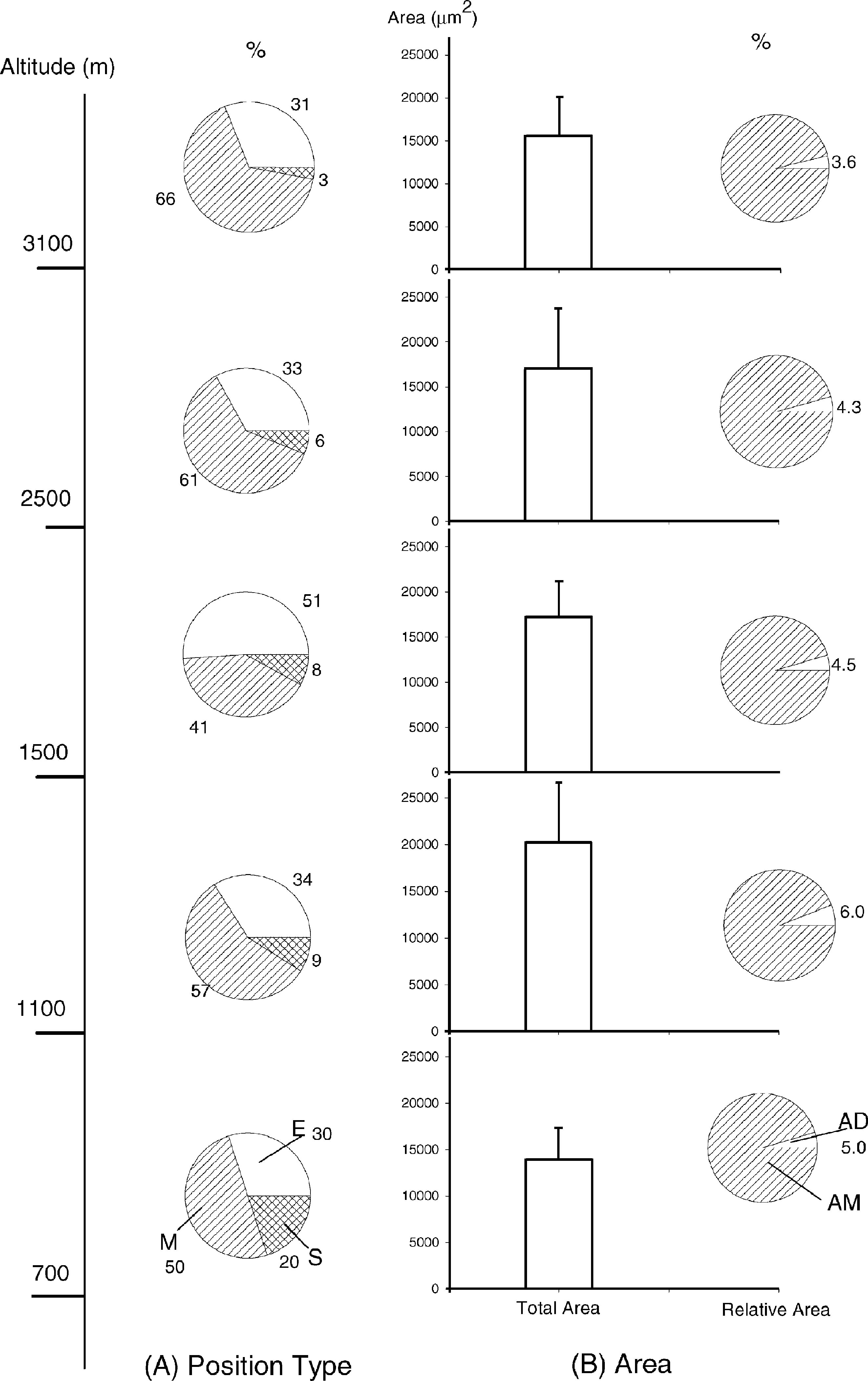
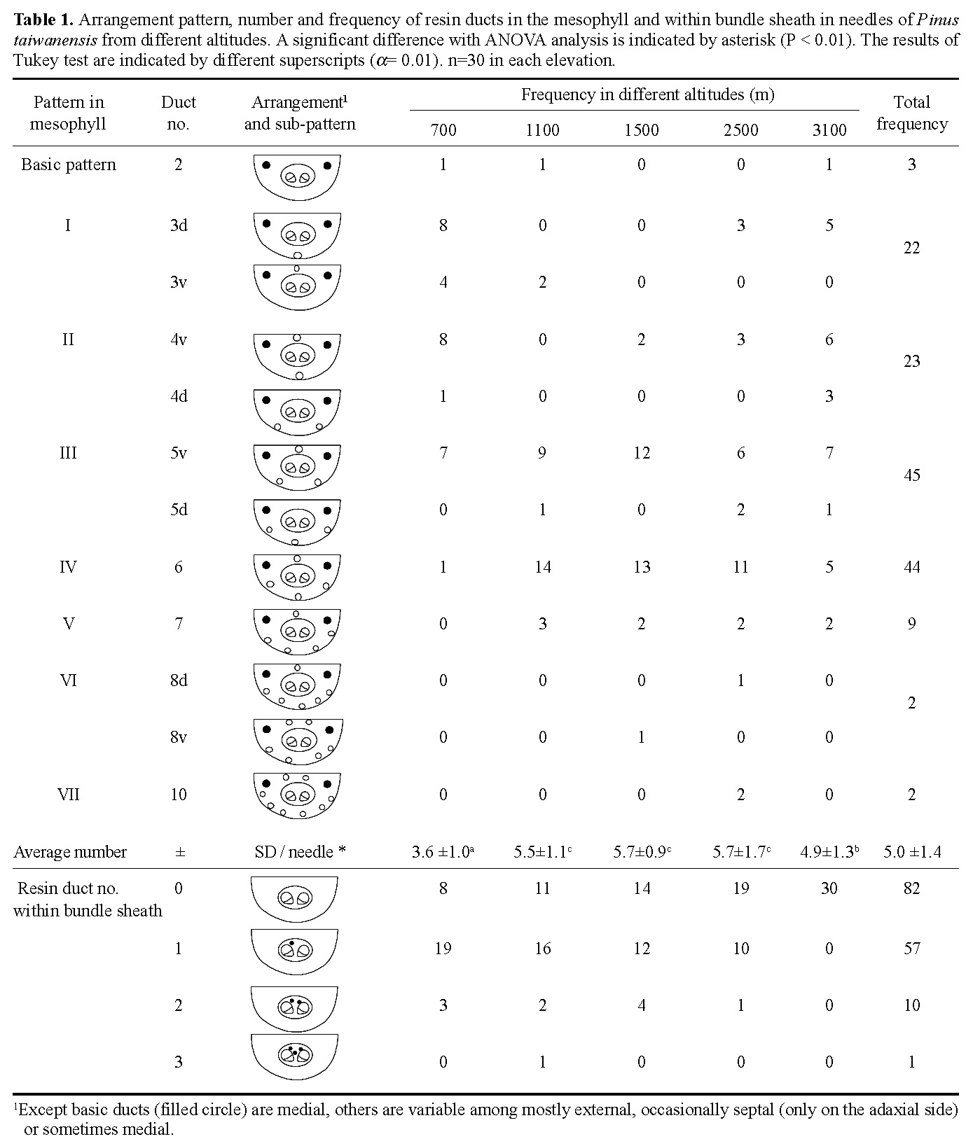
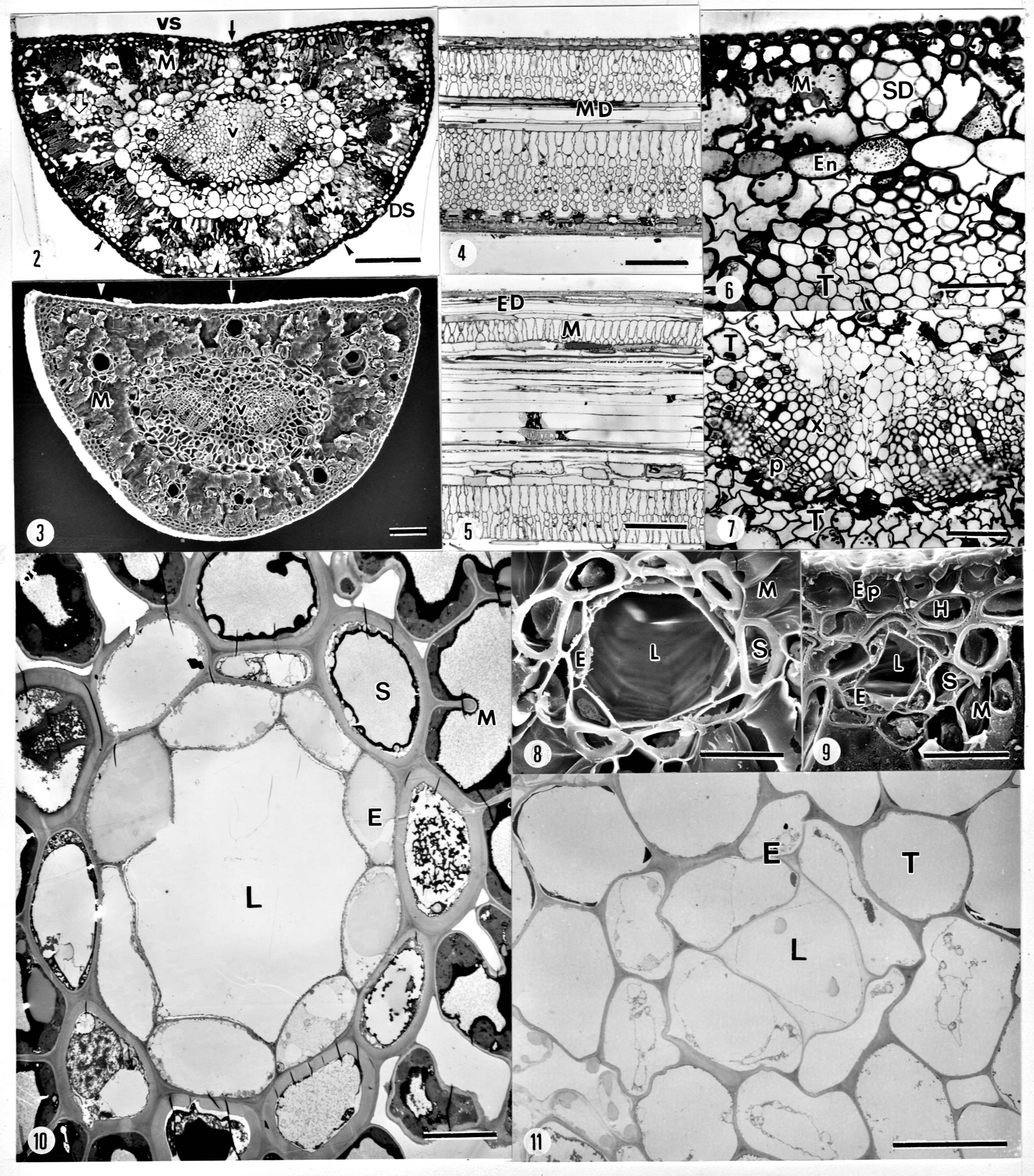
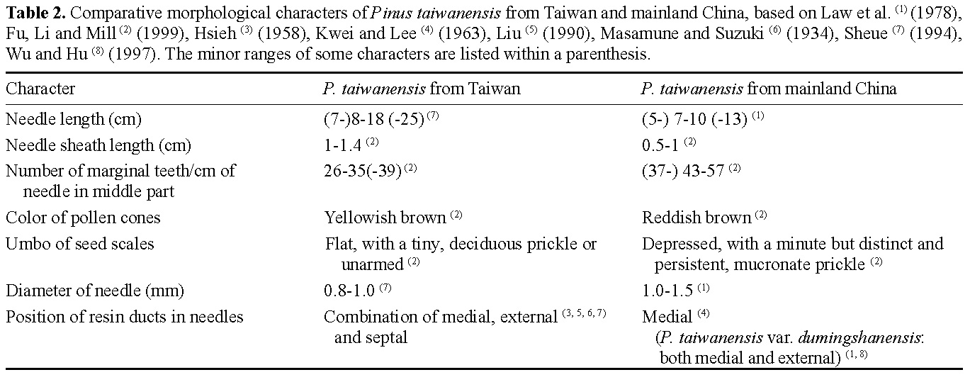
Abstract. The variation of resin ducts in needles of Pinus taiwanensis along an altitude gradient (700 m- 3,100 m) in central Taiwan is revealed for the first time in this study, including position type, number, size, arrangement pattern and anatomical structural variation. Three types of resin ducts-medial, external and septal-were found according to their relative positions in the mesophyll. All needles sampled showed combinations of these three resin duct types. Mostly, 3-7 resin ducts were found in the mesophyll per needle, and the number of resin ducts was significantly greater at mid altitudes than at high and low altitudes. Septal ducts decreased in number with an increase in altitude; however, needles from mid altitudes had more external ducts. Eight patterns (basic and patterns I-VII) of resin ducts in the mesophyll of needles were delineated to present the arrangement variations. In addition to the primary ducts found in the mesophyll, we found the rare bundle sheath ducts in some needles, and their frequencies decreased obviously with an increase in altitude. The consistent combination feature of medial, external, and septal ducts in needles of P. taiwanensis from Taiwan could be easily distinguished from those with medial ducts from mainland China. However, this difference has unrecognized before, and it should be considered in future taxonomic work.

Keywords: Altitudinal variation; Needle; Resin duct; Pinus taiwanensis.

Introduction

In conifers, the resin duct is a common structure of the plant body. It may be present both in the primary and secondary tissues; but it seems to occur more consistently in the former. Previous research has examined the structure, distribution, and development of resin ducts in the Pinaceae (Hanes, 1927; Mergen and Echols, 1955; Werker and Fahn, 1969; Lapasha and Wheeler, 1990; Wu and Hu, 1997).

Napp-Zinn (1966) classified four types of resin ducts according to their position in the needles of Pinus: (1) ducts in contact with the hypodermis (i.e., external); (2) ducts surrounded by chlorenchyma (i.e., medial); (3) ducts in contact with the bundle sheath (i.e., endodermis) in the chlorenchyma (i.e., endonal); and (4) ducts inside the bundle sheath. Although this last type is rare, P. halepensis Mill. is an example of a species possessing bundle sheath resin ducts (Werker and Fahn, 1969). Biswas and Johri (1997) described four similar resin ducts in the mesophyll (medial, external, endonal, and septal) but did not mention Napp-Zinn's fourth type of duct, the one inside the bundle sheath. Most species of Pinus contain one or two types of resin ducts in the needles (Biswas and Johri, 1997).

The resin duct is an important character, applied in classifying the Pineaceae and particularly in distinguishing Pinus species (Law et al., 1978; Li and Keng, 1994; Wu and Hu, 1997; Richardson, 1998; Fu et al., 1999; Boratyńska and Bobowicz, 2001). It is even used to differentiate hybrids (Kormutak et al., 1993). The number and position of resin ducts in needles may vary considerably and interspecifically in pines. Nevertheless, for the identification of pine species, the number of resin canals in the needles is of no particular importance, except for a few species that normally only have two or three canals; however, the relative position of the ducts in the needle may be used as an aid in identification (Harlow, 1931).

Four species of pines are native to Taiwan. Pinus taiwanensis Hayata of these native species has the widest distribution, ranging from altitudes of 680 m to 3,100 m in central and southern Taiwan (Sheue, 1994). Whether P. taiwanensis is endemic to Taiwan or also occurs in mainland China is hotly debated and depends on clarification of the taxonomic status of P. hwangshanensis Hsia (now treated as a synonym of P. taiwanensis in Flora of China, Fu et al., 1999) (Wu, 1956; Critchfield and Little, 1966; Cheng et al., 1975; Law et al., 1978; Silba, 1984; Li, 1997; Richardson, 1998; Fu et al., 1999). Furthermore, the discrepancy of feature of resin ducts in needles of P. taiwanensis between Taiwan and mainland China is detectable. Resin ducts in needles of so-called P. taiwanensis from mainland China are medial (Kwei and Lee, 1963; Law et al., 1978; Li, 1997; Wu and Hu, 1997) while

*Corresponding author. Tel: 886-2-23630231 ext. 2364; Fax: 886-2-23918940; E-mail: linglong@ccms.ntu.edu.tw


Botanical Bulletin of Academia Sinica, Vol. 44, 2003

ducts in needles of P. taiwanensis from Taiwan are a combination of external and medial (Masamune and Suzuki, 1934; Hsieh, 1958; Liu, 1990; Sheue, 1994; Sheue et al., 2000). Almost all taxonomists failed to recognize this simple difference in relative position of resin ducts in P. taiwanensis needles between specimens collected from mainland China and Taiwan (Wu, 1956; Cheng et al., 1975; Law et al., 1978; Li, 1997; Fu et al., 1999).

In this study, we were interested in the stability of relative duct position and the variation of resin ducts in needles of the widely distributional species, P. taiwanensis, from Taiwan. Sheue et al. (2000) compared the variations of 28 characters of needle structures along an elevational gradient, mainly describing the variation of resin duct number. We investigate and compare the variations of resin ducts in needles of P. taiwanensis along an elevation gradient, including number, position type, pattern, and size of resin ducts. The results will be useful to clarify the discrepancy of resin duct types in needles of P. taiwanensis between Taiwan and mainland China.

Materials and Methods

Needles of Pinus taiwanensis were collected from trees growing at different altitudes (700 m, 1,100 m, 1,500 m, 2,500 m and 3,100 m), which represent the elevational range where P. taiwanensis naturally grows in the watershed of the Tachia River in central Taiwan. Due to deforestation, samples could not be gathered from an altitude of 2,000 m. Three branchlets from different aspects were removed at 2-5 m height from ten individuals ranging in age from 10-15 years at each elevation (n = 10). One of the one-year-old needles was randomly sampled from each branchlet of the same individual and cut at the center for observation of embedded in Spurr's-resin (Sheue, 1994). Semi-thin (1 µm) and thin-sections (75 nm) were made by Ultracut E microtome, stained with 0.1% Toluidine blue, and double stained with lead citrate and uranium acetate, respectively (Kuo-Huang and Chen, 1999). These sections were observed with a Leitz Diaplan microscope or with a Hitachi H 600 transmission electron microscope. Areas of both resin duct and mesophyll were measured with microscopic images using PC-meter software. Some needles were free hand sectioned for investigation under a scanning electron microscope after being critical point dried and coated with gold. Voucher specimens were deposited in the Herbarium of the Department of Botany (TAI) at the National Taiwan University. Data were analyzed by Excel 8 (Microsoft Office 97), and statistical figures were made by Sigma Plot 2000 software.

Results

We observed two kinds of resin ducts in needles of P. taiwanensis: primary and secondary. Primary ducts were found in the mesophyll (medial, external, and septal types), and secondary ducts were found inside the bundle sheath (i.e., vascular tissue). Unlike the mesophyll ducts, the

bundle sheath resin ducts were found only in some needles below 3,100 m in altitude.

Type of Resin Duct According to its Relative Position in the Mesophyll

Three types of resin ducts were found in the mesophyll of needles of P. taiwanensis: external (Figures 2, 3, 5), medial (Figures 2, 3, 4), and septal (Figures 2, 6). To indicate the position more accurately, a new (septal) type of resin duct in the needles of P. taiwanensis is described. All the needles showed combinations of these three types; for example, each needle had two (external and medial, septal and medial) or all three types of resin ducts. Furthermore, the resin ducts of P. taiwanensis needles were never all-external or all-septal exclusively. Very occasionally, if a needle had only two or three ducts, they may all have been medial.

Generally speaking, two medial ducts occurred constantly on the ventral surface of a needle (Figures 2-4; Table 1). Therefore, we defined these two easily recognized ducts as the basic ducts in needles of P. taiwanensis (the filled circles in Table 1). The other duct positions seemed to be more variable: mostly external, occasionally septal, or sometimes medial.

Comparing the average number and percentage of these three types of resin duct at different elevations, septal resin ducts clearly decline from 0.8 (20%) at 700 m to 0.1 (3%) at 3,100 m altitude (Figure 1A). The number of medial resin ducts were highest at elevations of 1,100 m, 2,500 m and 3,100 m (three, ranging from 57-66% as compared to 41% at 1,500 m and 50% at 700 m). The needles from the middle elevation (1,500 m) had more external ducts (three, 51%).

Variation of Resin Duct Pattern in the Mesophyll

In needles of P. taiwanensis, resin ducts in the mesophyll showed a great diversity, in terms of number and relative position. We delineated one basic pattern and seven derived patterns, I to VII, based more on the number than on the position (Table 1). A needle with only two medial basic ducts in the mesophyll was defined as the basic pattern, and these two ducts could be found in each needle of P. taiwanensis at any elevation. Pattern I had one more resin duct than the basic pattern, and pattern II had two more resin ducts than the basic pattern in the mesophyll, and so on. Depending upon the ventral or dorsal location, two sub-patterns, v and d, were defined in patterns I, II, III and VI (Table 1). From the frequency of resin ducts of each sub-pattern, it seems that most of the ducts are dispersed evenly in a needle, rather than clustered near the same surface (Table 1). For example, in pattern I, the third duct was frequently located more in the dorsal surface (Pattern Id) than in the ventral surface to line up linearly (Pattern Iv). These similar results were also obvious in sub-patterns of II and III.

The patterns of resin ducts of the lowest (700 m) elevation were mostly patterns of Id, IIv and IIIv, i.e. with 3, 4, and 5 resin ducts (Table 1, Figure 2). However, ducts


Sheue et al. — Resin ducts in Pinus needles

in needles from middle elevations (1,100 m, 1,500 m and 2,500 m) appeared mainly in patterns of IIIv and IV, i.e. with 5 and 6 resin ducts (Figure 3). Only needles from the highest (3,100 m) elevation showed a more diverse duct pattern, including four dominant patterns (i.e. I to VI). For all elevations combined, resin duct patterns III and IV were most common.

Variation of Number and Size of Resin Duct in the Mesophyll

The number of resin ducts per needle of P. taiwanensis ranges from 2 to 10, mostly between 3 to 6, with an average of 5±1.4 (SD) (Table 1). The fewest number of resin

ducts per needle was found in needles from 700 m elevation, 3.6±1.0. The next lowest average number of ducts was at 3,100 m and was 4.9±1.3. The needles of mid altitudes (1,100 m, 1,500 m and 2,500 m) had the most resin duct per needle. The numbers of resin ducts among different elevations were significantly different, and needles could be divided into three groups (Table 1), i.e., low elevation (700 m), mid elevation (1,100 m, 1,500 m and 2,500 m), and high elevation (3,100 m).

We also compared the variation of resin ducts in number of needles taken from the same height of the same trees at different elevations (ten needles from one tree and five trees from an elevation). It showed that the number of resin

Figure 1. Variations in relative position and area of resin ducts in the mesophyll of Pinus taiwanensis needles along an altitudinal gradient. (A) Variation of duct position type, E: external, M: medial, S: septal type. (B) Variation of average area of resin duct, including the average total area and relative total area in the mesophyll. Error bars represent SD. AD: relative total area of ducts in the mesophyll; AM: relative total area of mesophyll. n =30 in each altitude except (A): n =10.


Botanical Bulletin of Academia Sinica, Vol. 44, 2003

ducts of needles were quite constant or occasionally fluctuated (only ±1, rarely 2) in duct number from the same individual (data not shown in the table).

The size of resin ducts can also vary within a needle. In the same needle, the largest resin duct (about 50 µm in diameter) might be three times the size of the smallest

(Figures 8, 9). Figure 1B shows the average total area of resin ducts (including sheath cells, epithelial cells, and lumen) and the average relative duct area (% area of duct in mesophyll) per needle of different altitudes. As shown in Figure 1 and Table 1, although the needles of 1,500 m and 2,500 m altitude had more resin ducts than those of


Sheue et al. — Resin ducts in Pinus needles

Figures 2- 11. Resin ducts in needles of Pinus taiwanensis. 2: Cross section of needle from low elevation (700 m) with one septal duct (arrow), two medial ducts (open arrow) and three external ducts (arrowhead). Note the shorter distance of the vascular bundle to the ventral surface in this needle than the distances from mid (Figure 3) or high altitudes. 3: Cross section of a needle from mid elevation (1,500 m), with four medial and two external ducts. The arrowhead and arrow show the longitudinal section of the medial and external ducts in Figures 4 and 5, respectively. 4: One of the basic pattern ducts (medial type) surrounded by mesophyll. 5: The external duct contacting with hypodermis. 6: One septal resin duct in ventral surface and one bundle sheath resin duct (arrow) in transfussion tissue nearing to endodermis. 7: Two bundle sheath resin ducts (arrows) neighboring to xylem. 8: One of the basic resin ducts (medial) in the mesophyll, with larger lumen, showing the thick-walled sheath cells and thin-walled epithelial cells. 9: An external duct, in contact with hypodermis, usually with smaller lumen. 10: Ultrastructure of a resin duct in the mesophyll consisting of sheath cells and epithelial cells, with rich cytoplasm inside cells. 11: Ultrastructure of a bundle sheath resin duct, consisting of a layer of somewhat collapsed epithelial cells only. Scale bars: 2-5 = 50 µm; 6-7 = 20 µm; 8-9 = 25 µm; 10-11= 10 µm. Key to lettering: DS: dorsal surface; E: epithelial cell; ED: external resin duct; En: endodermis; Ep: epidermis; H: hypodermis; M: mesophyll; MD: medial resin duct; L: lumen; P: phloem; S: sheath cell; SD: septal resin duct; T: transfusion tissue; V: vascular bundle; VS: ventral surface; X: xylem.


Botanical Bulletin of Academia Sinica, Vol. 44, 2003

other elevations, needles of 1,100 m had the greatest total and relative duct area (20275 µm2, 6.0%). The needles of 3,100 m altitude (with the second lowest number of resin ducts = 4.9) showed the least relative area of resin ducts (3.6%) while the needles from 700 m altitude (with the lowest number of resin ducts= 3.6, total duct area = 13910 µm2) had the second largest relative area of resin ducts (5.0%) (Table 1, Figure 1B).

In addition, we measured the area of two lumens of the basic ducts (called basic lumen for abbreviation) in each needle. This average basic lumen areas in the needles along an altitude from 700 m, 1,100 m, 1,500 m, 2,500 m and 3,100 m were 2068, 3023, 1633, 2074 and 2221 µm2. At the same time, the basic lumen area was positively related to the total area of resin ducts (Sheue, 1994). We found that the total area of ducts in a needle was usually determined by the size of the two large basic ducts.

Resin Duct Within Bundle Sheath

Additional resin ducts existed within the bundle sheath in some of the needles in P. taiwanensis, excluding those from 3,100 m altitude (Table 1, Figures 6, 7). Most commonly, we found one resin duct near the xylem of one of the vascular strands or among the tracheids of the transfusion tissue between the two vascular strands toward the ventral surface (Figures 6); rarely did we observe two or three resin ducts in the bundle sheath (Figures 7). In contrast to mesophyll resin duct patterns, the frequencies of such resin ducts appearing inside the bundle sheath decreased obviously in accordance with increasing elevation (Table 1).

Structural Variation of Resin Ducts in the Mesophyll and Within the Vascular Bundle

Consistent with the resin ducts in the mesophyll of pine needles of other Pinus species, two types of cells constituted the resin ducts of P. taiwanensis: thick-walled sheath cell and thin-walled epithelial cell (Figures 8-10). A lumen was formed as a circle surrounded by 6-10 lens-like epithelial cells and 7-11 sheath cells, depending on its size (20-55 µm in diameter). Comparing the structure of resin ducts in the mesophyll and inside the bundle sheath, the latter was smaller, usually with 4-5 epithelial cells only, 5-10 µm in diameter, and devoid of sheath cells (Figures 6, 7, 11). We examined six resin ducts in the bundle sheath and ten resin ducts in the mesophyll with a transmission electron microscope. Epithelial cells of resin ducts in the mesophyll were richer in cytoplasm than those inside vascular tissue (Figure 10). Resin ducts there are possibly somewhat collapsed and lack organelles (Figure 11).

Discussion

The variation of resin ducts of P. taiwanensis needles at different elevations is detailed here for the first time, and we include three position types, eight arrangement patterns, number and size, and two kinds of resin ducts (i.e., primary ducts in the mesophyll and secondary ducts in

the bundle sheath). It was apparent that the number of resin ducts and the number of primary and secondary ducts varied in different needles of P. taiwanensis.

Almost all the needles of P. taiwanensis showed a combination of three position types of resin ducts: medial, external, and septal. The septal type was first described in this species by this study using the classification of Biswas and Johri (1997). We speculate that the two medial basic ducts may be controlled genetically, and that the other variable ones are more susceptible to environmental factors. The number of septal ducts decreased with an increase in elevation. This phenomenon showed a high negative correlation with the distance of the vascular bundle to the adaxial surface (Sheue, 1994; Sheue et al., 2000). For example, needles from 700 m altitude, with the shortest distance (16.1 µm) of vascular bundle to adaxial surface, had more septal ducts. Others needles with greater vascular bundle to adaxial surface distances (17.55 µm at 1,100 m to 21.3 µm at 3,100 m altitude) had fewer septal ducts. The greater number of external ducts at 1,500 m may be correlated with the greater number of resin ducts at this elevation. Nevertheless, it was reported that the same duct could pass from one location type to another in different portions of the needle of P. halepensis (Werker and Fahn, 1969).

In addition to counting the number of ducts, we calculated the total resin duct area and the two basic lumen areas in each needle from five altitudes. The numbers of resin ducts in needles were significantly greater at mid altitudes (1,100 m, 1,500 m and 2,500 m) than at the higher (3100 m) and lower altitudes (700 m), but the needles from 1,100 m had the greatest total area and basic lumen area of resin duct. This pattern was similar to what Hengxiao et al. (1999) discovered: resin ducts were significantly wider in trees at the middle altitude than those at other elevations in P. yunnanensis Franchet. Our results suggested that the greatest number of resin ducts did not imply the greatest total and relative area of resin ducts. It was related to the changes of leaf structure in areas of leaf cross-section and mesophyll along an altitude gradient (Sheue, 1994, Sheue et al., 2000).

The number of resin ducts in a plant body is probably influenced by several genetic and environmental factors, including the height and age of the tree, nutrition, sunlight, radiation, temperature, wind, freezing, fire, insect attack, and phytohormones (Helmers, 1943; White and Beals, 1963; Fahn and Benayoun, 1976; Telewski et al., 1983; Skuterud et al., 1994; Jokela et al., 1998; Hengxiao et al., 1999; Telewski et al., 1999). Moreover, it has been reported that low temperature (4°C) will prohibit the development of resin duct in the root of P. halepensis seedling (Fahn and Benayoun, 1976). Whether high temperatures have the same effect is not yet known. Telewski et al. (1999) indicated that long-term CO2 enrichment had no significant effect on the density of resin canals in woods. However, the primary duct is less susceptible to external factors than the secondary duct (Fahn, 1990). Our results revealed that needles from mid altitude have more resin ducts (primary)


Sheue et al. — Resin ducts in Pinus needles

surface. Its smaller size and complex vascular tissue structure might be the reason it eluded detection before. The presence of fewer cytoplasm and organelles in epithelial cells of ducts within the bundle sheath may hint a weaker function for resin secretion as compared to those ducts in the mesophyll. We also suggest that only the resin ducts in the mesophyll are useful for species identification since the presence of resin ducts within the bundle sheath is not consistent.

It was reported that P. taiwanensis from mainland China only has medial resin ducts in its needles (Kwei and Lee, 1963; Li, 1997; Wu and Hu, 1997; Richardson, 1998). However, we confirmed that needles of P. taiwanensis from Taiwan possessed a combination of external, medial, and septal ducts. Agreeing with Harlow (1931), we strongly believe that the position type, not the number, of resin ducts in needles could be a valuable diagnostic character for discerning the materials in P. taiwanensis of Taiwan from mainland China. Although most of the taxonomists failed to recognize this difference before (Wu, 1956; Cheng et al., 1975; Law et al., 1978; Li, 1997; Fu et al., 1999), it should be considered in future taxonomic work.

Nevertheless, Mill, one of the authors of the Flora of China, based on several characters of needle and cone, preferred to treat all materials from mainland China as P. hwangshanensis, and materials from Taiwan as P. taiwanensis (see Table 2). Li (1997) pointed out that needles of P. taiwanensis from mainland China have fewer resin ducts (3-8) than those from Taiwan (6-7). However, we found that the needles of P. taiwanensis from Taiwan have 3-8 (rarely, as much as 10) resin ducts, not as Li (1997) reported previously. On the contrary, the results showed that the way to differentiate resin ducts in needles of P. taiwanensis in Taiwan from those in mainland China is to note the relative position of ducts, not the number. On the other hand, it was reported that P. taiwanensis var. damingshanensis from mainland China had medial and external ducts in its needles (Law et al., 1978) similar to those of P. taiwanensis from Taiwan. Accordingly, it requires further comprehensive study to elucidate the taxonomic sta

in the mesophyll while resin ducts inside the bundle sheath (secondary) decrease in accordance with increasing elevation. Variations in the number of these ducts in needles should result from genetic and environmental factors due to the habitat. With increased elevation, many physical factors indeed change gradually, such as temperature, pressure, UV, wind and rainfall. Furthermore, Su (1984) observed that a level of prevalent cloud and a zone of temperature inversion appeared in the areas of mid elevations in the watershed of the Tachia River, where we sampled the needles for this study. Whether these climatic factors also play a role affecting the duct number in needles of P. taiwanensis requires further study.

Napp-Zinn (1966) described the classification of resin ducts in leaves and indicated one type of duct located inside the bundle sheath; however, ducts of this type are rare. Džaparidze mentioned 11 species of Pinus with this type, P. halepensis being one example (c. f. Werker and Fahn, 1969). Pinus taiwanensis is another example from Taiwan of a species with such resin ducts located within a bundle sheath. As Werker and Fahn (1969) stated, this is one kind of secondary duct, continuing with the ducts of the secondary xylem of the blachyblast's axis and extending from the brachyblast axis into the needle, and it is narrower than other types in the mesophyll. These secondary needle ducts terminate a short distance above the needle base and are capped by a few layers of parenchyma cells (Werker and Fahn, 1969). However, these ducts are present in the middle part (about 7~11 cm away from the leaf base) of needles in P. taiwanensis, not a short distance above the base as Werker and Fahn observed in P. halepensis (Werker and Fahn, 1969). It is peculiar that none of these ducts were found in needles from 3,100 m altitude and that the frequency clearly decreased with increasing elevation. This kind of duct may appear in the metaxylem of the vascular strand or the transfusion tissue toward the ventral surface in P. halepensis (Werker and Fahn, 1969), but it was only found in the transfusion tissue in needles of P. taiwanensis, either near the xylem or among the tracheids between the two vascular strands toward the ventral


Botanical Bulletin of Academia Sinica, Vol. 44, 2003

sawfly (Hym. Diprionidae) performance. J. Appl. Entomol. 123: 465-471.

Hsieh, W.C. 1958. The anatomy of coniferous leaves in Taiwan. J. Agric. Forestry 7: 53-100.

Jokela, A., T. Sarjala, and S. Huttunen. 1998. The structure and hardening status of Scots pine needle at different potassium availability levels. Trees-Structure and Function 12: 490-498.

Kuo-Huang, L.L. and S.J. Chen. 1999. Subcellular localization of calcium in the crystal-forming sclereids of Nymphaea tetragona Georgi. Taiwania 44: 520-528.

Kormutak, A., R. Matusova, A. Szmidt, and D. Lindgren. 1993. Karyological, anatomical and restriction fragment length polymorphism characteristics of the interspecific hybrid Pinus banksiana × Pinus contorta. Biologia (Bratislava) 48: 95-100.

Kwei, Y.L. and C.L. Lee. 1963. Anatomical studies of the leaf structure of Chinese pines. Acta Bot. Sin. 11: 44- 58. Pl. I-V.

Lapasha, C.A. and E.A. Wheeler. 1990. Resin canals in Pinus taeda longitudinal canal lengths and interconnections between longitudinal and radial canals. Int. Assoc. Wood Anat. Bull. 11: 227-238.

Law, Y.W., W.C. Cheng, L.K. Fu, C.D. Chu, and C.S. Chao. 1978. Pinus. In Cheng W. C. and L. K. Fu (eds.), Flora Reipublicae Popularis Sinicae. Tomus 7, Gymnospermae. Science Press, Beijing, pp. 204-281.

Li, H.-L. and H. Keng. 1994. Pinaceae. In Editorial Committee of the Flora of Taiwan, Flora of Taiwan. vol. I Pteridophyta and Gymnospermae, 2nd edn. Taipei, pp. 567-581.

Li, D.Z. 1997. A reassessment of Pinus subgen. Pinus in China. Edinb. J. Bot. 54: 337-349.

Liu, C.C. 1990. Leaf comparative morphology of Pinaceae. MS Thesis, National Taiwan University, Taipei, Taiwan.

Liu, Y.C., F.Y. Lu, and C.H. Ou. 1994. Trees of Taiwan. Monographic publication No.7, College of Agriculture, National Chung-Shing University, Taiwan.

Masamune, G. and S. Suzuki. 1934. Vergleichende Anatomie der Blätten der Kiefer in Taiwan. Trans. Nat. Hist. Soc. Formosa 24: 391-396.

Mergen, E. and R.M. Echols. 1955. Number and size of radial resin ducts in slash Pine. Science 121: 306-307.

Napp-Zinn, K. 1966. Anatomie des Blattes. I. Blattanatomie der Gymnospermen. Berlin—Nikolassee: Gebrüdder Borntraeger.

Richardson, D.M. 1998. Ecology and Biogeography of Pinus. Cambridge University press.

Sheue, C.R. 1994. Structural variation of Pinus taiwanensis Hay. needles along an altitudinal gradient. MS Thesis, National Taiwan University, Taipei, Taiwan.

Sheue, C.R., Y.P. Yang, and L.L. Kuo-Huang. 2000. Structural variation of the needles of Pinus taiwanensis Hay. along an elevational gradient. Taiwan J. Forest Sci. 15: 337-349.

Silba, J. 1984. An international census of the Coniferae. I. Phytologia Memoirs 7: 47-59.

Skuterud, L., N.I. Goltsova, R. Nćumann, T. Silleland, and T. Lindmo. 1994. Histological changes in Pinus sylvestris L. in the proximal-zone around the Chernobyl power plant. Sci. Total Environ. 157: 387-397.

Su, H.J. 1984. Studies on the climate and vegetation types of

tus of this so-called P. taiwanensis (P. hwangshanensis) in mainland China.

Based on the results from this study, the relative position of resin ducts in needles of P. taiwanensis could be a diagnostic character to distinguish it from other morphologically similar species, such as P. massoniana Lamb. (with external ducts only) (Kwei and Lee, 1963; Li, 1997; Wu and Hu, 1997; Richardson, 1998) and P. luchuensis Mayr (with 2 medial ducts, rarely 3) (Liu et al., 1994, Li, 1997). The so-called P. massoniana, growing in central Taiwan on hills under 600 m, showed the same anatomical characters of needle structures and resin ducts as P. taiwanensis (Sheue and Yang, unpublished). It seems that the distribution of P. massoniana in Taiwan is questionable. More studies focusing on these three affinity species, P. luchuensis, P. hwangshanensis and P. taiwanensis, using different approaches are still necessary to resolve a long-standing taxonomic controversy.

Acknowledgments. The authors thank Drs. C. H. Tsou, S. T. Chiu, H. Y. Liu, V. Sarafis and T. P. Lin for helpful guidance and comments on the manuscript, Dr. S. H. Wang, M. Y. Chen for helping the statistical analysis and figure presentation, Miss C. Y. Lin for assistance in operating electron microscope, and Mr. Y. L. Huang for help with material collection from the field.

Literature Cited

Biswas, C. and B.M. Johri. 1997. The Gymnosperms. Springer-Verlag. New York.

Boratyńska, K. and M.A. Bobowicz. 2001. Pinus uncinata Ramond taxonomy based on needle characters. Plant Syst. Evol. 277: 183-194.

Cheng, W.C., L.K. Fu, and C.Y. Cheng. 1975. Gymnospermae Sinicae. Acta Phytotaxon. Sin. 13: 56-89. Pl. 1-66.

Critchfield, W.D. and E.L.J.R. Little. 1966. Geographic distribution of the pines of the world. U. S. Department of Agriculture Forest Service Miscellaneous Publication No. 991. Washington D.C.

Fahn, A. 1990. Plant Anatomy, 4th edn. Pergamon press, Oxford, England.

Fahn, A. and J. Benayoun. 1976. Ultrastructure of resin ducts in Pinus halepensis development, possible sites of resin synthesis, and mode of its elimination from the protoplast. Ann. Bot. 40: 857-863.

Fu, L., N. Li, and R.R. Mill. 1999. Pinaceae. In Z. Y. Wu and P. H. Raven (eds.), Flora of China, vol. 4, Cycadaceae through Fagaceae. Science Press, Beijing and Missouri Botanical Garden Press, St. Louis, pp. 11-52.

Hanes, C.S. 1927. Resin canals in seedling conifers. Bot. J. Linn. Soc. 47: 613-616.

Harlow, W.M. 1931. The identification of the pines of the United States, native and introduced, by needles structure. New York States College of Forestry Technical Publication 32.

Helmers, A.E. 1943. Ecological anatomy of ponderosa pine needles. Amer. Midl. Nat. 29: 55-71.

Hengxiao, G., J.D. McMillin, M.R. Wagner, J. Zhou, Z. Zhou, and X. Xu. 1999. Altitudinal variation in foliar chemistry and anatomy of yunnan Pine, Pinus yunnanensis, and pine


Sheue et al. — Resin ducts in Pinus needles

the natural forests in Taiwan (II): Altitudinal vegetation zones in relation to temperature gradient. Quart. J. Chinese For. 17: 57-73.

Telewski, F.W., R.T. Swanson, B.R. Strain, and J.M. Burns, 1999. Wood properties and ring width response to long-term atomspheric CO2 enrichment in field-grown loblolly pine (Pinus taeda L.). Plant Cell Environ. 22: 213-219.

Telewski, F.W., A.H. Wakefield, and M.J. Jaffe. 1983. Computer-assisted image analysis of tissues of ethrel-treated Pinus taeda seedlings. Plant Physiol. 72: 177-181.

Werker, E. and A. Fahn. 1969. Resin ducts of Pinus halepensis Mill. _ their structure, development and pattern of arrangement. Bot. J. Linn. Soc. 62: 379-411.

White, J.B. and H.O. Beals. 1963. Variation in number of resin canals per needle in pond pine. Bot. Gaz. 124: 251-253.

Wu, C.L. 1956. The taxonomic revision and phytogeographical study of Chinese pines. Acta Phytotaxon. Sin. 5: 131- 162. Pl. 24-27.

Wu, H. and Z.H. Hu. 1997. Comparative anatomy of resin ducts of the Pinaceae. Trees 11: 135-143.