Bot. Bull. Acad. Sin. (2004) 45: 69-85

Jane —Ultrastructure of endosperm development in Arundo formosana

Ultrastructure of endosperm development in Arundo formosana Hack. (Poaceae) from differentiation to maturity

Wann-Neng Jane

Electron Microscopy Lab, Institute of Botany, Academia Sinica, Taipei, Taiwan

(Received January 7, 2003; Accepted August 27, 2003)

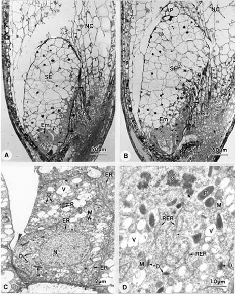
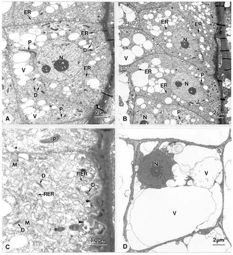
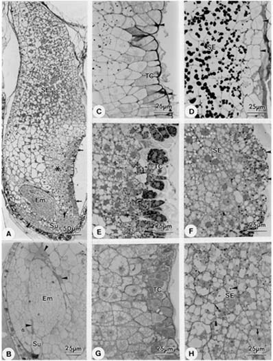
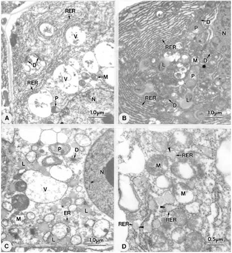
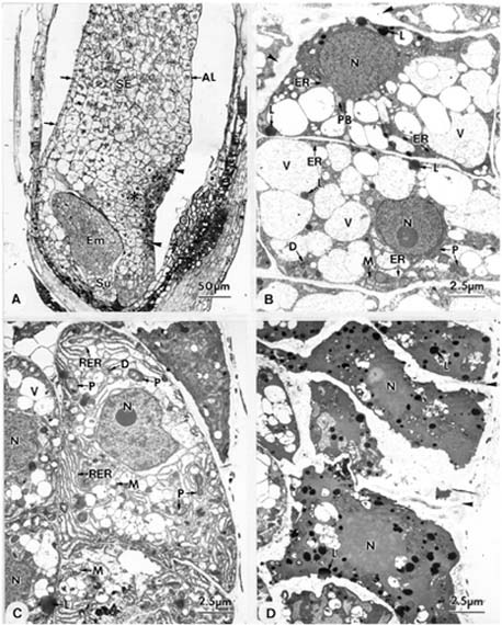
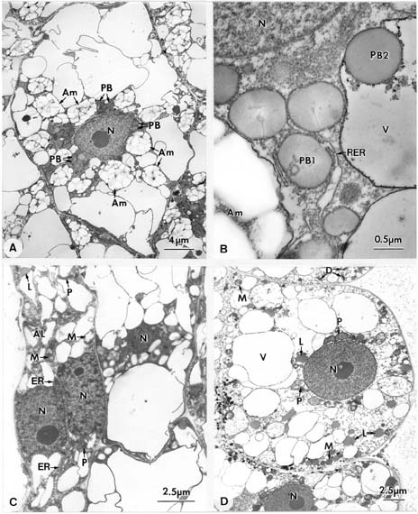
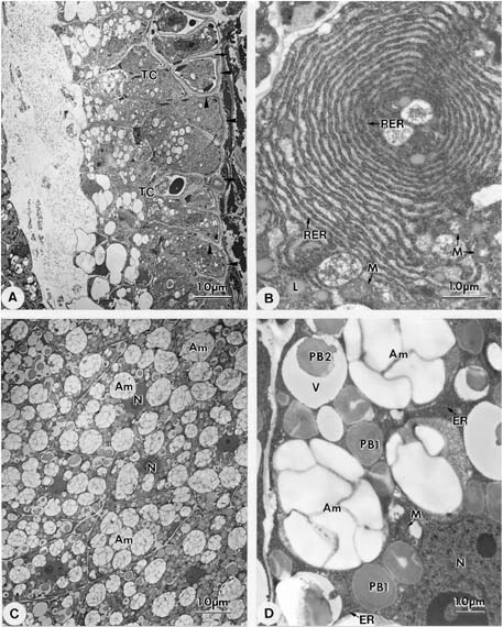
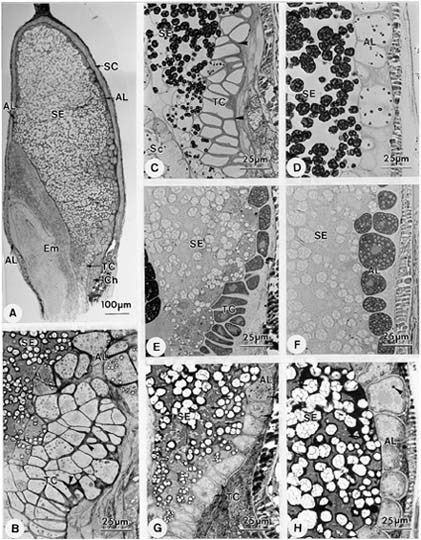
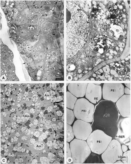
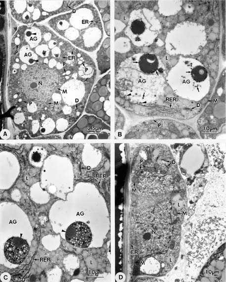
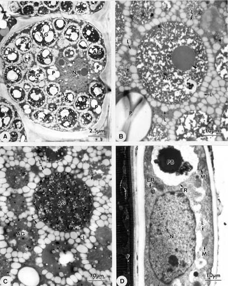
Abstract. Endosperm development of Arundo formosana Hack. is examined ultrastructurally and histochemically from differentiation to seed maturity. The differentiated endosperm contains four major cell types: the cells of the embryo surrounding region, transfer cells, aleurone layer, and starchy endosperm. After cellularization, cells of the embryo surrounding region and transfer cells contain dense cytoplasm and large quantities of rough endoplasmic reticulum (RER) and many dictyosomes. In the transfer cells, the wall ingrowths are present on the walls adjacent to the nucellus. The embryo surrounding region is distributed on the ventral side and surrounds the suspensor. After leaf primordium initiation, most cells of the embryo surrounding region have degenerated, but the outermost layer of the ventral side becomes part of the aleurone layer. The transfer cells differentiate into outer and inner cells. The outer transfer cells contain electron-dense cytoplasm, many oil bodies, large quantities of RER, and dictyosomes. These cells appear PAS-positive, especially in the thickening walls. The inner transfer cells have electron-transparent cytoplasm, some small vacuoles, organelles, and oil bodies. The outer transfer cells are finally compressed and degenerated. The inner cells contain PAS-positive thickening walls, many oil bodies, protein bodies, and RER at maturity. The starchy endosperm cells gradually accumulate reserves (mainly proteins and starch) during development, and are filled with amyloplasts and protein bodies at maturity. The protein bodies are of two types that originate in the RER and are then stored in the cisternal lumen of RER, and vacuoles, respectively. The outermost layer of the endosperm becomes the aleurone layer, except for the transfer cell region. The contents of the aleurone cells are initially similar to other starchy endosperm cells, and are finally filled with aleurone grains surrounded by many small lipid bodies. The aleurone grains are initiated from ER and accumulate in vacuoles.

Keywords: Aleurone grain; Aleurone layer; Arundo formosana Hack.; Embryo surrounding region; Endosperm development; Protein body; Starchy endosperm; Transfer cell.

Introduction

A general model for the development of cereal endosperm (Olsen et al., 1995; Olsen et al., 1998; Olsen et al., 1999) recognizes four major stages: syncytial, cellularization, differentiation, and maturation. After cellularization, the endosperm differentiates into two cell types in Triticum (Smart and O'Brien, 1983) and Zea (Schel et al., 1984), one with dense cytoplasm and the other with large vacuoles. Smart and O'Brien (1983) termed the former modified endosperm and the later general endosperm. Large quantities of RER are characteristic of the modified endosperm. The wall ingrowths occur in the placentochalazal region of Zea (Schel et al., 1984), but not in that of Triticum (Smart and O'Brien, 1983). Beyond the absorption and transport function of the endosperm, a high degree of synthesis also takes place in the modified endosperm (Schel et al., 1984).

According to the mechanisms of nuclear endosperm cellularization and development in cereals, Olsen et al.

(1999) and Olsen (2001) suggested that the differentiated cereal endosperm is composed of four cell-types: the embryo surrounding region, transfer cells, the starchy endosperm, and the aleurone layer. The underlying genetic programs for cell fate specification probably originated as independent genetic programs (Olsen, 2001).

In a previous paper, I described the embryo development of Arundo in detail (Jane, 1999). In this study, I will observe endosperm development from differentiation to maturation and discuss the relationship of endosperm and embryo development.

Material and Methods

Grains (caryopses) of Arundo formosana Hack., an endemic grass in Taiwan, were collected from Pingshi and Wulai of Taipei county. The voucher specimens, identification number 229386-229391, are deposited in the Herbarium of Department of Botany, National Taiwan University. Whole grains were fixed in 2.5% glutaraldehyde in 0.1 M sodium phosphate buffer, pH 7.2 at 4°C overnight. After three 20 min buffer rinses, the grains were postfixed in 1% OsO4 in the same buffer for 4 h at room temperature and then rinsed in three 20 min changes of buffer. The

*Corresponding author. Tel: 886-2-27899590 ext.110; Fax: 886- 2-27827954; E-mail: wnjane@gate.sinica.edu.tw


Botanical Bulletin of Academia Sinica, Vol. 45, 2004

grains were dehydrated in an acetone series, embedded in Spurr's resin (Spurr, 1969), and sectioned with a Leica Ultracut E ultramicrotome. Semi-thin sections (1 µm) for light microscopy were placed on slides and stained with 0.1% toluidine blue for 1 min at 60°C on a hot plate. Histochemical tests included 0.3% Sudan Black B in 70% alcohol for lipids (Bronner, 1975), periodic acid-Schiff (PAS) reaction for polysaccharides (Gahan, 1984), and 1% Coomassie Brilliant Blue in 7% acetic acid for proteins (Gahan, 1984). Thin sections on grids were stained with uranyl acetate and lead citrate (Reynolds, 1963) and studied with Philips CM 100 or JEM 1200 EX II transmission electron microscope at 80 KV.

Results

After cellularization, most of the endosperm cells are occupied by large vacuoles except for those surrounding the embryo and in the placentochalazal region. The endosperm surrounding the embryo and distributed in the placentochalazal region contains abundant cellular contents classified into embryo surrounding region and transfer cell region, respectively (Figure 1A). The endosperm with large vacuoles is starchy endosperm. Following development, the embryo surrounding region and the transfer cell region gradually increase in cell number and cell layer (Figure 1B). The cells of the embryo surrounding region (Figure 1C-D) and the transfer cells (Figure 2A-B) display dense cytoplasm, large nuclei, an abundance of ER, and many dictyosomes. In these cells, the mitochondria have well-developed cristae. The plastids are devoid of starch grains, and the dictyosomes display many small vesicles (Figure 1D, 2C). In the transfer cells, the wall ingrowths are present on the walls adjacent to the nucellus (Figure 2C) and the tangential walls. The degree of wall ingrowths increases from lower cells (Figure 2A) to upper cells (Figure 2B). In the starchy endosperm cells, most of the cell lumen is occupied by vacuoles, and most of the organelles are distributed in the perinuclear region (Figure 2D).

In subsequent development, the embryo surrounding region and the transfer cell region appear differentiated in the cell constitution, and the cells in the middle region of the starchy endosperm begin to accumulate starch. When an embryo has differentiated into embryo proper and suspensor, the embryo surrounding region with an abundance of cell contents is mainly distributed on the ventral side and in the surrounding suspensor (Figure 3A). The endosperm cells surrounding the embryo proper are compressed and broken, and some regions appear PAS-positive (Figure 3B). Two parts of the transfer cell region can be distinguished: the outermost layer and the inner cells (Figure 3A). The transfer cells of the outermost layer appear PAS-positive, especially the thickening walls (Figure 3C), and contain many lipid bodies (Figure 3E) and an abundance of proteins (Figure 3G). Compared to the outermost layer, the inner transfer cells contain less polysaccharide and proteins (Figure 3C-G), but have some lipid bodies (Figure 3E). The starchy endosperm accumulates some

storage reserves, including starch grains (Figure 3D), lipid bodies (Figure 3F), and protein bodies (Figure 3H). The starch grains are stored in plastids forming amyloplasts, and the protein bodies are formed in cytoplasm and vacuoles (Figure 3H). The accumulation of the storage reserves is mainly in the upper half of the starchy endosperm (Figure 3A). The first reserve formed is starch, followed by lipids, and finally proteins. The outermost layer of the endoperm, except for the transfer cell region, is aleurone initials. The starch grains and protein bodies are seldom found in aleurone initials (Figure 3D), in which some lipid bodies are present (Figure 3F). In general, the aleurone initials in the dorsal side are larger than those in the ventral side (Figure 3A). The aleurone initials undergo periclinal division, and the outermost cells become aleurone cells, and the inner cells become starchy endosperm cells (Figure 3D).

Large quantities of RER and dictyosomes are characteristic of cells in the embryo surrounding region (Figure 4A) and in the outermost layer of the transfer cell region (Figure 4B). In these cells, RER is usually arranged parallel with the cell wall. The cells in the outermost layer of the transfer cell region have electron-dense cytoplasm (Figure 4B). Many lipid bodies, some plastids and mitochondria are also present in theses cells (Figure 4B). The inner cells of the transfer cell region have electron-transparent cytoplasm containing fewer RER, dictyosomes, plastids, mitochondria, lipid bodies, and more small vacuoles (Figure 4C) than the outermost cells. The upper half of the starchy endosperm contains many storage reserves, especially starch grains. In these cells, the protein bodies are initiated in the cisterae of RER (Figure 4D).

When the embryo undergoes coleoptile initiation, the aleurone layer is apparent in the outermost layer of the endosperm, except for the transfer cell region (Figure 5A). The vacuolation of the cells of the embryo surrounding region increases, especially in the ventral region. Some cells of the embryo surrounding region in the dorsal region still contain large quantities of RER (Figure 5C). The cytoplasm of the outer cells of the transfer cell region appear denser (Figure 5A) than in the previous stage. The outer cells of the transfer cell region contain electron-dense contents, many lipid bodies, and thickening walls (Figure 5D). In these cells, the organelles are difficult to distinguish. In the inner cells of the transfer cell region, some plastids, mitochondria, dictyosomes, ER, protein bodies, and many vacuoles are present in the cytoplasm (Figure 5B).

In the starchy endosperm cells, each nucleus is surrounded by many large amyloplasts and protein bodies (Figure 6A). The protein bodies have two types: Type I (PB1) is enlarged in the cisteral lumen of RER, and type II (PB2) is accumulated in vacuoles (Figure 6B). The ventral aleurone cells (Figure 6C) are smaller than the dorsal ones (Figure 6D). The aleurone cells contain some organelles and lipid bodies, but fewer starch grains (Figure 6C, 6D).

In subsequent development, most cells of the embryo surrounding region are degenerated, but the outermost


Jane —Ultrastructure of endosperm development in Arundo formosana

Figure 1. Longitudinal section of developing caryopses. A, The cells of the embryo surrounding region (') and the transfer cell region (†) are rich in cell contents. The cells of the starchy endosperm are occupied by large vacuoles; B, The embryo surrounding region ('), the transfer cell region (†) and the starchy endosperm increase in cell number during development; C, The cells of the embryo surrounding region in the dorsal region contain many organelles, including large nucleus, plastids, mitochondria, small vacuoles, ER, and dictyosomes; D, The cell of the embryo surrounding region in the ventral region showing cell contents. The plastids contain dense matrix, and the mitochondria have well-developed cristae. The dictyosomes are active in producing vesicles. AP: Antipodal cell; C: Chromosome; Ch: Chalaza; D: Dictyosome; Em: Embryo; ER: Endoplasmic reticulum; M: Mitochondrion; N: Nucleus; NC: Nucellus; P: Plastid; RER: Rough endoplasmic reticulum; SE: Starchy endosperm; V: Vacuole.


Botanical Bulletin of Academia Sinica, Vol. 45, 2004

layer of the ventral region differentiates itself into a part of the aleurone layer. The outer cells of the transfer cell region are also compressed and degenerated, but the inner cells develop thick walls (Figure 7A). These thickening walls are PAS-positive. These cells contain large quantities of RER and dictyosomes. In these cells, RER sometimes appears arranged in whorled stacks (Figure 7B).

The number of lipid bodies increases, and the plastids do not contain starch grains. Some electron-dense materials accumulate in the vacuoles. In the starchy endosperm cells, the amyloplasts enlarge, and the protein bodies increase and enlarge. The cells are gradually filled by amyloplasts and protein bodies (Figure 7C). The nucleus is usually centrally located. Some RER and mitochondria are present

Figure 2. Longitudinal section of a developing caryopsis. A, The transfer cells close to embryo contain many organelles including large nucleus, plastids, mitochondria, small vacuoles, ER, and dictyosomes; B, The transfer cells showing cellular contents. The cytoplasm is rich in organelles, especially large quantities of ER; C, The transfer cells showing RER, dictyosomes, plastids, and mitochondria. Many ingrowth walls (†) are present on endosperm walls adjacent to nucellus; D, A starchy endosperm cell showing cellular contents. The nucleus is close to cell wall, and most of the cell lumen is occupied by large vacuoles. D: Dictyosome; ER: Endoplasmic reticulum; M: Mitochondrion; N: Nucleus; P: Plastid; RER: Rough endoplasmic reticulum; V: Vacuole.


Jane —Ultrastructure of endosperm development in Arundo formosana

Figure 3. Longitudinal section of a developing caryopsis. A, The embryo surrounding region (†) is distributed in the ventral region and surrounds the suspensor. The transfer cell region consists of outer (') and inner (Ý) parts. The starchy endosperm accumulates some storage reserves; B, Caryopses section stained with PAS shows that the compressed and broken cells of the endosperm are PAS-positive (†); C, Caryopses section stained with PAS showing polysaccharide distribution. The transfer cells in the outermost layer appear PAS-positive, especially the thichening walls (†); D, Caryopses section stained with PAS showing polysaccharide distribution. The cells of the outermost layer of endosperm (aleurone initial) seldom contain starch grains. The starchy endosperm have many starch grains. The aleurone cells (†) in the dorsal region are initiated by periclinal divisions of the cells of the outermost layer; E, Caryopses section stained with Sudan Black B showing lipid distribution. In the transfer cells, lipid bodies in outer cells greatly outnumber those in inner cells. Some lipid bodies can be also seen in starchy endosperm cells; F, Caryopses section stained with Sudan Black B showing lipid distribution. Lipid bodies in aleurone initials (†) outnumber those in starchy endosperm cells; G, Caryopses section stained with Coomassie brilliant blue showing protein distribution. The outer cells of the transfer cells contain large amounts of proteins, and the inner cells have few proteins; H, Caryopses section stained with Coomassie brilliant blue showing protein distribution. The starchy endosperm cells contain some protein bodies. Some protein bodies are in cytoplasm ('), and some are in vacuoles (†). Em: Embryo; SE: Starchy endosperm; Su: Suspensor; TC: Transfer cell.


Botanical Bulletin of Academia Sinica, Vol. 45, 2004

mainly consists of transfer cells, aleurone layer, and starchy endosperm (Figure 8A). The transfer cells have one to three cell layers (Figure 8B). The thickening walls of the transfer cells appear PAS-positive (Figure 8C). In the transfer cells, there are many lipid bodies (Figure 8E) and protein bodies (Figure 8G), but fewer starch grains (Figure 8C). The aleurone layer has one to several cell layers (Figure 8A). The aleurone cells have some ring structures

in the cytoplasm (Figure 7D). Two types of protein bodies are enlarged in cisterae of RER and vacuoles, respectively (Figure 7D). The protein bodies in the cisterae of RER are not uniform and appear light in their central region.

At maturity, the caryopsis contains the seed coat, the embryo and the endosperm (Figure 8A). The endosperm

Figure 4. A, The cells of the embryo surrounding region adjacent to suspensor contain large quantities of RER, many dictyosomes, and small vacuoles. B, An outer cell of the transfer cell showing cellular contents. The cytoplasm is very dense and contains large quantities of RER, many dictyosomes, and lipid bodies; C, The inner cells of the transfer cell showing cellular contents. The cytoplasm contains some plastids, mitochondria, dicytosomes, ER, lipid bodies, and many small vacuoles; D, A starchy endosperm cell in the middle region showing protein bodies (†) initiated in the cisterae of RER. D: Dictyosome; ER: Endoplasmic reticulum; L: Lipid body; M: Mitochondrion; N: Nucleus; P: Plastid; RER: Rough endoplasmic reticulum; V: Vacuole.


Jane —Ultrastructure of endosperm development in Arundo formosana

Figure 5. Longitudinal section of a developing caryopsis during coleoptile initiation. A, The vacuolation of the embryo surrounding region increases, especially on ventral side. The outer cells of the transfer cell region (†) contain dense cytoplasm and the inner cells (Ý) is high vacuolation. The aleurone cells (') of the ventral region are initiated by periclinal divisions of the cells in the outermost layer of the endosperm. The starchy endosperm accumulates more storage reserves; B, The inner cells of the transfer cells showing cellular contents. Many vacuoles, some plastids, mitochondria, dictyosomes, ER, and lipid bodies are present in the cells. Some protein bodies can be found in the cistae of ER. The cell walls thicken (†); C, The cells of the embryo surrounding region showing cellular contents. Large quantities of RER are present in cytoplasm; D, The outer cells of the transfer cells showing cellular contents. The cytoplasm with many lipid bodies is very electron-dense, and the thick walls (†) are electron-lucent. AL: Aleurone layer; Ch: Chalaza; D: Dictyosome; Em: Embryo; ER: Endoplasmic reticulum; L: Lipid body; M: Mitochondrion; N: Nucleus; P: Plastid; RER: Rough endoplasmic reticulum; SE: Starchy endosperm; Su: Suspensor; V: Vacuole.


Botanical Bulletin of Academia Sinica, Vol. 45, 2004

Figure 6. Longitudinal section of a developing caryopsis. A, The starchy endosperm cells showing cellular contents. Many amyloplasts and protein bodies surround the nucleus; B, Type I protein bodies (PB1) enlarge in the cisternae of RER. Type II protein bodies (PB2) accumulate in vacuoles; C, A aleurone cell is initiated in the ventral region by periclinal division. The aleurone cell contains some organelles and lipid bodies; D, The aleurone cells in the dorsal region showing cellular contents. Many lipid bodies and vacuoles are present in the cytoplasm. Am: Amyloplast; AL: Aleurone layer; D: Dictyosome; ER: Endoplasmic reticulum; L: Lipid body; M: Mitochondrion; N: Nucleus; P: Plastid; PB: Protein body; PB1: Type I of protein body; PB2: Type II of protein body; RER: Rough endoplasmic reticulum; V: Vacuole.


Jane —Ultrastructure of endosperm development in Arundo formosana

Figure 7. Longitudinal section of a pre-mature caryopsis. A, The transfer cells showing the outer radial and outer tangential wall thickenings (†). The outer cells of the transfer cells (') in foregoing stages are degenerated and compressed; B, A transfer cell contains large quantities of RER that appear in a circular arrangement; C, The starchy endosperm cells are almost filled with amyloplasts and protein bodies; D, A starchy endosperm cell showing cellular contents. Note two types of protein bodies present. Am: Amyloplast; ER: Endoplasmic reticulum; L: Lipid body; M: Mitochondrion; N: Nucleus; PB1: Type I of protein body; PB2: Type II of protein body; RER: Rough endoplasmic reticulum; TC: Transfer cell; V: Vacuole.


Botanical Bulletin of Academia Sinica, Vol. 45, 2004

Figure 8. Longitudinal section of a mature caryopsis. A, The mature caryopsis contains the seed coat, the embryo, and the endosperm. The endosperm mainly consists of the transfer cells, the aleurone layer, and the starchy endosperm; B, The dorsal region of the endosperm showing transfer cells, aleurone layer, and starchy endosperm. The transfer cells contain thickening walls ('); C, Caryopsis section stained with PAS showing polysaccharide distribution. The transfer cells seldom contain starch, but the thickening walls show PAS-positive (†); D, Caryopsis section stained with PAS showing polysaccharide distribution. The aleurone cells contain some small starch grains. The starchy endosperm has many large starch grains; E, Caryopsis section stained with Sudan Black B showing lipid distribution. The transfer cells contain many lipid bodies seldom found in the starchy endosperm cells; F, Caryopsis section stained with Sudan Black B showing lipid distribution. The aleurone cells contain many lipid bodies seldom found in starchy endosperm cells; G, Caryopsis section stained with Coomassie brilliant blue showing protein distribution. The starchy endosperm has large quantities of protein bodies ('). The aleurone cells also contain many protein bodies (†). The transfer cells have much less protein than the starchy endosperm or aleurone layer; H, Caryopsis section stained with Coomassie brilliant blue showing protein distribution. The starchy endosperm has large quantities of protein bodies ('). The evident protein particles (†) can be found in aleurone cells. AL: Aleurone layer; Ch: Chalaza; Em: Embryo; SC: Seed coat; SE: Starchy endosperm; TC: Transfer cell.


Jane —Ultrastructure of endosperm development in Arundo formosana

that are aleurone grains (Figure 8B). The aleurone cells contain abundant lipid bodies (Figure 8F), protein bodies (Figure 8H), and some starch grains (Figure 8D). The starchy endosperm is filled with storage reserves, including starch grains (Figure 8C-D) and protein bodies (Figure 8G-H), but fewer lipid bodies (Figure 8E-F).

The transfer cells have thickened outer radial and outer tangential cell walls (Figure 9A) in which many plasmodesmata are present (Figure 9B). The transfer cells contain abundant RER that is often perinuclear in distribution. The number of lipid bodies noticeably increases, and the vacuoles contain electron-dense materials that are protein bodies (Figure 9B). The plastids contain a light matrix and small starch grains. The mitochondria have well-developed cristae. The starchy endosperm cells are filled by large amyloplasts and protein bodies and contain dense cytoplasm (Figure 9C). Two types of protein bodies are evident by morphology. Type II protein body appears much denser than type I (Figure 9D). Many plasmodesmata are present between the two starchy endosperm cells.

The aleurone layer is the outermost layer of the endosperm except for the transfer cell region. When the embryo undergoes coleoptile initiation, the outermost cells of the endosperm divide periclinally, and the outer cells form the primary aleurone layer (Figure 5A). During development, the cell number of the primary aleurone layer is increased by anticlinal divisions. The aleurone cells in the ventral region are smaller than those in the dorsal region. The aleurone cells contain some vacuoles and lipid bodies. In cytoplasm, some RER is present but there are fewer dictyosomes (Figure 6C-D). The mitodrondria have simple internal structures. Some plasmodesmata are present in the cell walls.

In following development, the aleurone cells still contain many vacuoles. However, the vacuoles of the aleurone cells on the dorsal or ventral regions contain electron-dense particles that form aleurone grains (Figure 10A). The aleurone grains are situated typically at the lateral sides of the vacuoles (Figure 10B-C). Some aleurone grains contain dense and light small particles (Figure 10C). Some are more compact, and dense small particles are on the surfaces (Figure 10B). Others contain spherical protein-carbohydrate bodies (Figure 10C). The small particles are derived from RER and dictyosomes (Figure 10B). Besides vacuoles, the aleurone cells contain many lipid bodies and some organelles (Figure 10B, 10C). The plastids have small starch grains and are distributed in the perinuclear regions. Some RER surrounds the vacuoles and some dictyosomes are present in the cytoplasm. The ventral aleurone cells surrounding the embryo contain an abundance of organelles (Figure 10D). The plastids do not have starch grains, and some oil bodies are present.

At maturity, the aleurone cells are almost filled by many aleurone grains (Figure 11A). The aleurone grains surrounded by many lipid bodies are characteristic of the aleurone cells (Figure 11A) except for the regions adjacent to the transfer cells and surrounding the embryo. Some aleurone grains contain loose ground substances and protein-

carbohydrate bodies (Figure 11B), and some contain more compact ground substances (Figure 11C). Most of the organelles are distributed in the perinuclear regions and the plastids contain small starch grains (Figure 11A). The ventral region surrounding the embryo has uni-layer and discontinued aleurone cells. These cells appear elongated (Figure 11D), and contain some organelles and protein bodies, but few lipid bodies (Figure 11D). The plastids have no starch grains.

Discussion

In line with the mechanisms of nuclear endosperm cellularization and development in cereals, Olsen et al. (1999) and Olsen (2001) suggested that the cereal differentiated endosperm is composed of four cell-types: the embryo surrounding region, transfer cells, the starchy endosperm, and the aleurone layer. In our study, Arundo's endosperm fully conforms to this interpretation.

After cellularization, the Arundo endosperm has two distinctive regions. The cells with dense cytoplasm are distributed in the embryo surrounding region and in the transfer cell region, and cells containing large vacuoles are the starchy endosperm that occupies most of the endosperm. The transfer cells adjacent to the chalaza have many ingrowth walls. These two regions expand during development. Smart and O'Brien (1983) coined the term modified endosperm to describe the highly active endosperm cells lining the embryo in Triticum. Large quantities of RER are characteristic of the modified endosperm. In cellular contents, the cells of the embryo surrounding region and the transfer cells of Arundo are very similar to the modified endosperm of Triticum. However, these authors do not mention the presence of wall ingrowths in the endosperm at all, but only in nucellar epidermis cells at later developmental stages. In Arundo and Zea (Schel et al., 1984), wall ingrowths of the endosperm cells occur in the placentochalazal region.

In Arundo, the embryo surrounding region is always present until leaf primordium initiation. These cells with dense cytoplasm surround the embryo at early development, and later are distributed in the ventral region and surround the suspensor when the embryo differentiates into embryo axis and suspensor. As in Triticum (Smart and O'Brien, 1983) and Zea (Schel et al., 1984), the embryo surrounding region in Arundo may produce some materials to support embryo development because large quantities of RER and dicytosomes are present. However, the cells of the embryo surrounding region gradually degenerate during later development in Arundo except for the outermost layer of the ventral region that differentiates into a part of the aleurone layer.

In cereals, the endosperm transfer cells characterized by prominent wall ingrowths develop over the vascular tissue, where they facilitate transport of photosynthate into the endosperm (Olsen, 2001). Like Zea (Schel et al., 1984), there are many ingrowth walls in the placentochalazal region adjacent to the chalazal nucellus during early development


Botanical Bulletin of Academia Sinica, Vol. 45, 2004

Figure 9. Longitudinal section of a mature caryopsis. A, The transfer cells contain thickening outer radial and outer tangential walls (†). The endosperm adjacent to the scutellum is degenerated ('); B, The transfer cells showing cellular contents. Some plastids with small starch grains, mitochondria, RER, protein bodies, and lipid bodies are present in the cytoplasm; C, The starchy endosperm cells are filled with amyloplasts and protein bodies; D, Two types of protein bodies occur in a starchy endosperm cell. Type II appears much denser than Type I. Am: Amyloplast; L: Lipid body; M: Mitochondrion; N: Nucleus; P: Plastid; PB: Protein body; PB1: Type I of protein body; PB2: Type II of protein body; Pl: Plasmodesma; RER: Rough endoplasmic reticulum; Sc: Scutellum.


Jane —Ultrastructure of endosperm development in Arundo formosana

Figure 10. Longitudinal section of a pre-mature caryopsis showing aleurone layer. A, The aleurone cells in the ventral region showing cellular contents. Many organelles and lipid bodies are present in the cytoplasm. The plastids contain small starch grains, and the aleurone grains are formed in vacuoles (†); B, A portion of the aleurone cells in the ventral region showing cellular contents. Some vesicles (†) can be seen in the periphery of vacuoles. Many electron dense particles (') are present in vacuoles and accumulate together; C, A portion of an aleurone cell in the dorsal region showing cellular contents. Spherical particles (†) are present in the aleurone grains; D, An aleurone cell in the ventral region surrounding the embryo showing cellular contents. Some plastids, mitochondria, dictyosomes, ER, and lipid bodies are present in the cytoplasm. AG: Aleurone grain; D: Dictyosome; ER: Endoplasmic reticulum; L: Lipid body; M: Mitochondrion; N: Nucleus; P: Plastid; Pl: Plasmodesma; RER: Rough endoplasmic reticulum.


Botanical Bulletin of Academia Sinica, Vol. 45, 2004

Figure 11. Longitudinal section of a mature caryopsis showing aleurone layer. A, The aleurone cells in the dorsal region showing many aleurone grains surrounded by lipid bodies. The plastids contain small starch grains; B, Spherical protein carbohydrate body (†) is present in an aleurone grain. Many oil bodies surround the aleurone grains; C, The aleurone grains contain compact ground substances; D, A portion of an aleurone cell in the ventral region surrounding the embryo showing cellular contents. Some mitochondria, ER, protein bodies, and lipid bodies are present in the cytoplasm. AG: Aleurone grain; ER: Endoplasmic reticulum; L: Lipid body; M: Mitochondrion; N: Nucleus; P: Plastid; PB: Protein body.


Jane —Ultrastructure of endosperm development in Arundo formosana

of Arundo's endosperm. In Zea (Schel et al., 1984), two to three cell layers of endosperm cells have wall ingrowths in a gradient decreasing toward the interior of the endosperm. Only one cell layer is present in Arundo. Schel et al. (1984) suggested that the transfer cells transport nutrients to the embryo beside secreting materials in Zea.

In subsequent development, the transfer cells of Arundo gradually differentiate into two cell types: the outer cells with dense cytoplasm and the inner cells with many small vacuoles. The transfer cells differentiate into two different regions with various functions never described in other Poaceae. In Arundo, the outer cells of the transfer cell region contain very dense cytoplasm, in which many lipids and proteins are present, as well as large quantities of RER. During development, the ingrowth walls in the outer portion of the transfer cell region are covered by thickening cell walls that appear PAS-positive. The inner cells have many small vacuoles and some organelles. The outer transfer cells gradually degenerate during development. The inner transfer cells gradually accumulate lipid bodies and protein bodies in cytoplasm and develop thickening cell walls that appear PAS-positive. RER and dictyosomes are more developed compared to the foregoing stage. At maturity, the cellular contents of the Arundo transfer cells are very similar to the groove aleurone layer of Triticum (Morrison et al., 1978). In Arundo, the transfer cells have two suspected functions at different stages. In the early embryo stage, the transfer cells synthesize and transport nutrients to support embryo development. In the later embryo stage, the transfer cells can accumulate nutrients and may become a part of the aleurone layer.

During early development, the Arundo starchy endosperm occupies most of the endosperm and proliferates by cell division. Like other angiosperms (Bhatnagar and Sawhney, 1980), the Arundo starchy endosperm begins to accumulate storage reserves at the later globular embryo stage. The accumulation and distribution of the storage reserves—including starch, lipids, and proteins—are variable. Starch first appears in the middle region and accumulates toward the two poles. Most lipid bodies are distributed in the peripheral region. Proteins appear late and their accumulation is similar to starch.

During development of Arundo's starchy endosperm, the storage reserves are further from the embryo than other regions. The endosperm surrounding the embryo is degenerated, especially the elongated regions of the scutellum. This condition can also be found in Triticum (Smart and O'Brien, 1983). The degenerated region may result from compression by the developing scutellum. The cellular contents of the degenerated region appear PAS-positive. In early development of Solanum seed (Briggs, 1993a, b), a special region called the zone of separation and secretion (ZSS) is distinguishable in the endosperm. This region expands during development, and these cells contain a large number of lipid bodies. The ZSS facilitates the growth of the embryo through the rest of the endosperm.

In Poaceae, a main focus of endosperm study is the formation of protein bodies, which seem to have various origin. In general, the protein bodies of Poaceae starchy endosperm have two forms: One is initiated and enlarged in the cistae of RER, and dictyosomes may be involved in their enlargement; the other is initiated from RER or/and dictyosomes, and is accumulated and enlarged in vacuoles. In Arundo, two types of protein bodies in the starchy endosperm are initiated in the cisterae of RER, enlarge in the cisteral lumen of RER, and are stored in the vacuoles. Oryza has three types of protein bodies (Bechtel and Juliano, 1980). The large, spherical protein bodies are initiated in the cisterae of RER and distributed in the central region of the endosperm (Harris and Juliano, 1977; Bechtel and Pomeranz, 1978; Bechtel and Juliano, 1980). Two other types, the crystalline and small-spherical protein bodies, are initiated in the dictyosomes and cistae of RER, respectively. These two types of protein bodies are only distributed in the subaleurone region. In Arundo's general endosperm, the dictyosomes are seldom present and may not be involved in the initiation and enlargement of the protein bodies. In Zea (Khoo and Wolf, 1970; Larkins and Hurkman, 1978), the protein bodies are initiated in the cistae of RER and enlarge, and dictyosomes are also involved in protein body enlargement by producing vesicles. Researchers do not agree about the initiation and enlargement of Triticum protein bodies. Some have suggested that RER is directly involved in it (Campbell et al., 1981). Some have suggested that only dictyosomes are involved (Bechtel et al., 1982; Parker and Hawes, 1982; Kim et al., 1988). Still others have suggested RER and dictyosomes are all involved.

Like most Poaceae, the outermost layer of the Arundo endosperm develops into the aleurone layer that covers the endosperm, excluding the transfer cell region. In the endosperm of all cereals, immediately after completion of endosperm cellularization, aleurone cell differentiation is initiated in the proximal part of the endosperm above the maternal vascular bundle (Olsen et al., 1998). The aleurone grains are characteristic of the aleurone cells. In Poaceae, the structure and contents of the aleurone grains show variation. Some only contain protein matrix, such as in Oryza (Bechtel and Pomeranz, 1977), Zea (Kyle and Styles, 1977), Triticum (Morrison et al., 1978), Pennisetum and Sorghum (Zelezank and Varriano-Marston, 1982); some contain globoids besides matrix, such as in Setaria (Rost and Lersten, 1970) and Hordeum (Jacobsen et al., 1971; Bethke, et al., 1998); some contain protein-carbohydrate bodies (also called protein crystalloids) besides matrix, such as in Hordeum (Jacobsen et al., 1971; Bethke et al., 1998) and Triticum (Morrison et al., 1975). In Arundo, the aleurone grains do not contain globoids, but some contain protein-carbohydrate bodies. Like most Poaceae, the aleurone grains of Arundo are surrounded by many small lipid bodies that form many ring structures. The aleurone cells of some grasses, such as Setaria (Rost and Lersten, 1970), Echinochloa (Zee and O'Brien, 1971), Pennisetum (Fussell


Botanical Bulletin of Academia Sinica, Vol. 45, 2004

veloping wheat grains. Aust. J. Plant Physiol. 8: 5-19.

Corner, E.J.H. 1976. The Seeds of Dicotyledrons. Vols 1 and 2. pp. 311 and 552. Cambridge University Press, Cambridge.

Fussel, L.K. and D.M. Dwarte. 1980. Structural changes of the grains associated with black region formation in Pennisetum americanum. J. Exp. Bot. 31: 645-654.

Gahan, P.B. 1984. Plant histochemistry and cytochemistry: an introduction. Academic Press Inc.

Groot, E.P. and L.A. van Caeseele. 1993. The development of the aleurone in canloa (Brassica napus). Can. J. Bot. 71: 1193-1201.

Harris, N. and B.O. Juliano. 1977. Ultrastructure of endosperm protein bodies in developing rice grains differing in protein contents. Ann. Bot. 41: 1-5.

Jacobsen, J.V., R.B. Knox, and N.A. Pyliotis. 1971. The structure and composition of aleurone grains in the barley aleurone layer. Planta 101: 189-209.

Jane, W.N. 1999. Ultrastructure of embryo development in Arundo formosana Hack. (Poaceae). Int. J. Plant Sci. 160: 46-63.

Khoo, U. and M.J. Wolf. 1970. Origin and development of protein granules in maize endosperm. Amer. J. Bot. 57: 1042-1050.

Kim, W.T., V.R. Franceschi, H.B. Krishnan, and T.W. Okita. 1988. Formation of wheat protein bodies: involvement of the golgi apparatus in gliadin transport. Planta 176: 173-182.

Kyle, D.J. and E.D. Styles. 1977. Development of aleurone and subaleurone in maize. Planta 137: 185-193.

Larkins, B.A. and W.J. Hurkman. 1978. Synthesis and deposition of zein in protein bodies of maize endosperm. Plant Physiol. 62: 256-263.

Morrison, I.N., J. Kuo, and T.P. O`Brien. 1975. Histochemistry and fine structure of developing wheat aleurone cells. Planta 123: 105-116.

Morrison, I.N., T.P. O'Brien and J. Kuo. 1978. Initial cellularization and differentiation of the aleurone cells in the ventral region of the developing wheat grain. Planta 140: 19-30.

Olsen, O.-A. 2001. Endosperm development: cellularization and cell fate specification. Annu. Rev. Plant Physiol. Plant Mol. Biol. 52: 233-267.

Olsen, O.-A., R.C. Brown, and B.E. Lemmon. 1995. Pattern and process of wall formation in developing endosperm.. BioEssays 17: 803-812.

Olsen, O.-A., B.E. Lemmon, and R.C. Brown. 1998. A model for aleurone cell development. Trends Plant Sci. 3: 168-169.

Olsen, O.-A., C. Linnestad, and S.E. Nichols. 1999. Developmental biology of the cereal endosperm. Trends Plant Sci. 4: 253-257.

Parker, M.L. and C.R. Hawes. 1982. The golgi apparatus in developing endosperm of wheat (Triticum aestivum L.). Planta 154: 277-283.

Reynolds, E.S. 1963. The use of lead citrate at high pH as an electron-opaque stain in electron microscopy. J. Cell Biol. 55: 541-552.

Rost, T.L. and N.R. Lersten. 1970. Transfer aleurone cells in Setaria lutescens (Gramineae). Protoplasma 71: 403-408.

Schel, J.H.N., H. Kieft, and A.A.M. van Lammeren. 1984. Interactions between embryo and endosperm during early de

and Dwarte, 1980) and Zea (Schel et al., 1984), contain ingrowth walls adjacent to the chalaza called aleurone transfer cells.

Besides Poaceae, the aleurone layers can also be found in seeds of several families, e.g. in Brassicaceae (Corner, 1976). However, the initiation of the aleurone layer appears different. In Brassica (Groot and van Caeseele, 1993), the aleurone layer is developed from the outermost layer of the endosperm. However, in Sinapis (Bergfeld and Schopfer, 1986), the aleurone layer is initiated from the inner integument and distributed in the seed coat, which would make it "perisperm." The aleurone cells are also present on the seed coat of Glycine (Yaklich et al., 1992).

In this study, I describe in detail the development of the four parts of Arundo endosperm. This study confirms Olsen's interpretation (2001), but the development of Arundo endosperm shows some differences from other grasses. The most special structure is the transfer cells that differentiate to two regions and have different supposed functions. The function of the transfer cells in early development is secretion and transport based on abundant RER and ingrowth walls; the function in later development is secretion and storage because of abundant RER and reserves.

Literature Cited

Bechtel, D.B., R.L. Grains, and Y. Pomeranz. 1982. Early stages in wheat endosperm formation and protein body initiation. Ann. Bot. 50: 507-518.

Bechtel, D.B. and B.O. Juliano. 1980. Formation of protein bodies in the endosperm of rice (Oryza sativa L.): A reinvestigation. Ann. Bot. 45: 503-509.

Bechtel, D.B. and Y. Pomeranz. 1977. Ultrastructure of the mature ungerminated rice (Oryza sativa) caryopsis. The caryopsis caot and aleurone cells. Amer. J. Bot. 64: 966-973.

Bechtel, D.B. and Y. Pomeranz. 1978. Ultrastructure of the mature ungerminated rice (Oryza sativa) caryopsis. The germ. Amer. J. Bot. 65: 75-85.

Bergfeld, R. and P. Schopfer. 1986. Differentiation of a functional aleurone layer within the seed coat of Sinapsis alba L. Ann. Bot. 57: 25-33.

Bethke, P.C., S.J. Swanson, S. Hillmer, and R.L. Rost. 1998. From storage compartment to lytic organelles: the metamorphosis of the aleurone protein storage vacuole. Ann. Bot. 82: 399-411.

Bhatnagar, S.P. and V. Sawhney. 1980. Endosperm- Its morphology, ultrastructure, and histochemistry. Intl. Rev. Cytol. 73: 55-102.

Briggs, C.L. 1993a. Endosperm development in Solanum nigrum L. Formatiom and distribution of lipid bodies. Ann. Bot. 72: 295-301.

Briggs, C.L. 1993b. Endosperm development in Solanum nigrum L. Formation of the zone of separation and secretion. Ann. Bot. 72: 303-313.

Bronner, R. 1975. Simultaneous demonstration of lipids and starch in plant tissues. Stain Technol. 50: 1-4.

Campbell, W.P., J.W. Lee, T.P. O`Brien, and M.G. Smart. 1981. Endosperm morphology and protein body formation in de


Jane —Ultrastructure of endosperm development in Arundo formosana

velopmental stages of maize caryopses (Zea mays). Can. J. Bot. 62: 2842-2853.

Smart, M.G. and T.P. O'Brien. 1983. The development of the wheat embryo in relation to the neighbouring tissues. Protoplasma 114: 1-13.

Spurr, A.R. 1969. A low-viscosity epoxy resin embedding medium for electron microscopy. J Ultrastruct. Rev. 26: 31-43.

Yaklich, R.W., E.L. Vigil, E.F. Erbe, and W.P. Wergin. 1992. The

fine structure of aleurone cells in the soybean seed coat. Protoplasma 167: 108-119.

Zee, S.Y. and T.P. O'Brien. 1971. Aleurone transfer cells and other structural features of the spikelets of millet. Aust. J. Biol. Sci. 24: 391-395.

Zeleznak, K. and E. Varriano-Marston. 1982. Pearl millet (Pennisetum americanum (L.) Leeke) and grain sorghum (Sorghum bicolor (L.) Moench) ultrastructure. Amer. J. Bot. 69: 1306-1313.