Bot. Bull. Acad. Sin. (2004) 45: 101-109

Su et al. — Expression analyses of RP10

Expression analyses of a rice 10 kDa sulfur-rich prolamin gene

Pai-Hsiang Su1 , Su-May Yu2, and Ching-San Chen1,3,*

1Institute of Botany, Academia Sinica, Nankang, Taipei 115, Taiwan

2Institute of Molecular Biology, Academia Sinica, Nankang, Taipei 115, Taiwan

3Department of Agricultural Chemistry, National Taiwan University, Taipei 106, Taiwan

(Received October 15, 2003; Accepted January 6, 2004)

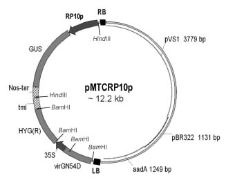
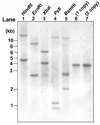
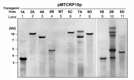
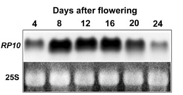
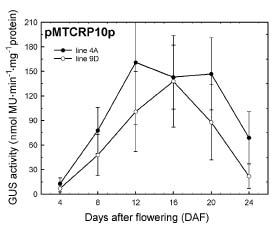
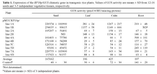
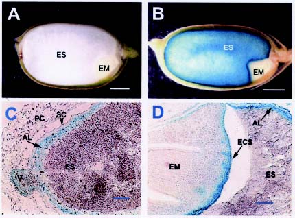
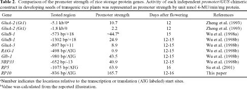
Abstract. Expression analyses of genes encoding sulfur-rich 10 kDa rice prolamins have not been reported to date although the entire genome of the model monocot plant has been sequenced in recent years. We isolated a 10 kDa rice prolamin gene, RP10, by genomic PCR, and the gene was registered in GenBank under the accession number AF294580. Genomic Southern-blot and bioinformatic study revealed that genes encoding RP10 homologes (class IV rice prolamins) were composed of two to three copies per haploid genome in Japonica cultivars Tainung 67 and Nipponbare. To elucidate the temporal and spatial expression of RP10, we introduced a chimeric gene that consisted of the 836 bp upstream sequence of RP10 and the coding regions of b-D-glucuronidase (GUS) into rice via Agrobacterium tumefaciens mediated transformation. The expression levels of GUS followed the accumulation pattern of endogenous RP10 transcript in parallel, indicating that the expression of GUS did reflect the endogenous temporal control of RP10 gene. Maximal GUS activity was reached at 12~20 days after flowering (DAF) in maturing seeds. Histochemical analysis showed that the specific expression of GUS in seeds was not restricted to the endosperm cells, but also occurred in the vascular bundle and epithelial cells of scutellum. In comparison with promoters of other rice storage protein genes, RP10 promoter exhibited a high expression level, with a long plateau period. Our studies suggest that RP10 promoter could be potentially useful for over-expression of foreign genes in transgenic rice seeds.

Keywords: Epithelial cells of scutellum; Oryza sativa; Prolamin RP10; Promoter; Seed-specificity; Storage proteins; Transformation.

Abbreviations: DAF, days after flowering; GUS, b-glucuronidase; 4-MU, 4-methylumbelliferone; PB-I, protein body I; X-Gluc, 5-bromo-4-chloro-3-indolyl-b-D-glucuronide.

Introduction

Prolamin is one of the major seed storage proteins in rice grains (Krishnan and White, 1995). It consists of three polypeptide groups, 10, 13, and 16 kDa, which all accumulate in the protein body I (PB-I) localized in the starchy endosperm. Each group has further micro-heterogeneity resulting from the corresponding gene subfamily (Tanaka et al., 1980; Ogawa et al., 1987). Analysis of amino acid composition revealed that the 13 kDa prolamin peptides, the major group, are rich in glutamic acid, glutamine, and leucine, but are poor in methionine, cysteine, and lysine. In contrast to the 13 kDa prolamin, the 10 and 16 kDa prolamins are rich in sulfur-containing amino acids. The 10 kDa prolamin, in particular, contains a large amount of methionine and cysteine (Hibino et al., 1989).

Ultrastructural analysis by electron microscopy shows that PB-I is a spherical concentric ring structure with a dense center (Bechtel and Juliano, 1980; Ogawa et al., 1987). In light of the staining mechanism, the dense center and dotted area were suggested to be the deposition

site of reducing substances, possibly contributed by 10 and/or 16 kDa sulfur-rich prolamins (Hibino et al., 1989). Research on the digestibility of rice grain revealed that the dense center of PB-I was resistant to pepsin treatment and indigestible by the human digestive tract (Bechtel and Pomeranz, 1978; Tanaka et al., 1978). Therefore, the isolation and characterization of the 10 kDa prolamin polypeptides and genes are important for understanding the formation of PB-I and genetic engineering to improve the digestibility of rice grain.

The cDNA clone lRP10, isolated from the japonica rice cultivar Nipponbare and encoding the 10 kDa sulfur-rich prolamin, was first reported by Masumura et al. (1989). A polypeptide sequence composed of 134 amino acids was deduced from the nucleotide sequence. After removing the signal peptide, the mature polypeptide contains 20% methionine and 10% cysteine (Masumura et al., 1989). Thereafter, several genes encoding the 10 kDa sulfur-rich prolamin peptides were cloned from different rice cultivars (Masumura et al., 1989; Feng et al., 1990; Wang et al., 1994; You et al., 1995). Comparison of these cloned genes revealed that nucleotide and deduced amino acid sequences share more than 95% and 93% homology, respectively. Based on the unique amino acid composition, which is rich in sulfur-containing amino acids, some workers have at

*Corresponding author. Tel: +886-2-27899590 ext. 251; Fax: +886-2-27858936; E-mail: chingsan@gate.sinica.edu.tw


Botanical Bulletin of Academia Sinica, Vol. 45, 2004

tempted to express the 10 kDa prolamin gene in heterologous plants to improve their nutritional quality (Wang et al., 1994; Yu and Ao, 1997). To date, however, the molecular details concerning the copy number, expression pattern, and promoter activity of the 10 kDa rice prolamin genes have not been reported.

In this paper, a genomic clone, RP10, encoding a rice 10 kDa prolamin polypeptide was isolated by genomic PCR. Southern-blot analysis combined with data mining of paralogs in whole genome sequences of rice suggested that genes encoding 10 kDa prolamins were composed of two to three copies per haploid genome. Transgenic analyses showed the seed-specifically expressed b-glucuronidase (GUS) driven by RP10 promoter was not restricted to the endosperm cells, but also occurred in the scutellar epithelial cells. This is the first report that a rice prolamin gene promoter was active in the embryo tissue of transgenic rice. Temporal expression of RP10 burst out earlier than the other classes of prolamin genes, and lasted for longer periods at high levels, indicating the RP10 promoter is potentially useful in over-expressing the gene of interest in rice seeds.

Materials and Methods

PCR Cloning and Characterization of RP10 Gene

Based on the published rice 10 kDa prolamin gene sequence (Feng et al., 1990), two specific primers, RP10p-S (5'-CCCAAGCTTGTGATCGCTGCACTGGATA-3') and RP10t2-AS (5'-CGGAATTCAAATAGGGATATGTTAA-3'), were designed and used to amplify the candidate genomic fragment from the Japonica rice cultivar Tainung 67 by PCR with the pfu DNA polymerase. After resolving the PCR products on agarose gel, a desired band of 1.5 kb was recovered by DNA gel extraction kit and subcloned to pBlueScript II SK(-) vector (Stratagene, USA). The recombinant plasmids were sequenced using ABI773 (PE Co., USA) autosequencer. An isolated gene encoding 10 kDa rice prolamin was named RP10.

To characterize the copy numbers and the transcript accumulation patterns of RP10 in rice, two specific primers, RP10-S (5'-ATATTGTCTACACCATCTGG-3') and RP10-AS (5'-ACACCTTATTACTAGAGTAC-3'), were designed and used to amplify the digoxigenin (DIG)-labeled RP10 coding sequence as probe by PCR method.

For Southern-blot analysis, genomic DNA was isolated from leaf tissues by the CTAB method (Dolye and Dolye, 1987). After restriction endonuclease digestion, DNA was electrophoretically separated and transferred to an Immobilon Ny+ filter membrane (Millipore, Bedford, MA). The resulting membrane was hybridized with DIG-labeled RP10 coding sequence.

For RNA analysis, developing rice seeds of different stages were harvested and stored in liquid nitrogen. Total RNA was isolated from approximately 100 seeds at 4, 8, 12, 16, 20 and 24 days after flowering as described by Ecker and Davis (1987). Approximately 25 mg RNA was resolved

by electrophoresis on 1.2% agarose gels containing formaldehyde, blotted to nylon plus membranes, and hybridized with DIG-labeled RP10 coding sequences.

Labeling, hybridization, and washing procedures were performed according to the instruction manual of the DIG labeling and Luminescent Detection kits (Roche Molecular Biochemicals, Germany).

Plasmid Construction and Agrobacterium Transformation

The 5' flanking region from -836 to -1, relative to the translation initiation site of RP10, was amplified in vitro by polymerase chain reaction and transcriptionally fused to the GUS reporter gene. A 3 kb fragment containing the RP10p/GUS chimeric gene and nopaline synthase (nos) terminator in the pBlueScript based vector was further excised with restriction enzyme HindIII and was ligated into a HindIII excised binary vector pMTC510 (Su et al., 2001). The recombinant plasmid was designated pMTCRP10p (Figure 1) and transformed to Agrobacterium tumefaciens strain EHA101 by the freeze-thaw method (An et al., 1988).

Plant Transformation

Scutellum-derived calli from matured seeds of a rice cultivar Tainung 67 were infected with Agrobacterium strain EHA101 (pMTCRP10p) as described by Toki (1997). After 3-d co-cultivation, the calli were washed and transferred onto N6D medium (Toki, 1997) containing 200 mg/L ticarcillin and 50 mg/L hygromycin for 4 weeks. Colonies of cells that proliferated on the selection medium were then plated onto regeneration medium containing 100 mg/L ticarcillin and 50 mg/L hygromycin. Regenerated plants were eventually transferred to soil in pots and grown to maturity in a greenhouse. Genomic Southern-blot probed

Figure 1. Schematic diagram of the binary transformation vector pMTCRP10p. LB and RB, left and right border sequences of T-DNA; 35S, CaMV 35S promoter; GUS, b-glucuronidase; HYG(R), hygromycin resistant gene; Nos-ter, nopaline synthase terminator; RP10p, RP10 promoter; tml, tml terminator; virGN54D, mutated virG gene.


Su et al. — Expression analyses of RP10

with a DIG-labeled GUS-coding region confirmed the transgenic events.

GUS Analysis

GUS activity was quantified by the fluorometric method used by Jefferson et al. (1987). Protein content was determined by the method of Bradford (1976) using bovine serum albumin as a standard. In consideration of the segregation manner of the introduced GUS gene cassette in R1 seeds, at least 16 maturing seeds of transgenic rice were collected and analyzed. The enzyme activity of GUS-expressed seeds was averaged to represent the quantitative value of GUS activity in R1 seeds of each individual rice plant.

For histochemical analysis, hand-cut sections of tissues were fixed with 0.2% glutaraldehyde (in 50 mM Na-phosphate buffer, pH 7.0) for 10 min and then were visualized in 100 mM Na-phosphate buffer (pH 7.0) containing 1 mM 5-bromo-4-chloro-3-indolyl glucuronide (X-Gluc) and 20% methanol at 37°C. After GUS staining, tissues were incubated in 70% ethanol at 60°C to remove pigments. Parts of stained samples were further embedded with paraffin wax for microtomic sectioning. Sections of about 20 mm were sliced with manual microtome and photographed under light microscopy.

Results

Cloning and Characterization of the Gene Encoding Rice 10 kDa Prolamin

As described in Materials and Methods, we gained a series of candidate clones encoding 10 kDa rice prolamin from rice Japonica cultivar Tainung 67. Four clones were randomly selected for DNA sequencing. Among them, three showed identical sequences, and the fourth had two base variations from its counterparts. Whether the discrepancies were due to PCR errors or another homologous gene (paralog) was uncertain. The sequence of the three identical clones was subjected to the Basic Local Alignment Search Tool (BLAST) with the non-redundant nucleotide database in GeneBank. Its coding region exhibited 97% to 100% identity with the other published rice 10 kDa prolamin genes (accession# L36604, L36605, X15231, X17074, X81970, X84649). The sequence was designated RP10 and submitted to the GenBank database under the accession number AF294580. Noticeably, RP10 showed nearly 100% identity to the 10 kDa prolamin gene (accession# X17074) published by Feng et al. (1990), with only two base pair differences located outside the coding region. The 1540 bp RP10 genomic fragment contains a 405 bp intronless open reading frame (from start codon ATG to stop codon TGA), encoding a 134 amino acid polypeptide, containing 20% methionine and 10% cysteine in mature protein.

The copy numbers of genes encoding 10 kDa rice prolamin have not been reported to date. In this paper, high stringent genomic Southern-blot was adopted to analyze

the composition of RP10. As shown in Figure 2, genomic DNA individually digested with three hexamer restriction enzymes, HindIII, EcoRI and XbaI, which do not recognize the RP10 coding region, showed two dominant hybridized bands in each lane, indicating that rice genome contained at least two copies of RP10 homologs per haploid genome. Noticeably, genomic DNA restricted with XbaI hybridized an additional weak band of nearly 3 kb. To investigate the possibility that a paralog with lower similarity might exist, we searched all rice genome sequences (HTGS collections in GenBank) by BLAST. Results showed that two highly homologous genes located on chromosome 3 (accession# AC099043) and chromosome 11 (accession# AC133248), respectively, shared 100% and 92% similarity with the full length RP10 sequence. Surprisingly, two genomic fragments with relatively less similarity (82%~83%) to the RP10 sequence were also found on chromosome 3 (accession# AC099043). One of them contains an intact open reading frame, encoding a 136 amino acid polypeptide, and the other has no intact open reading frame with the introduction of an in-frame stop codon. After analysis of the restriction enzyme sites in silica, each fragment could be cut into an approximate 2.7 kb length by XbaI, but could not be cut into the same molecular weights by HindIII or EcoRI. This may explain why genomic Southern-blot cut by XbaI showed a weak

Figure 2. Analysis of RP10 homologes in rice genome by Southern-blot. The genomic blot was digested by restriction enzymes as shown in each lane and was hybridized with DIG-labeled RP10 coding sequence in standard hybridization buffer at 68°C as suggested by the DIG system manual. Lane 6 and 7 represent the copy number standards per haploid genome.


Botanical Bulletin of Academia Sinica, Vol. 45, 2004

Figure 3. Genomic Southern-blot of rice transformed with Agrobacterium EHA101 harboring pMTCRP10p. Genomic DNAs were digested with BamHI, and probed with GUS-coding sequence. NT, non-transformed control.

band of nearly 3 kb under high stringency conditions, but not with HindIII or EcoRI digestion. These results suggest that genes encoding 10 kDa rice prolamins belong to a simple gene family, composed of two to three copies per haploid genome in Japonica cultivars Tainung 67 and Nipponbare. Each copy of RP10 homologous gene has a 100% matched corresponding EST sequence (accession# AU163802, AU163754, and AU174520, respectively), indicating that all of them may be functional genes.

Transgenic Rice Plants Were Confirmed by Genomic Southern-Blot

Total DNAs were extracted from putative transformants selected with hygromycim-containing medium. Analysis of Southern hybridization confirmed 10 independent transgenic rice plants, designated pMTCRP10p / line 1A, 2A, 4A, 4B, 5C, 7A, 9D, 1B, 2B, and 5D (Figure 3). Their transgene insertion events were quite simple and without cluster insertion, except for line 2B, which also showed morphological variations on the grain shape and reduced seed set. Most transformants harbored a single inserted transgene and exhibited normal phenotype and fertility.

The Expression Level of GUS Reporter Gene Followed the Accumulation Pattern of RP10 mRNA in Parallel

Developing rice seeds at different stages were harvested to analyze their transcript accumulations. As shown in Figure 4, mRNA of RP10 homologes obviously accumulated early at 4 days after flowering (DAF) and rapidly reached

the plateau between 8 and 16 DAF. Then, RP10-related mRNA declined from 20 to 24 DAF. The GUS reporter gene showed remarkable activity at 4 DAF, and activity surged thereafter (Figure 5). Maximal GUS expression level was reached at 12 DAF and lasted until 20 DAF. The temporal expression level of GUS reporter gene paralleled the accumulation of RP10-related transcripts, indicating that the expression pattern of GUS did reflect the temporal control of endogenous RP10 gene.

RP10 Promoter Showed Strong Promoter Activity in Rice Seeds

GUS activity driven by RP10 promoter was quantified fluorometrically in different tissues of plants. As shown

Figure 4. The transcript accumulation of RP10 homologes in developing rice seeds. The Northern blots were probed with DIG-labeled RP10 coding sequences. 25S, 25S rRNA visualized by UV exposure after ethidium bromide staining.


Su et al. — Expression analyses of RP10

in Table 1, seeds at 12~16 DAF from each independent transgenic plant showed high levels of GUS activity. Their specific activity varied from 93,181 to 254,635 pmol 4-MU/min/mg protein and averaged 165,662 pmol 4-MU/min/mg protein.

Higher GUS activities, relative to control, were detected in the leaves of pMTCRP10p/line 1B and in the culms of pMTCRP10p/line 1A, 1B and 2B (Table 1). One possible explanation is that the measured GUS activity in vegetative tissues may be the consequence of position effects. However, the GUS expression levels in seeds of these lines were more than 173-fold that of any vegetative tissues. These results suggest that the 836 bp RP10 promoter basically confers seed-specific activity.

GUS Showed an Evident Activity in the Peripheral Layers of Endosperm

Localization of GUS was further analyzed histo-chemically. GUS enzyme showed an evident activity in all endosperm cells, with stronger expression in the peripheral layers (Figure 6B). Fine structure of the peripheral endosperm layers was visualized under light microcopy after dissecting stained specimens with microtome. As shown in Figure 6C and D, blue stained areas were not restricted to the cells of aleurone layers, but also occupied the vascular bundle and epithelial cells of scutellum, closely adjacent to the a1eurone cells. It was noted that the blue indigo dye precipitated in inner endosperm cells was washed away during the specimen's preparation.

Discussion

Rice prolamin polypeptides exhibit quite complex polymorphism on SDS-PAGE and 2-D gel (Shyur et al., 1994). Their corresponding genes fall into four groups, class I, II, III and IV, based on the nucleotide sequence similarity and the amino acid composition of the deduced polypeptide sequences (Kim and Okita, 1988; Feng et al., 1990; Shyur and Chen, 1993). The same class shares more than 82% similarity in coding regions. Classes I, II and III, exhibit similarities from 58% to 83% with each other (pair-wise alignment). The unique class IV, which encodes 10 kDa rice prolamin polypeptides, only showed a 32% to 36% similarity with the other classes. Analysis of genome composition has revealed that class I and III prolamin genes were composed of dozens to hundred of copies per haploid genome depending the cultivars tested (Kim and Okita, 1988; Shyur and Chen, 1993). Masumura et al. (1990) reported that class II prolamin genes contained about 7 copies per haploid genome assayed by high stringency

Figure 5. Temporal expression pattern of the RP10p/GUS chimeric gene during rice seed development. Each point is the mean of 12~16 rice seeds. Bars represent standard error.


Botanical Bulletin of Academia Sinica, Vol. 45, 2004

Figure 6. Histochemical analysis of GUS expression in transgenic rice. Hand-cut sections were incubated in 5-bromo-4-chloro-3-indolyl-b-D-glucuronide (X-Gluc) solution at 37°C for 1~2 h. (A, B) Longitudinal-section of seeds from the non-transformed control and pMTCRP10p/line 7A, respectively. (C, D) Cross-sections of seeds of pMTCRP10p/line 7A were dissected by microtome after GUS staining. Bars represent 1 mm in (A, B) and 0.1 mm in (C,D). AL, aleurone layer; ECS, epithelial cells of scutellum; EM, embryo; ES, endosperm; PC, pericarp; SC, seed coat; V, vascular tissue.

Southern hybridization. These results reflect the observed complexity of prolamin polypeptides on 2-D gels (Shyur et al., 1994). The copy numbers of class IV prolamin genes have not been reported to date. In this paper, we have shown that a relatively simple gene family, composed of two to three copies per haploid genome, encodes the 10 kDa rice prolamins.

Northern-blot analysis showed that the transcripts of RP10 homologes plateaued at 8 DAF and remained at a high level to 16 DAF. In a previous study, we demonstrated that mRNA of RP5 homologes (class III prolamin) rapidly increased after 8 DAF and peaked at 12~16 DAF (Su et al., 2001). The steady state mRNA levels of class I (Prol 14) and class II (Prol 17) prolamins continue to increase throughout seed development and attain maximal levels at 20~25 DAF (Kim et al., 1993). The non-coordinated expression patterns of these prolamin genes suggest that different classes of rice prolamin genes may have different regulatory machineries. Accumulation of the RP10 transcript seems to reach its plateau earlier and remain at the maximal level much longer than the other classes of prolamin genes during seed development.

Almost all seed storage proteins are composed of gene families, and the determination of spatial and temporal expression of a single gene is difficult (Pickardt et al., 1998). The introduction and analysis of a promoter/reporter fusion gene in a transgenic system provides an alternative strategy. In this report, we show that the temporal expression pattern of the GUS reporter gene paralleled that of the endogenous RP10 transcript (Figures 4 and 5), indicating that the 836 bp RP10 promoter is sufficient to confer the temporal expression as the endogenous RP10 gene. Histochemical analysis showed that RP10p/GUS chimeric gene was expressed at a high level in transgenic rice seeds. The expression of GUS was not restricted in endosperm cells, but also in the vascular bundle and epithelial cells of scutellum. This is the first report that a rice prolamin gene promoter was active in the embryo tissue of transgenic rice.

The use of plants as bioreactors for foreign gene expression has increased recently (Gallie, 1998). Cereal grains have been considered as molecular farms for the production of recombinant proteins and novel metabolites. Consequently, potentially useful promoters, which could drive a desirable level of gene expression with tissue-


Su et al. — Expression analyses of RP10

specificity, are in demand of late. A number of rice storage protein gene promoters have been evaluated recently. The rice globulin has been postulated as the most abundant seed storage protein encoded by a single gene, and the globulin promoter could be the strongest among all seed storage protein genes in rice (Hwang et al., 2001; Hwang et al., 2002). However, comparing the promoter strength among rice storage protein genes, in Table 2, RP10 promoter could be one of the strongest (Zheng et al., 1993; Wu et al., 1998a,b; Su et al., 2001). Note that Hwang et al. (2001) adopted a transient expression assay to evaluate the promoter activity in rice grains. In their study, immature endosperm tissues harvested from 7 to 9 days after pollination showed the highest GUS expression of all of the promoter/reporter constructs tested. At 7~9 days after pollination, indeed, the expression levels of many storage protein genes have not peaked (Kim et al., 1993; Mitsukawa et al., 1999; Su et al., 2001). Furthermore, it is well known that the bulky seed storage proteins are deposited in the outmost layers of endosperm at middle to later developmental stages of maturing seeds. Thus, the use of stable transformation for studying the gene regulation of seed storage proteins seems more reliable.

The segregation of antibiotic-resistant traits in R1 progeny of pMTCRP10p line 1A and 4A followed the 3:1 (resistant to sensitive) ratio with the x2-test. This indicated that the single-copy transgene inserted transformants followed the classic Mendelian inheritance. Furthermore, the RP10p/GUS chimeric gene was expressed in the R2 seeds from all transgenic R1 progeny plants and remained at higher levels than those measured in the R1 seeds (data not shown). Thus, RP10 promoter seems an excellent candidate for expressing the gene of interest at higher levels in rice seeds in view of its seed-specificity, high expression level, long expression periods at plateau stage, and inheritability.

Acknowledgements. This work was supported by Grant NSC-89-2311-B-001-020 from the National Science Council, Republic of China. We thank Dr. Mei-Chu Chung at the Institute of Botany, Academia Sinica for help with the microtomic section.

Literature Cited

An, G., P.R. Ebert, A. Mitra, and S.B. Ha. 1988. Binary vector. In S.B. Gelvin, R.A. Schilperoort, and D.P.S. Verma (eds.), Plant Molecular Biology Manual. Kluwer Academic Publishers, The Netherlands, pp. A3: 1-19.

Bechtel, D.B. and B.O. Juliano. 1980. Formation of protein bodies in the starchy endosperm of rice (Oryza sativa L.): a re-investigation. Ann. Bot. 45: 503-509.

Bechtel, D.B. and Y. Pomeranz. 1978. Ultrastructure of the mature ungerminated rice (Oryza sativa) caryopsis. The starchy endosperm. Amer. J. Bot. 65: 684-691.

Bradford, M.M. 1976. A rapid and sensitive method for the quantification of microgram quantities of protein utilizing the principle of protein-dye binding. Anal. Biochem. 72: 248-254.

Doyle, J.J. and J.J. Doyle. 1987. A rapid DNA isolation procedure for small quantities of fresh leaf tissue. Phytochem. Bull. 19: 11-15.

Ecker, J.R. and R.W. Davis. 1987. Plant defense genes are regulated by ethylene. Proc. Natl. Acad. Sci. USA 84: 5202-5206.

Feng, G., L. Wen, J.K. Huang, B.S. Shorrosh, S. Muthukrishnan, and G.R. Reeck. 1990. Nucleotide sequence of a cloned rice genomic DNA fragment that encodes a 10 kDa prolamin polypeptide. Nucleic Acids Res. 18: 3.

Gallie, D.R. 1998. Controlling gene expression in transgenics. Curr. Opin. Plant Biol. 1: 166-172.

Hibino, T., K. Kidzu, T, Masumura, K. Ohtsuki, K. Tanaka, M. Kawabata, and S. Fujii. 1989. Amino acid composition of rice prolamin polypeptides. Agric. Biol. Chem. 53: 513-518.

Hwang, Y.S., D. Yang, C. McCullar, and N. Huang. 2001. Evaluation of expression cassettes in developing rice endosperm using a transient expression assay. Plant Sci. 161: 1107-1116.

Hwang, Y.S., D. Yang, C. McCullar, L. Wu, L. Chen, P. Pham, S. Nandi, and N. Huang. 2002. Analysis of the rice endosperm-specific globulin promoter in transformed rice cells. Plant Cell Rep. 20: 842-847.

Jefferson, R.A., T.A. Kavanagh, and M.W. Bevan. 1987. GUS fusions: b-glucuronidase as a sensitive and versatile gene fusion marker in higher plants. EMBO J. 6: 3901-3907.


Botanical Bulletin of Academia Sinica, Vol. 45, 2004

Kim, W.T., X. Li, and T.W. Okita. 1993. Expression of storage protein multigene families in developing rice endosperm. Plant Cell Physiol. 34: 595-603.

Kim, W.T. and T.W. Okita. 1988. Structure, expression, and heterogeneity of the rice seed prolamins. Plant Physiol. 88: 649-655.

Krishnan, H.B. and J.A. White. 1995. Morphometric analysis of rice seed protein bodies: implication for a significant contribution of prolamine to the total protein content of rice endosperm. Plant Physiol. 109: 1491-1495.

Masumura, T., T. Hibino, K. Kidzu, N. Mitsukawa, K. Tanaka, and S. Fujii. 1990. Cloning and characterization of a cDNA encoding a rice 13 kDa prolamin. Mol. Gen. Genet. 221: 1-7.

Masumura, T., D. Shibata, T. Hibino, T. Kato, K. Kawabe, G. Takeba, K. Tanaka, and S. Fujii. 1989. cDNA cloning of an mRNA encoding a sulfur-rich 10 kDa prolamin polypeptide in rice seeds. Plant Mol. Biol. 12: 123-130.

Mitsukawa, N., R. Konishi, M. Uchiki, T. Masumura, and K. Tanaka. 1999. Molecular cloning and characterization of a cysteine-rich 16.6-kDa prolamin in rice seeds. Biosci. Biotech. Biochem. 63: 1851-1858.

Ogawa, M., T. Kumamaru, H. Satoh, N. Iwata, T. Omura, Z. Kasai, and K. Tanaka. 1987. Purification of protein body-I of rice seed and its polypeptide composition. Plant Cell Physiol. 28: 1517-1527.

Pickardt, T., B. Ziervogel, V. Schade, L. Ohl, H. Bäumlein, and M. Meixner. 1998. Developmental-regulation and tissue-specific expression of two different seed promoter GUS-fusions in transgenic lines of Vicia narbonensis. J. Plant Physiol. 152: 621-629.

Shyur, L.F. and C.S. Chen. 1993. Rice prolamins: heterogeneity of cDNAs and synthesis of precursors. Bot. Bull. Acad. Sin. 34: 143-154.

Shyur, L.F., T.N. Wen, and C.S. Chen. 1994. Purification and characterization of rice prolamins. Bot. Bull. Acad. Sin. 35:

65-71.

Su, P.H., S.M. Yu, and C.S. Chen. 2001. Spatial and temporal expression of a rice prolamin gene RP5 promoter in transgenic tobacco and rice. J. Plant Physiol. 158: 247-254.

Tanaka, K., T. Sumigoto, M. Ogawa, and Z. Kasai. 1980. Isolation and characterization of two types of protein bodies in the rice endosperm. Agric. Biol. Chem. 44: 1633-1639.

Tanaka, Y., A.P. Resurreccion, B.O. Juliano, and D.B. Bechtel. 1978. Properties of whole and undigested fraction of protein bodies of milled rice. Agric. Biol. Chem. 42: 2015-2023.

Toki, S. 1997. Rapid and efficient Agrobacterium-mediated transformation in rice. Plant Mol. Biol. Rep. 15: 16-21.

Wang, G.L., Z.X. Qian, B.X. Liu, M. Ma, and Z.P. Lin. 1994. Cloning and sequencing of the gene encoding 10 kD prolamin of rice and transformation in Lotus corniculatus. Acta Bot. Sin. 36: 351-357.

Wu, C.Y., T. Adachi, T. Hatano, H. Washida, A. Suzuki, and F. Takaiwa. 1998a. Promoters of rice seed storage protein genes direct endosperm-specific gene expression in transgenic rice. Plant Cell Physiol. 39: 885-889.

Wu, C.Y., A. Suzuki, H. Washida, and F. Takaiwa. 1998b. The GCN4 motif in a rice glutelin gene is essential for endosperm-specific gene expression and is activated by Opaque-2 in transgenic rice plants. Plant J. 14: 673-683.

You, L.R., M. Xie, L.J. Qu, X.W. Liang, H.Y. Gu, and Z.L. Chen. 1995. Molecular cloning and sequence analysis of a gene encoding rice 10 kD prolamin. Acta Bot. Sin. 37: 507-513.

Yu, J.J. and G.M. Ao. 1997. Expression of 10 kD sulfur-rich prolamin gene of rice in potato. Acta Bot. Sin. 39: 329-334.

Zheng, Z., Y. Kawagoe, S. Xiao, Z. Li, T. Okita, T.L. Hau, A. Lin, and N. Murai. 1993. 5' distal and proximal cis-acting regulator elements are required for developmental control of a rice seed storage protein glutelin gene. Plant J. 4: 357-366.


Su et al. — Expression analyses of RP10