Bot. Bull. Acad. Sin. (2004) 45: 111-118

Chen et al. — Purification and characterization of carboxymethyl cellulase

Purification and characterization of carboxymethyl cellulase from Sinorhizobium fredii

Po-Jui Chen1, Tao-Chun Wei2, Yao-Tsung Chang2, and Liang-Ping Lin2,*

1Department of Agricultural Chemistry, National Taiwan University, Taipei 106, Taiwan, Republic of China

2Institute of Microbiology and Biochemistry, National Taiwan University, Taipei 106, Taiwan, Republic of China

(Received July 12, 2001; Accepted January 13, 2004)

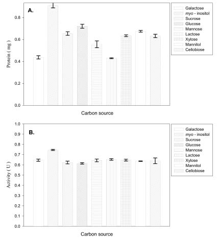
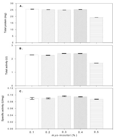
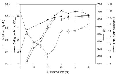
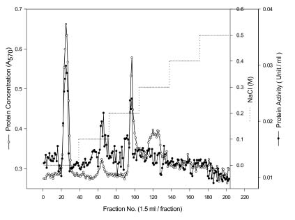
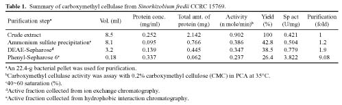
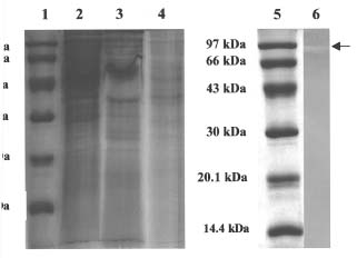
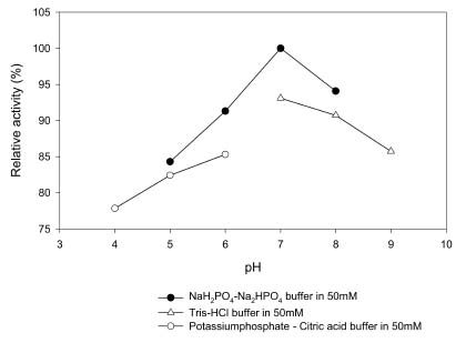
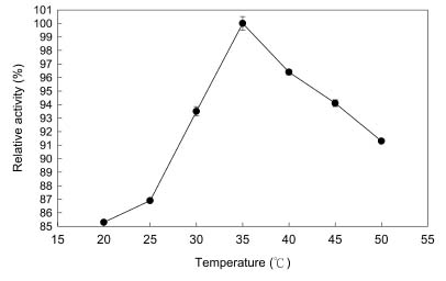
Abstract. Carboxymethyl cellulase (CMCase) is the key enzyme used by bacteria to decompose the plant root hair-wall during symbiosis. In this study, CMCase activities were routinely assayed using carboxymethyl cellulose (CMC) as a substrate. Coupled with the catalytic reaction of BCA (2,2'-bicinchoninic acid) solution, the color developed at 570 nm was measured to determine the enzyme activity. Sinorhizobium fredii CCRC 15769 was utilized to produce CMCase for this study. The optimum growth conditions of the cultural medium were studied in order to obtain sufficient cells for further CMCase purification, and further, for large-scale fermentation production. BIII medium containing 0.3% myo-inositol proved to be a suitable carbon source for bacterial cell growth and specific activity of CMCase. A 22.4 g wet weight of cells can be harvested from 3.5 liters of fermented medium through continuous centrifugation. The cells were suspended in 50 mM potassium phosphate-citric acid (PCA) buffer at pH 5.2 and disintegrated through ultra-sonication to obtain crude extract. Then, certain precipitates were collected at 40-60% ammonium sulfate saturation via ammonium sulfate fractionation of the crude extract. The supernatant obtained following centrifugation was loaded onto a DEAE Sepharose anion-exchange column, and the active fractions were collected and dialyzed against 10 mM Tris-HCl buffered at pH 7.4. For further purification, the dialyzed fraction was loaded onto a Phenyl-Sepharose column. The active fractions were dialyzed and then assayed by SDS-PAGE activity stain to confirm that it contained CMCase activity at the fraction of 94 kDa. The characterizations of CMCase were demonstrated as follows: the optimal temperature and pH were 35°C and 7.0, respectively. The purification fold was 9.08, and the recovery yield was 26.4%, and the specific activity was 3.822 U mg-1.

Keywords: Carboxymethyl cellulase; Sinorhizobium fredii; Protein extracts.

Abbreviations: CMCase, carboxymethyl cellulase; BCA, 2,2'-bicinchoninic acid; PCA, potassium phosphate-citric acid.

Introduction

Rhizobia are gram-negative bacteria that can induce morphologically distinct nodules on leguminous plant roots. The symbiotic interaction results in the inclusion of highly differentiated bacterial cells in host plant nodule cells, where they reduce atmospheric dinitrogen to ammonia, which is then utilized by the host plant. Establishment of an intracellular symbiosis between rhizobia and plants, which produces dinitrogen fixation, requires mutual recognition and signaling, penetration of the host cell, and finally nodulation and survival of bacteria in bacteroids. Carboxymethyl cellulase is probably involved in all of the infection stages.

The precise mechanism by which Rhizobium infects temperate legumes successfully remains unknown. Nutman (1956) proposed that rhizobia invade root hairs via an invagination process of the plant cell wall, which implies that the bacteria do not penetrate the root cell. Ljunggren and

Fahraeus (1961) proposed that polygalacturonase production by plant root cells, induced by homologous strains of Rhizobium, results in an increase in plant cell wall softening at the infection site on the root hair, thus allowing the bacteria to penetrate the cell membrane and initiate an infection thread. Mateos et al. (1992) proposed that the enzyme from Rhizobium leguminosarum bv. trifolii degraded both carboxymethyl cellulose and polypectate model substrate. This produced a localized degradation that completely traversed the root hair wall and facilitated direct bacterial penetration.

Electron microscopic studies of the infection process confirm a localized degradation of the root hair wall, indicating involvement of cell wall hydrolytic enzymes in the penetration process (Higashi and Abe, 1980; Callaham and Torrey, 1981; Ridge and Rolfe, 1985; Turgeon and Bauer, 1985). We are concerned with the possibility that the wall-degradation enzymes involved in the process are either associated with the bacteria or are locally induced in the plant by bacterial components.

Infection between rhizobia and legume roots is a delicately balanced process (Dazzo and Hubbell, 1982). If the infected root hair did not remain intact and viable, the in

*Corresponding author. Tel: 886-2-23635523; Fax: 886-2-23626455; E-mail: m046@ccms.ntu.edu.tw


Botanical Bulletin of Academia Sinica, Vol. 45, 2004

mM Tris-HCl buffer (pH 7.4) and precipitated with ammonium sulfate at 40 to 60% saturation for 1 h with gentle stirring. The precipitated proteins were recovered by centrifugation at 8,000 g for 30 min, and were dialyzed against 10 mM Tris-HCl buffer (pH 7.4) supplemented with 0.15 M NaCl. The insoluble residue was removed after dialysis by centrifugation at 8,000 g for 30 min.

Double-Layer Plate Assay

A modification of the Saleh-Rastin et al. (1991) plate assay was employed herein. To detect the crude enzyme activity, a bottom layer that contained 15 ml of 0.7% (w/v) agarose and 50 mM potassium phosphate-citric acid buffer (pH 5.2) was overlaid with 5 ml of 0.2% (w/v) carboxymethyl cellulose (CMC) and 0.5% (w/v) agarose. The plates were inoculated with 40 µl protein extract and were incubated at 30°C for 20 h. In order to detect carboxymethyl cellulase activity, plates were flooded with 0.1% (w/v) Congo red solution for 30 min and then rinsed several times with 1 M NaCl. This procedure revealed distinct hydrolysis regions.

Purification of CMCase from Crude Protein Extract

A two-step chromatographic procedure was employed to purify CMCase from a crude protein extract. Carboxymethyl cellulose was employed as the substrate to monitor carboxymethyl cellulase activity. The experiments were performed at room temperature.

(i)Ion-exchange chromatography on DEAE-sepharose. Approximately 8.5 ml of clear crude extract was loaded onto a DEAE-Sepharose Fast Flow anion-exchange column (Pharmacia; 17.5 by 1.6 cm; 1 ml min-1) previously equilibrated in 50 mM Tris-HCl buffer (pH 7.4). The carboxymethyl cellulase was eluted in 50 mM Tris-HCl buffer (pH 7.4) with a 0.1 to 0.5 M NaCl gradient. Fractions (1.5 ml) were collected and tested for carboxymethyl cellulase activity. Active fractions were pooled, dialyzed, and equilibrated with 50 mM Tris-HCl buffer (pH 7.4).

(ii)Hydrophobic chromatography on phenyl-sepharose. Carboxymethyl cellulase active fractions were loaded onto a Phenyl-Sepharose 6 column (Pharmacia; 11.5 by 1.6 cm; 1 ml min-1) previously equilibrated in 50 mM Tris-HCl buffer (pH 7.4) supplemented with 40% (w/v) ammonium sulfate. Carboxymethyl cellulase was eluted first via 50 mM Tris-HCl buffer (pH 7.4) supplemented with 40% (w/v) ammonium sulfate and then with a 30 to 0% ammonium sulfate gradient. Active fractions were pooled, dialyzed and concentrated with a Centricon plus-20 membrane (Millipore, USA) to a final volume of approximately 0.2 ml.

Assay for CMCase Activity

CMCase activities were assayed using sodium salts of CMC in PCA buffer 0.2% (w/v) as substrate. The reaction mixture contained 0.2 ml of substrate, 0.2 ml of enzyme solution, and 0.4 ml of PCA buffer. This mixture was incubated at 30°C for 5 h under shirring. A 2,2'-

fection would cease. Furthermore, wall-degrading enzymes would affect the infection, and the activities would remain limited and localized at the site of infection to avoid destroying the root hair. However, there is no direct evidence for the involvement of hydrolytic enzymes in the infection process, and little is known about this enzyme in Rhizobium (Martinez-Molina and Olivares, 1982; Al-Mallah et al., 1987; Chaliflour and Benhamou, 1989). Mateos et al. (1992) developed sensitive assays to detect cellulolytic enzymes that were produced by various wild-type strains of Rhizobium leguminosarium bv. trifolii. The enzyme was cell-bound and commonly produced by wild-type strains isolated from various geographical regions. It contained at least two carboxymethyl cellulase (CMCase) isozymes. Growth in defined B-INOS medium supplemented with carboxymethyl cellulose (CMC) or by flavone 7,4'-dihydroxyflavone that activates expression of pSym nodulation genes failed to affect the cell-bound enzyme in sonicated cell extracts (Mateos et al., 1992; Jimenez-Zurdo et al., 1996). Recently, Mateos et al. (2001) suggested a complementary function of rhizobial cell-bound hydrolytic enzymes exists in the initial host infection.

These experimental results explain the molecular mechanisms implied in symbiosis and in detecting the polysaccharide-degrading enzyme by rhizobia. Furthermore, they support the hypothesis of hydrolytic enzyme involvement in the symbiosis. Therefore, a carboxymethyl cellulase that is constitutively expressed in Sinorhizobium fredii sp. strain CCRC 15769 was purified and characterized herein.

Materials and Methods

Microorganism and Culture Maintenance

Rhizobium fredii RO (CCRC 15769) was isolated from indigenous soil in Taiwan and stored in yeast-mannitol agar at 4°C. The bacteria were maintained on BIII agar : 3 g myo-inositol l-1, 1.1 g sodium glutamate l-1, 0.23 g K2HPO4 l-1, 0.1 g MgSO4·7H2O l-1, 1 ml trace element l-1 (containing CaCl2, H3BO3, FeSO4·7H2O, CoSO4·6H2O, CuSO4·5H2O, MnCl2·4H2O, ZnSO4·7H2O, Na2MoO4), nitrilotriacetic acid 1 ml, vitamin l-1 (containing p-aminobenzoic acid, biotin, calcium pantothenate, inositol, pyridoxine-HCl, thiamine-HCl, riboflavin ), 20 g agar l-1; pH 7.0. They were also cultured aerobically in BIII broth at 28°C with agitation. For protein purification, bacteria were grown at 28°C in a 5-liter fermentor (Mitsuwa Biosystem Inc, Tokyo, Japan) in BIII medium with stirring and gentle aeration. In the late exponential growth phase, rhizobia were harvested by centrifugation at 15,000 g for 15 min. A 22.4-g bacterial pellet was recovered from the cultures and stored at 4°C.

Crude Protein Extraction

All extraction steps were performed at 4°C. A stored bacterial pellet (22.4 g) was resuspended in 25 ml of 50 mM potassium phosphate-citric acid buffer (PCA; pH5.2), sonicated in six cycles of 30-min bursts at 50 W with microprobe (Virsonic, USA), and centrifuged at 8,000 g for 30 min. The supernatant was resuspended in 15 ml of 50


Chen et al. — Purification and characterization of carboxymethyl cellulase

bicinchoninic acid (BCA) method was used to measure the product released (Waffenschmidt and Jaenicke, 1987). Samples of 0.2 ml were mixed with 0.8 ml distilled water and 1 ml of BCA reagent in tubes, which were capped and heated to 100°C for 15 min. Samples were then cooled at room temperature for 10 min, and their absorbance was measured at 570 nm.

Analytical Methods

Bradford's method (Bradford, 1976), which used the Bio-Rad assay reagent and bovine serum albumin (BSA) as the standard, determined protein contents. Based on the Laemmli method (1970), sodium dodecyl sulfate (SDS)-polyacrylamide gel electrophoresis (PAGE) was carried out on 12.5% polyacrylamide gels in the presence of b-mercaptoethanol with Bio-Rad apparatus. The gels were stained with Coomassie blue R250. The low-molecular-weight protein markers and active stain protein markers were purchased from Bio-Rad (Hercules, CA, USA). SDS-PAGE on 10% polyacrylamide gels with 0.05% (w/v) SDS was employed to detect carboxymethyl cellulase activity and 20 µg of bovine serum albumin ml-1 was used to enhance renaturation of electrophoresed enzymes. Enzymes were mixed with sample buffer (62 mM Tris-HCl at pH 6.8, supplemented with 10% glycerol, 0.025% bromophenol blue, and 2% SDS) in a 5:1 (v/v) ratio without heating. Following electrophoresis, the proteins were renatured via incubation for 6 h in two changes of 10 mM PCA buffer at pH 5.2. The running gel, which had been renatured and incubated in a moist chamber at 30°C for 12 h was placed on agarose overlay that contained 0.2% (w/v) CMC and 0.5% (w/v) agarose. Then, the overlay was flooded with 0.1% Congo red solution for 30 min, rinsed in 1 M NaCl, and photographed.

Results and Discussion

Effect of Carbon Sources on CMCase Production

Cultures were grown in BIII medium (pH 7.0) that contained 0.5% (w/v) of various carbon sources at 28°C for 12 h, and the CMCase activities of protein extract were assayed (Figure 1). Maximum activity (0.76 U) was detected in cultures that contained 0.5% (w/v) myo-inositol as the growth carbon source. Cultures containing 0.5% (w/v) glucose as the growth carbon source presented the minimum CMCase activity (0.62 U). Furthermore, cultures were grown in BIII medium (pH 7.0) that contained 0.1 to 0.5% (w/v) myo-inositol concentrations at 28°C for 12 h, and the CMCase activities of protein extract were also assayed (Figure 2). Maximum activity (100% representing specific activity of 0.096 U/mg) was detected in cultures that contained 0.3% (w/v) myo-inositol. However, the original concentration of 0.5% (w/v) myo-inositol presented alternate findings. That is, only 0.88 U mg-1 of the specific activity appeared in cultures. Unlike in previous investigations (Fukumori et al., 1985; Kawai et al., 1988; Shikata et al., 1990), the CMCase activity in Bacillus sp. was detected (100% representing specific activity of 1.34 U/mg) in cul

Figure 1. Effect of different carbon source medium on carboxymethyl cellulase production and activity from Sinorhizobium fredii sp. Strain CCRC 15769 (A) protein (B) activity. Crude extract protein (mg) was assayed from 200 ml cultures. One unit of enzyme activity (U) is the amount releasing 1nmole of reducing sugar per min at 35°C.

Figure 2. Effect of the myo-inositol content in medium on (A) CM-cellulase production; (B) CM-cellulase activity; and (C) CM-cellulase specific activity. Crude extarct protein (mg) was assayed from 200 ml cultures. One unit of enzyme activity (U) is the amount releasing 1 nmole of reducing sugar per min at 35°C. Specific activity: (B) total activity / (A) total protein.


Botanical Bulletin of Academia Sinica, Vol. 45, 2004

tures that contained 1% (w/v) CMC as the growth substrate. Cultures that contained Avicel presented only 50% of CMCase activity. Robson and Chambliss (1984) reported that the presence of metabolized carbohydrates in the growth medium stimulated the production of cellulolytic activity. Furthermore, Mateos et al. (1992) confirmed that growth in B-INOS broth supplemented with 0.2% (w/v) CMC or polypectate substrates failed to alter the specific activities of the corresponding depolymerizing enzymes in the sonicated cell fraction of Rhizobium leguminosarum bv. trifolii ANU843, thus indicating no evidence for substrate induction.

Culture Conditions and CMCase Activities Performance

The Sinorhizobium fredii sp. strain CCRC 15769 was cultured in BIII medium. Growth and CMCase production required neutral conditions (pH 7.0). CMCase activity was measured during growth in the modified medium, in which the enzymatic activity increased simultaneously with culture time. The maximal activity occurred in the late exponential growth phase, following 40 h of cultivation (roughly 0.7 U). Culture pH decreased rapidly in the exponential growth phase at 16 h of cultivation, but increased following the middle exponential growth phase. The pH value in the final culture time (early stationary growth phase) was approximately 6.92 (Figure 3). Protein extracts from Sinorhizobium fredii sp. strain CCRRC 15769 were assayed to determine their ability to hydrolyze polysaccharides (e.g. CMC) at various enzymatic reaction times (Figure 4). High levels of CMCase activity were detected after 3 h enzymatic reaction. In the original enzymatic reaction (5 h) lower levels of CMCase activity were detected. This indicated that the protein extracts from Sinorhizobium fredii sp. strain CCRRC 15769 had passed the optimum enzymatic reaction time (3 h). Industries are concerned with rapidly growing cellulolytic bacteria. Some strains, such as Bacillus are able to produce CMCase in a short time, roughly 12 h of cultivation (Chan and Au, 1987; Sharma et

al., 1990). However, generally, maximal production in other Bacillus strains can be achieved only after 2 or 3 days (Robson and Chambliss, 1984; Kawai et al., 1988; Ito, 1989).

Preliminary Location of CMCase

The double-layer plate assay in sonicated cell extracts of Sinorhizobium fredii sp. strain CCRC 15769, as well as in other cell treatments, detected CMCase activities during the extraction process (Figure 5). The enzyme activities were detected within 20 h of incubation. No activity was found in either the supernatant of cell extracts or in the extracellular supernatant of culture media following centrifuging. CMCase activity occured in the pellet of cell extracts and formed a concentric circle zone on the upper

Figure 4. Contribution and CM-cellulase activity of the different cell fraction or treatment at different enzyme reaction time from Sinorhizobium fredii strain CCRC 15769 using ultra-sonication method. One unit of enzyme activity (U) is the amount releasing 1 nmole of reducing sugar per min at 30°C.

Figure 5. CM-cellulase activities of 20 incubation time and fractions from Sinorhizobium fredii RO detected by the double-layer plate assay. (A) blank; (B) cellulase from Aspergillus niger; (C) supernatant of the cell extract after centrifuging; (D) extracellular supernatant after centrifuging; (E) pellet of the cell extract after centrifuging.

Figure 3. Growth and CM-cellulase production in culture of Sinorhizobium fredii CCRC 15769. The values and the error bar reported means of triplicate samples derived from equivalent numbers of cells.


Chen et al. — Purification and characterization of carboxymethyl cellulase

Figure 6. The elution profile of DEAE-Sepharose Fast Flow anion exchange chromatography. The dialyzed enzyme solution collected from 40~60 % saturation of ammonium sulfate was loaded onto a DEAE- Sepharose Fast Flow column (1.6 × 17.5 cm) pre-equilibrated with 50 mM Tris-HCl buffer at pH 7.4. After the column had been washed with two bed volumes of the same buffer, the protein was eluted with a stepwise increasing gradient of NaCl from 0.1 M to 0.5 M. The active fractions 22-29, 62-68 and 94-102 were collected and dialyzed against 50 mM Tris-HCl buffer at pH 7.4.

Figure 7. The elution profile of Phenyl Sepharose 6 Fast Flow hydrophobic interaction chromatography. The dialyzed enzyme solution collected from anion exchange chromatography was applied to a Phenyl Sepharose 6 Fast Flow column (1.6 × 11.5 cm) pre-equilibrated with 50 mM Tris-HCl buffer at pH 7.4 containing ammonium sulfate at 40% saturation. After the column had been washed with three bed volumes of the same buffer, the protein was eluted with a stepwise decreasing gradient from 40% to 0%. The active fraction 84-87 was collected and dialyzed against 50 mM Tris-HCl at pH 7.4.

extracts of pelleted cells produced by sonication or treated with lysozyme-EDTA. Our study also corroborated the finding that CMCase activity in Sinorhizobium fredii sp. strain CCRC 15769 was detected in cell pellet extracts.

Purification of CMCase

The crude protein was first precipitated with a 40 to 60% ammonium sulfate saturation. Then a two-step chromatography procedure was employed to purify the collected CMCase activity fractions. Following concentration, DEAE-Sepharose anion exchange chromatography purified the enzyme partially (Figure 6). Due to the low volume of pooled fractions (62-68) and (94-102), the pooled fractions (22-29) CMCase activities were collected for the next purification step. The final hydrophobic interaction chromatography (Figure 7) step yielded 62 µg of electrophoretically pure CMCase from cell pellet (Table 1).

layer of the agar plates. Mateos et al. (1992; 2001) reported that CMCase activity was cell-bound and is commonly produced by diverse wild-type Rhizobium leguminosarum bv. trifolii strain. Our study also confirms these experimental results and further indicates that various rhizobia also commonly produce cellulolytic enzymes. The sensitive BCA reducing sugar assay was employed to investigate the cell-bound or the extracellular distribution of CMCase, polygalacturonase, and b-galactosidase activities associated with rhizobia grown in the absence or presence of inducer of flavone, which activates nodulation gene expression (Peters et al., 1986). Notably, b-galactosidase was included since it is cell-bound and often associated with cellulase in both aerobic and anaerobic bacteria (Rapp and Beermann, 1991). Polygalacturonase activity was only detected in the extracts of sonicated cells, while both CMCase and b-galactosidase activities were detected in


Botanical Bulletin of Academia Sinica, Vol. 45, 2004

The enzyme was purified approximately 9.1-fold. Its specific activity and yield were 3.82 U mg-1 and 26.4%, respectively. An activity gel overlay detection method was employed to estimate the relative molecular weight (Mr) of the CMCase. In addition, 12.5% SDS-PAGE in the presence with b-mercaptoethanol clearly indicated that the polypeptide chains did not contain any interchain disulfide bridges (Figure 8A). SDS-PAGE resolved the purified CMCase of Sinorhizobium fredii sp. strain CCRC 15769, and the activity stain overlay technique detected it (Figure 8B). The purified CMCase appeared to be a monomer. That is, in comparison to the low-molecular-weight standards of Bio-Rad electrophoresis within SDS revealed a single band at a molecular weight of 94,000. No other CMCase isozyme was detected under these conditions. CMCase purification from the culture supernatant of alkaline Bacillus sp. strain HC-1 at pH 9.5 had a yield of 10.6% and specific activity of 5.7 U/mg (Khyami-Horani, 1991). Chatterjee and Sanwal (1999) also purified CMCase from Cuscuta reflexa with 42-fold purification and had an approximate 6% recovery rate. Native PAGE and antigen-antibody cross reactivity determined that the purified enzyme was homogeneous. Berthelot and Delmotte (1999) purified a novel a-glucosidase, which performed an essential function in degrading of the plant cell wall from protein extracts of Rhizobium sp. strain USDA 4280. Glucosidases are involved in the stage of infection between plant root hairs and rhizobia.

Effect of pH on Activity and Stability

The enzyme hydrolyzed CMC in the pH range of 4.0 to pH 9.0, with maximum activity occurring at pH 7.0 (Figure 9). The enzyme's stability was investigated in buffer solutions of various pH values. Purified enzyme solution (30 µg in 100 µl) was mixed with 100 µl of the appropriate buffer and incubated at 4°C for 24 h, after the BCA assay method had examined residual activities. The enzyme was stable over the pH range of 6.0 to 9.0 within these conditions. However, it had maximum stability at pH 7.0 in 50 mM phosphate buffer and retained 75% of its activity at pH 4.0 in 50 mM potassium phosphate buffer (Figure 9). Alkaline pH value (pH 12) also affected the CMCase activity in Bacillus sp. by 50%. Furthermore, the CMCase in Bacillus sp. is alkaline, which renders it suitable for use as an effective laundry detergent additive (Khyami-Horani, 1991).

Effect of Temperature on Activity and Stability

The enzyme was assayed at various temperatures in 50 mM phosphate buffer (pH 7.0). Although it had maximal activity at 35°C, it retained 96% of it at 40°C (Figure 10). The thermal stability of the enzyme indicated that the activity was stable at 30 to 45°C, kinetic was low at 20 to 25°C.

Acknowledgements. This work was supported by a grant from the National Science Council, Taipei 106, Taiwan, Republic of China.

A B

Figure 8. (A) 12.5% Polyacrylamide SDS-PAGE of carboxymethyl cellulase from Sinorhizobium fredii RO. Lane 1, low-molecular-weight Bio-Rad standard markers; Lane 2, ammonium sulfate precipitate fraction (40%~60% saturation); Lane 3, DEAE-Sepharose fractions; Lane 4, Phenyl-Sepharose fractions; (B) 12.5% SDS-PAGE. Lane 5, low-molecular-weight Bio-Rad standard markers; Lane 6, enzymatic activity of purified carboxymethyl cellulase from Phenyl-Sepharose 6 column.

Figure 9. Effect of pH on enzyme activity and stability. The three different pH regions are examined for relative activities.

Figure 10. Effect of reaction temperature on enzyme activity and stability in 50 mM phosphate buffer at pH 7.0.


Chen et al. — Purification and characterization of carboxymethyl cellulase

Literature Cited

Al-Mallah, M., M. Davey, and E. Cocking. 1987. Enzymatic treatment of clover root hairs removes a barrier to Rhizobium-host specificity. Biotechnology 5: 1319-1322.

Berthelot, K. and F.M. Delmotte. 1999. Purification and characterization of an a-glucosidase from Rhizobium sp. (Robinia pseudoacacia L.) strain USDA 4280. Appl. Environ. Microbiol. 65: 2907-2911.

Bradford, M.M. 1976. A rapid and sensitive method for the quantitation of microgram quantities of protein utilizing the principle of protein-dye binding. Anal. Biochem. 72: 248-254.

Callaham, D. and J. Torrey. 1981. The structural basis for infection of root hairs of Trifolium repens by Rhizobium. Can. J. Microbiol. 59: 1647-1664.

Chalifour, F.P. and N. Benhamou. 1989. Indirect evidence for cellulase production by Rhizobium in pea root nodules during bacteroid differentiation: cytochemical aspects of cellulose breakdown in rhizobial droplets. Can. J. Microbiol. 35: 821-829.

Chan, K.Y. and K.S. Au. 1987. Studies on cellulase production by Bacillus subtilis. Antoine van Leeuwenhoek 53: 125-136.

Chatterjee, U. and G.G. Sanwal. 1999. Purification and properties of a protein from Lantana camara activating Cuscuta reflexa cellulase. Phytochemistry 52: 361-366.

Dazzo, F.B. and D.H. Hubbell. 1982. Control of root hair infection. In Nitrogen Fixation, Rhizobium. Broughton W (eds). Clarendon, Oxford. 2: 274-310.

Fukumori, F., T. Kudo, and K. Horikoshi. 1985. Purification and properties of a cellulase from alkalophilic Bacillus sp. No. 1139. J. Gen. Microbiol. 131: 129-135.

Higashi, S. and M. Abe. 1980. Scanning electron microscopy of Rhizobium trifolii infection sites on root hairs of white clover. Appl. Environ. Microbiol. 40: 1094-1099.

Ito, S., S. Shikata, K. Ozaki, K. Kawai, S. Okamoto, S. Inque, A. Takel, Y-I. Ohta, and T. Satoh. 1989. Alkaline cellulase for laundry detergents: Production by Bacillus sp. KSM-635 and enzymatic properties. Agri. Biol. Chem. 53: 1275-1281.

Jimenez-Zurdo, J.I. P.F. Mateos, F.B. Dazzo, and E. Martinez-Molina. 1996. Cell-bound cellulase and polygalacturonase production by Rhizobium and Bradyrhizobium species. Soil. Biol. Biochem. 28: 917-921.

Kawai, S., H. Okoshi, K. Ozaki, S. Shikata, K. Ara, and S. Ito. 1988. Neutrophilic Bacillus strain, KSM-522, that produces an alkaline carboxymethyl cellulase. Agri. Biol. Chem. 52: 1425-1431.

Khyami-Horani, H. 1991. Characterization and Cellulase Synthesis. In Some Thermotolerant Bacilli from Jordan. PhD Thesis. University of Heriot-Watt, Edinburgh, Scotland.

Laemmli, U.K. 1970. Cleavage of structural proteins during the assembly of the head of the bacteriophage T4. Nature 227: 680-685.

Ljunggren, H. and G. Fahraeus. 1961. The role of polygalacturonase in root-hair invasion by nodule bacteria. J. Gen. Microbiol. 26:521-528.

Mateos, P.F., J.I. Jimenez-Zurdo, J. Chen, A. Squartini, S.K. Haack, E. Martinez-Molina, D.H. Hubbell, and F.B. Dazzo. 1992. Cell-associated pectinolytic and cellulolytic enzymes

in Rhizobium leguminosarum biovar trifolii. Appl. Environ. Microbiol. 58: 1816-1822.

Mateos, P. F., D. L. Baker, M. Petersen, E. Velazquez, J. I. Jimenez, E. Martinez-Molina, A. Squartini, G. Orgambide, D. H. Hubbell, and F. B. Dazzo. 2001. Erosion of root epidermal cell walls by Rhizobium polysaccharide-degrading enzymes as related to primary host infection in the Rhizobium-legume symbiosis. Can. J. Microbiol. 47: 475-487.

Martinez-Molina, E. and J. Olivares. 1982. A note on evidence for involvement of pectolytic enzymes in the infection process of Medicago sativa by Rhizobium meliloti. J. Appl. Bacteriol. 52: 453-455.

Nutman, P.S. 1956. The influence of the legume in root nodule symbiosis. A comparative study of host determinants and functions. Biol. Rev. (Cambridge) 31: 109-151.

Peters, N.K., J.W. Frost, and S.R. Long. 1986. A plant flavone, luteolin, induces expression of Rhizobium meliloti nodulation genes. Science 233: 977-980.

Rapp, P. and A. Beermann. 1991. Bacterial cellulases. In C.H. Haigler and P.J. Weimer (eds.), Biosynthesis and Biodegradation of cellulose. Marcel Dekker, New York, pp. 535-597.

Ridge, R.W. and B.G. Rolfe. 1985. Rhizobium sp. degradation of legume root hair cell wall at the site of infection thread origin. Appl. Environ. Microbiol. 50: 717-720.

Robson, L.M. and G.H. Chambliss. 1984. Characterization of the cellulolytic activity of a Bacillus isolate. Appl. Environ. Microbiol. 47: 1039-1046.

Saleh-Rastin, N., M. Petersen, S. Coleman, and D.H. Hubbell. 1991. Rapid plate assay for hydrolytic enzymes of Rhizobium. In D. Keister and P. Gregon (eds.), The Rhizosphere and Plant Growth. Klewvier Academic, Dordrecht, pp. 188.

Sharma, P., J.K. Gupta, D.V. Vadehra, and D.K. Dube. 1990. Purification and properties of an endoglucanase from a Bacillus isolate. Enz. Microbial. Technol. 12: 132-137.

Shikata, S., K. Saeki, H. Okoshi, T. Yoshimatsu, K. Ozaki, S. Kawai, and S. Ito. 1990. Alkaline cellulase for laundry detergents: production by alkalophilic strains of Bacillus and some properties of crude enzymes. Agri. Biol. Chem. 52: 91-96.

Song, S.C. and L.P. Lin. 1999. The transition of Rhizobium fredii lipopolysaccharides induced by soybean root exudation. Bot. Bull. Acad. Sin. 40: 73-78.

Turgeon, B.G. and W.D. Bauer. 1985. Ultrastructure of infection-thread development during the infection of soybean by Rhizobium japonicum. Planta 163: 328-349.

Waffenschmidt, S. and L. Jaenicke. 1987. Assay of reducing sugars in the nanomole range with 2,2'-bicinchininate. Anal. Biochem. 165: 337-340.

Wei, G.J., S.C. Song, L.P. Lin, and G.R. Her. 1996. Structural studies of extracellular polysaccharide produced by Rhizobium fredii Tu6, a polysaccharide with nonasaccharide repeating units. Bot. Bull. Acad. Sin. 37: 127-131.

Wu, H.L., R.S. Hseu, and L.P. Lin. 2001. Identification of Chlorella spp. isolates using ribosomal DNA sequences. Bot. Bull. Acad. Sin. 42: 115-121.

Yang, F.L. and L.P. Lin. 1998. Cytostructure, lipopolysaccharides, and cell proteins analysis from Rhizobium fredii. Bot. Bull. Acad. Sin. 39: 261-267.


Botanical Bulletin of Academia Sinica, Vol. 45, 2004