Bot. Bull. Acad. Sin. (2004) 45: 143-147

Chou and Chang _ Seed germination of orchids

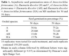
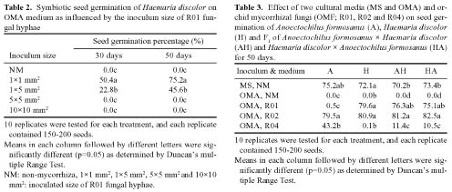
Asymbiotic and symbiotic seed germination of Anoectochilus formosanus and Haemaria discolor and their F1 hybrids

Ling-Chin Chou and Doris Chi-Ning Chang*

Department of Horticulture, National Taiwan University, Taipei, Taiwan 10617, ROC

(Received July 15, 2003; Accepted February 23, 2004)

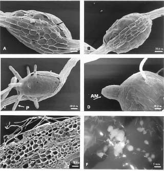
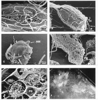
Abstract. Two orchid species Anoectochilus formosanus Hayata and Haemaria discolor var. dawsoniana, which have different flowering times in nature, were induced to flowering synchronously by controlling the culture temperature and light duration in the phytotrons. They were crossed, and the F1 hybrid seeds were collected and named as H. discolor × A. formosanus (HA) and A. formosanus × H. discolor (AH). It was found that fifty days after the asymbiotic culture in MS medium, the germination rates of all the four kinds of seed were higher than 70%. For the symbiotic seed germination on oatmeal agar (OMA) medium, only the R02, as compared to the R01 and R04 isolates of Rhizoctonia spp., had a more than 80% germination rate. SEM observation of various seed germination stages for the non-mycorrhizal and the mycorrhizal protocorms of A. formosanus showed that 70 days after seed sowing, the inoculated seeds developed to a more advanced stage than the non-inoculated control. Asymbiotic and symbiotic germination occurred after the uptake of water, and the seed coat was ruptured by the enlargement of embryo. Afterward the papilla, protocorm and apical meristem appeared. In the symbiotic germinated embryos, the fungal hyphae penetrated the protocorm and formed pelotons, which was the tolypophagy type of infection. No hypha infection was found in the asymbiotic germinated embryos.

Keywords: In vitro propagation; Orchid mycorrhizal fungi; Orchidaceae; Jewel orchid; Scanning electron microscopy; Seed germination.

Introduction

Both Haemaria discolor var. dawsoniana and Anoectochilus formosanus Hayata belong to orchidaceae and were perennial and medicinal herbs. The whole H. discolor plant has medicinal value in lung protection, and suffers little from disease or insect problems. It is also more heat resistant than A. formosanus, which is highly susceptible to diseases and is non-heat resistant. Anoectochilus formosanus is regarded as " the king of medicines " by aborigines in Taiwan because of its diverse pharmacological effects, such as liver protection, cancer prevention, blood sugar reducing for diabetes, and the treatment of cardiovascular diseases etc. (Kan, 1986; Shiau et al., 2002). Haemaria discolor blooms from late December to March (Chou, 1997) while A. formosanus generally blooms from August to November. In order to enhance the heat-tolerance and disease resistance of A. formosanus, the breeding of these two species of orchid plants was conducted. We hope that an F1 hybrid of H. discolor × A. formosanus and A. formosanus × H. discolor will be heat-tolerant and yet still maintainits medicinal value. Although F1 hybrids of Anoectochilus sikkimensis × Ludisia discolor have been reported (Belitsky and Bersenev, 1999), there are no reports concerning the symbiotic germination of the F1 hybrid seeds. In our previous studies, we have shown that

these two orchids could be propagated from seeds through symbiotic and asymbiotic germinations (Chou, 1997). Besides, we have found that the growth of these two orchids was stimulated by Rhizoctonia spp. (Chou, 1997; Tsai, 1997). The present study reports a feasible and effective method for the germination of A. formosanus, H. discolor and their F1 hybrid seeds through asymbiotic and symbiotic cultures. The asymbiotic and symbiotic germination processes are also investigated by scanning electron microscopy (SEM).

Materials and Methods

Plant Materials and Culture Conditions

Plants of H. discolor and A. formosanus were cultivated for eight months under 25/20°C day/night temperature in the phytotron. Then they were put into the phytotron at 23/18°C and a 16-h photoperiod of 3,000-4,000 Lux fluorescent light in mid May to induce blooming. In late October, both kinds of plants could flower synchronously. Between these two orchids, hand pollination was conducted. After pollination for 40 days, capsules of H. discolor (H), A. formosanus (A), H. discolor × A. formosanus (HA) and A. formosanus × H. discolor (AH) were collected. Capsules were surface-sterilized with a cotton ball swab moistened with 70% ethanol, followed by disinfesting under agitation for 15 min in 1% sodium hypochlorite, and were rinsed three times with sterile distilled water. About 200-300 seeds from disinfested capsules

*Corresponding author. Tel. & Fax: +886-2-23690482; E-mail:hyha@ccms.ntu.edu.tw


Botanical Bulletin of Academia Sinica, Vol. 45, 2004

were spread into each glass tube with solid medium and cultivated at 25°C under a 16-h photoperiod.

Seed Morphology and Viability

For counting the viability % of seeds, seeds freshly obtained from capsules were stained with 1% (V/V) 2, 3, 5-triphenyl tetrazolium chloride (TTC) in darkness overnight and then were observed by stereo microscopy. Red color in the embryo indicated seed viability.

Asymbiotic Germination

Seeds were sprinkled onto the surface of MS basal medium (Murashige and Skoog, 1962), prepared with 3 g l-1 tryptone, 30 g l-1 sucrose (Sigma), and 8 g l-1 agar (Sigma). The pH of the medium was adjusted to 5.2 before it was dispensed into glass tubes (2.5 cm diameter, 13 cm high) that contained 15 ml of nutrient solution. Only seeds with the appearance of papillaes were considered germinated.

Symbiotic Germination

Seeds were spread onto the surface of oatmeal agar medium (OMA; 2.5 g l-1 oat meal, and 8 g l-1 Sigma agar, pH of 5.2), 15 ml in each glass tube (2.5 cm diameter, 13 cm height) for one week, and then were inoculated with either a 1×1, 1×5, 5×5 or 10×10 mm2 block of water agar containing fungal hyphae of a single fungal isolate. Three Rhizoctonia spp. (R01, R02 and R04) orchid mycorrhizal fungi (OMF) isolated from wild grown orchids were used as inocula, and only one isolate was inoculated for each treatment. Those plates without fungal inoculation either on OMA or MS medium served as double controls.

Statistical Analysis

The germination rate was recorded after sowing for 30 and 50 days. Only those seeds with the appearance of papillaes were considered germinated. The analysis of variance (ANOVA) appropriate for the design was carried out to detect the significance of differences among the treatment means.

Scanning Electron Microscopy (SEM)

Seeds with various developed stages were collected and fixed with 2.5% glutaradehyde for 12 h, dehydrated by a series of ethyl alcohol from 40% to 100%, 10% increment for each step and 15 min each. The specimens were then dehydrated in 100% ethyl alcohol twice and finally two steps of acetone, 10 min each. The dehydrated materials were critical-point-dried by liquid CO2 in a Polaron Critical Point Drier, mounted on aluminum stubs, and then coated with gold for 90 sec by a Biorad SC502 ion coater. The samples were investigated by ABT-60 of SEM and recorded with digital image.

Results and Discussions

Anoectochilus formosanus and Haemaria discolor bloomed synchronously under 23/18°C and a 16-h photo

period of 3,000-4,000 Lux light intensity in the phytotron in late October. The seed viability test, as stained by TTC, showed that small embryos were present in most of the four orchid seeds, and the viability was 75-85%.

Asymbiotic Germination

On MS medium the four orchid (H, A, HA and AH) seeds reached 70-75% of germination rate within 50 days (Table 1). The higher strength of MS could promote the seed germination of H. discolor (Chou and Chang, 1999; Chang and Chou, 2001) and A. formosanus (Shiau et al., 1995; Shiau et al., 2002).

Symbiotic Germination in vitro

Seeds of most orchids in nature need OMF to germinate, and the symbiotic germination technique showed potential for propagation of some rare species (Clements and Ellyard, 1979; Hadley, 1982). Researchers have demonstrated that in vitro seed germination in some orchid species could easily be carried out with specific Rhizoctonia fungi isolated from orchid mycorrhizae. (Zelmer and Currah, 1997; Zettler, 1998; Vujanovic et al., 2000). Previous study showed that MS medium induced a rapid growth of orchid mycorrhizal fungi (OMF) that could lead to the death of seedlings in vitro (Chang and Chou, 2001). So an appropriate "inoculation medium," such as OMA, was required. OMA medium has given good symbiotic germination with OMF isolates of Sedacina vermifera (Warcup, 1981) and H. discolor (Chang and Chou, 2001). Among four sizes of R01 fungal mycelium blocks of agar, only those seeds inoculated with 1×1 or 1×5 mm2 of OMF were able to germinate, and if 5×5 or 10×10 mm2 was inoculated, then seed germination failed (Table 2). The four orchid seeds could not germinate without the presence of OMF on OMA medium. If inoculated with R01, R02 or R04 isolate 1×1 mm2 block of agar containing fungal mycelium, seeds of HA and AH could germinate 50 days after sowing on OMA medium while H. discolor could only germinate if the seeds were inoculated with R01 or R02 isolate. Anoectochilus formosanus seeds


Chou and Chang _ Seed germination of orchids

could only be stimulated by the inoculation of R02 or R04 isolate. For the four orchids, seeds inoculated with R02 OMF exhibited about a 80-83% germination percentage for 50 days after sowing on OMA medium (Table 3). R02 isolate also stimulated seed germination of Haemaria discolor var. dawsoniana (Chou and Chang, 1999; Chang and Chou, 2001). The result indicates that R02 isolate for orchidaceae is non-species-specific. Lee (2001) indicated that R02 might be Rhizoctonia callae.

In order to obtain higher germination rates for the studied four orchids, MS medium for asymbiotic germination, or symbiotic germination by inoculation with R02 isolates of OMF on 2.5 g l-1 OMA medium is recommended. However for most cultivators, the asymbiotic germination technique is easier than symbiotic germination. Whether the inoculation of OMF had any practical use is still under investigation.

Figure 1. SEM (A-E) and stereomicroscopic (F) photographs of the seeds of Anoectochilus formosanus Hayata in different asymbiotic ermination stages. A: After imbibition, the seed coat was broken ("); B: The embryo burst from the seed coat; C: The embryo formed papilla (P); D: Protocorm with an apical meristem (AM); E: Longitudinal section view of the asymbiotic seedling showing no hypha infection; F: Protocroms on MS medium after sowing for 70 days.


Botanical Bulletin of Academia Sinica, Vol. 45, 2004

Figure 2. SEM (A-E) and stereomicroscopic (F) photographs of the seeds of Anoectochilus formosanus Hayata in different symbiotic germination stages. A: Hyphae (H) penetrated the seed coat through a broken small hole; B: Embryo burst out of the seed coat first and then formed a protocorm with papilla (P). Note the presence of hyphae (H) on the protocorm; C: Protocorm with an apical meristem (AM) and papilla (P); D: Longitudinal section view of the mycorrhizal seedling in which OMF hyphae formed pelotons (PE) in cells of protocorm; E: Close up of pelotons; F: Seed germination and seedling growth inoculated with OMF on OMA medium after sowing for 70 days.

Observation of Seed Asymbiotic and Symbiotic Germination

Asymbiotic germination of A. formosanus as observed by scanning electron microscopy, showed that the seed contained a very rudimentary embryo covered with a seed coat. Seed germination occurred after the uptake of water, which resulted in swelling of the embryo, and then the seed coat was ruptured by the embryo (Figure 1A, B), and the papilla appeared on the one end of protocorm (Figure 1C). Furthermore, the seed developed to protocorm with apical meristem on the other end (Figure 1D). A longitudinal section of the asymbiotic germinated protocorm showed no fungal hyphae in the cortex cell (Figure 1E), but in symbiotic germinated seeds, fungal hyphae penetration of the seed coat was noted. The uptake of water enlarged the embryo and then ruptured the seed coat (Figure 2A). The embryo then burst from the seed coat. Papilla formed on the one end of protocorm. (Figure 2B), and there was an apical meristem on the other end (Figure 2C). The longitudinal section of protocorm showed fungal invasion hyphae, and coil and branching of the hyphae to form pelotons (Figure 2D, E), which was a tolypophagy type of infection (Burgeff, 1959). The seedling developmental processes were similar that of H. discolor, H. discolor × A. formosanus and A. formosanus × H. discolor seed germination. Seed germination in MS medium was slower than those in OMA medium with OMF inoculation (Figures 1F and 2F).

Literature Cited

Belitsky, V. and N.A. Bersenev. 1999. Jewel orchids. Orchids 68: 33-37.

Burgeff, H. 1959. Mycorrhiza of orchid. In C.L. Withner (eds.), The Orchids-a Scientific Survey. The Ronald Press Co. New York, pp. 316-395.

Chang, D.C.N. and L.C. Chou. 2001. Seed germination of Haemaria discolor var. dawsoniana and the use of mycorrhizae. Symbiosis 30: 29-40.

Chou, L.C. and D.C.N. Chang. 1999. The physiological study of seed germination and seedling growth of Haemaria discolor var. dawsoniana. Seed Nursery 1: 57-67.

Chou, L.C. l997. Mass Propagation of Haemaria discolor var. dowsoniana and the Use of Rhizoctonia spp. Master's thesis, Department of Horticulture, National Taiwan University, 105 pp.

Clements, M.A. and R.K. Ellyard. 1979. The symbiotic germination of Australian terrestrial orchids. Amer. Orchid Soc. Bull. 48: 810-815.

Hadley, G. 1982. Orchid mycorrhiza. In Arditti (ed.), Orchid Biology: Reviews and Perspective II Cornell University Press, Ithaca. New York, pp. 85-118.

Kan, W.S. 1986. Pharmaceutical Botany, 7th ed.; National Research Institute of Chinese Medicine: Taiwan, pp. 647.

Lee, G.C. 2001. Identification and improvement of production techniques for Anoectochilus formosanus Hayata and orchid mycorrhizal fungi. Ph.D. Dissertation, Department of Horticulture, National Taiwan University, 191 pp.


Chou and Chang _ Seed germination of orchids

Master's thesis, Department of Horticulture, National Taiwan University, Taipei, Taiwan, ROC. 80 pp.

Vujanovic, V., M. St-Arnaud, D. Barabe, and G. Thibeault. 2000. Viability testing of orchid seed and the promotion of colouration and germination. Ann. Bot. 86: 79-86.

Warcup, J.H. 1981. The mycorrhizal relationships of Australian orchids. New Phytol. 87: 371-381.

Zelmer, C.D. and R.S. Currah. 1997. Symbiotic germination of Spiraanthes lacera (Orchidaceae) with a naturally occurring endophyte. Lindleyana 12: 142-148.

Zettler, L.W. 1998. Propagation of the little culb-spur orchid (Platanthera clavellata) by symbiotic seed germination and its ecological implications. Environ. Exp. Bot. 39: 189-195.

Murashige, T. and F. Skoog. 1962. A revised medium for rapid growth and bioassays with tabacco tissue culture. Physiol. Plant. 15: 473-497.

Shiau, Y.J., A.P. Sagare, U.C. Chen, S.R. Yang, and H.S. Tasy. 2002. Conservation of Anoectochilus formosanus Hayata by artifical cross-pollination and in vitro culture of seeds. Bot. Bull. Acad. Sin. 43: 123-130.

Shiau, Y.J., U.C. Chen, C.Y. Lu., and H.S. Tsay. 1995. Tissue culture of Anoectochilus formosanus Hayata I. Studies on the improvement of seed germination. J. Agric. Res. China 44: 279-286.

Tsai, C.Y. l997. Effects of Rhizoctonia spp, and temperature on the growth of Anoectochilus formosanus Hayata plantlets.