Bot. Bull. Acad. Sin. (2004) 45: 171-178

Chen and Hsieh — Astrosphaeriella from Taiwan

Astrosphaeriella from Taiwan, including two new species

Chi-Yu Chen* and Wen-Hsui Hsieh

Department of Plant Pathology, National Chung Hsing University, Taichung 402, Taiwan, Republic of China

(Received October 2, 2003; Accepted January 9, 2004)

Abstract. A modified generic concept is adopted in the study of ascomycetes referred to Astrosphaeriella from Taiwan. Seven species, including two new species, are described and illustrated. A key to the Taiwan species is also provided. Those species with striate ascospores are found to constitute a distinct group within Astrosphaeriella and can readily be distinguished from the other species in Astrosphaeriella.

Keywords: Ascomycetes; Astrosphaeriella; Systematics; Taiwan.

Introduction

Astrosphaeriella Syd. and H. Syd. was reintroduced by Hawksworth (1981) to encompass four species with hemispherical to conical ascomata occurring on monocotyledonous hosts. The ascomata in three of the species, including the type species A. stellata (Pat.) Sacc., are in common erumpent to become superficial with host remnants around the base of ascomata. The other species, A. aosimensis Hino & Katum., with immersed clypeate ascomata is apparently close to species of Massarina Sacc. revised by Aptroot (1998). Aptroot (1998) also stated that no clear division could be made between these two genera apart from the different forms of pseudoparaphyses, characters de facto not readily discernable. Most recently, Massarina species with fusiform ascospores have been considered to belong to Lophiostoma on the basis of molecular analyses (Hyde et al., 2002; Liew et al., 2002). Trematosphaeria is another genus considered closely related to Astrosphaeriella by Boise (1985), Hawksworth and Boise (1985), and Hyde and Fröhlich (1997). Hawksworth and Boise (1985) sorted out ten species of Astrosphaeriella. Among these species, A. africana H. Hawks. and A. striaspora (E. Müll.) D. Hawksw. & Boise, resemble Trematosphaeria in the relative position of ascomata to the substrate and in the shape of asci and the color of ascospores. Furthermore, the characteristic striate ascospores possessed by these two species, together with the above characters shared with Trematosphaeria, make a clear distinction between these species and the other species in Astrosphaeriella. In addition to the many typical Astrosphaeriella species with superficially appearing ascomata flanked by host remnants, several further species were added to this genus by Hyde and Fröhlich (1997), including Massarina-like species and Trematosphaeria-like species with striate ascospores. It is beyond the scope of this study to re-evaluate the classi

fication of the entire genus. However, in the present study, a strict generic concept excluding Massarina-like species is adopted. Trematosphaeria-like species, possessing striate ascospores readily distinguishable from those of the real Trematosphaeria species, are best retained in Astrosphaeriella as a distinct group before further DNA analysis is done. Astrosphaeriella was defined as a monocotyledon-inhabiting genus. However, because of its strongly saprophytic nature, confining this genus to the specific host range seems unjustified. The two new species from Taiwan, A. macrospora and A. pallidipolaris, have striate ascospores. Astrosphaeriella pallidipolaris, although occurring on the dicotyledon, can be well-grouped with the other Trematosphaeria-like species accommodated in Astrosphaeriella by sharing common characters like the clypeate ascomata, the clavate asci, and the brown and striate ascospores.

Two species of Astrosphaeriella have previously been reported in Taiwan (Hsieh et al., 2000). In this study, one of the two reported species is corrected and five further species are added, including two new species. Specimens examined are deposited at TNM (National Museum of Natural Science) and NCHUPP (National Chung Hsing University).

Key to Species of Astrosphaeriella from Taiwan

1. Ascomata immersed, with a clypeus. Asci clavate. Ascospores with striate ornamentation 2

Ascomata superficial with ruptured host tissue surrounded. Asci cylindrical. Ascospores smooth 4

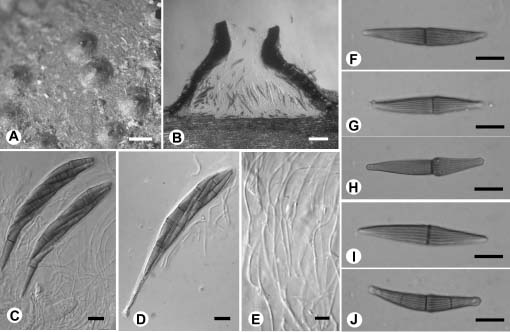
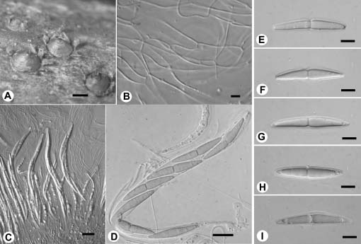
2. Ascospores brown, with paler end cells A. pallidipolaris

Ascospores evenly brown 3

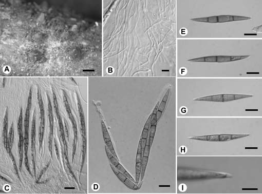
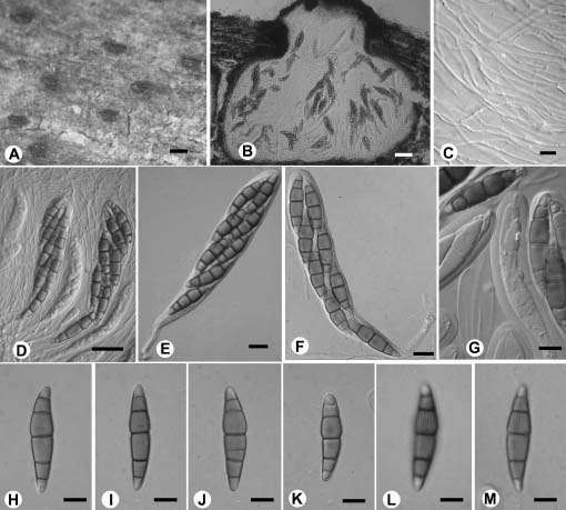
3. Ascospores 1- or occasionally 3-septate, 40-54 µm long A. africana

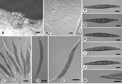
Ascospores 1-5-septate, 56-90 µm long A. macrospora

*Corresponding author. Tel: 04-22840730-347; E-mail chiyu86@yahoo.com.tw


Botanical Bulletin of Academia Sinica, Vol. 45, 2004

4. Ascospores 1-septate 5

Ascospores more than 1-septate 6

5. Ascospores with sheath A. stellata

Ascospores with appendages A. lenticularis

6. Ascospores 3-septate A. maquilingiana

Ascospores mostly 5-septate A. exorrhiza

Astrosphaeriella africana D. Hawksw. in D. Hawksw. and Boise, Sydowia 38: 116. 1985. (Figure 1)

Ascomata scattered, immersed, raising the overlying host tissue, hemispherical, 700-800 µm wide, 300-360 µm high, apex papillate, usually elongated to become a neck, flattened at base. Peridium laterally merging with the host tissue to form a shield-like clypeus, much reduced at base, exhibiting a palisade-like arrangement of cells at the rim margin. Asci clavate, 110-130 × 12-14 µm, long-stalked, stalk up to 40 µm long, 8-spored. Pseudoparaphyses embedded in gelatinous matrix, frequently branched and anastomosing, 1-1.5 µm wide. Ascospores fusiform, 40-54 × 5-7 µm, acute or occasionally swollen at ends, brown, striate, 1-septate, with septum usually slightly constricted, median or supra-median, broadest on the upper cell near the septum, occasionally becoming 3-septate, arranged bi- or triseriately in upper part of ascus and uniseriately below.

Specimens examined. Taichung Hsien: Kukuan, on indet. bamboo, 5 Apr. 2001, C.Y. Chen (NCHUPP c0105); Paileng, on Bambusa oldhamii, 19 Aug. 2001, C.Y. Chen (NCHUPP c0215).

Note. Astrosphaeriella africana has immersed hemispherical clypeate ascomata. It can readily be recognized by the protruded necks. It is interesting to note that on surface view the shape of ascomata does not appear conical for those Astrosphaeriella species with immersed, clypeate ascomata. This rounded (hemispherical appearance, disregarding the neck) instead of pointed (conical appearance) apex of ascomata provides further evidence to account for the fundamental difference between the species with immersed ascomata and the species with superficially appearing ascomata.

Astrosphaeriella exorrhiza Boise in D. Hawksw. and Boise, Sydowia 38: 117. 1985. (Figure 2)

Ascomata scattered, erumpent through the host tissue to become superficial, conical, 600-1000 µm wide, 500-800 µm high, apex non-papillate to slightly papillate, flattened at base, surrounded with tooth-like ruptured host tissue. Peridium at sides carbonaceous, composed of melanized and opaque cells, much reduced at base, with palisade-like cells at the rim margin. Asci cylindrical, 180-230 × 10-12 µm, sessile to short-stalked, 8-spored. Pseudoparaphyses embedded in gelatinous matrix, frequently branched and anastomosing, 1-1.5 µm wide. Ascospores ellipsoid to fusiform, 70-85 × 7-10 µm, acute at ends, dark brown, smooth, (3-)5(6)-septate, the first septum largely median and slightly constricted, bi- or triseriately arranged in ascus, occasionally with inconspicuous, up to 1 µm long, mucilaginous appendages at ends.

Figure 1. Astrosphaeriella africana. A, Ascomata on host; B, section of ascoma; C-D, Asci; E, Pseudoparaphyses; F-J. Ascospores. Scale bars, A=500 µm, B=100 µm, C-D=10 µm, E=5 µm, F-J=10 µm.


Chen and Hsieh — Astrosphaeriella from Taiwan

Specimens examined. Nantou Hsien: Tsuifeng, on Miscanthus, 23 Dec. 2001, C.Y. Chen (NCHUPP c0317); Meifeng, on Miscanthus, 15 Oct. 2002, C.Y. Chen (NCHUPP c0828). Taoyuan Hsien: Lalashan, on Miscanthus, 20 Oct. 1994, C.Y. Chen (NCHUPP 2320-1).

Note. The ascospore appendages of this species were not reported by Hawksworth and Boise (1985), though Hyde and Fröhlich (1997) described their presence. In Taiwan material appendages can only be found on some ascospores.

Astrosphaeriella lenticularis K.D. Hyde and J. Fröhl., Sydowia 50: 97. 1997. (Figure 3)

Ascomata scattered, erumpent through the host tissue to become superficial, conical, 650-1000 µm wide, 500-800 µm high, flattened at base, apex usually papillate, ruptured host tissue deciduous or remaining as tooth-like flange around the rim of ascoma. Peridium at sides carbonaceous, cells melanized and opaque, base of peridium much reduced, with palisade-like cells at the rim margin. Asci cylindrical, 180-220 × 11-14 µm, sessile to short stalked, 8-spored. Pseudoparaphyses embedded in gelatinous matrix, frequently branched and anastomosing, 1-1.5 µm wide. Ascospores fusiform, 38-50 × 7-9 µm, acute at ends, smooth, dark brown, slightly constricted at the median or slightly submedian septum, arranged uniseriately to partially biseriately in ascus, furnished with up to 3 µm long mucilaginous appendages at ends.

Specimens examined. Kaohsiung Hsien: Yakou, south cross road 149K, on Miscanthus, 11 Jun. 1995, C.Y. Chen (NCHUPP 2380). Chiai Hsien: Tataka-ambu, on Yushania niitakayamensis, 2 Mar. 2002, C.Y. Chen (NCHUPP c0714).

Note. Apart from the characteristically 1-septate and appendage-bearing ascospores, this species can be distinguished by its relatively large and pointed ascomata.

Astrosphaeriella macrospora C.Y. Chen and H.W. Hsieh, sp. nov. Holotypus: In caulibus emortuis Miscanthi, Nantou Hsien, Wushe, Pihu, 11 Feb. 1988, A. Sivanesan, TNM(F) 15219; isotypus: NCHUPP s383. (Figure 4)

Ascomata dispersa, immersa, hemisphaerica, 800-1000 µm lata et 360-420 µm alta, nigra, interdum collo usque 200 µm longo praedita. Paries ascomatis lateraliter cum textura hospitis commiscens, clypeo faciens. Asci clavati, 170-220 × 20-24 µm, pedicelli usque 60 µm longi, (4-7)8-spori. Pseudoparaphyses 1-1.5 µm crassae. Ascosporae fusiformes, 56-90 × 7-10 µm, brunneae, 1-5-septatae, striatae, superne 3-4 confertae in asco et inferne uniseriatae.

Ascomata scattered, immersed, raising the overlying host tissue to form hemispherical pustules, 800-1000 µm wide, 360-420 µm high, apex papillate or forming a neck up to 200 µm long, with flattened base. Peridium merging with the host tissue at sides to form a shield-like clypeus, poorly developed at base, cells at rim vertically oriented

Figure 2. Astrosphaeriella exorrhiza. A, Ascomata on host; B, Pseudoparaphyses; C-E, Asci; F-J, Ascospores; K, Appendage of ascospore. Scale bars, A=500 µm, B=5 µm, C-E=20 µm, F-J=10 µm, K=5 µm.


Botanical Bulletin of Academia Sinica, Vol. 45, 2004

Figure 3. Astrosphaeriella lenticularis. A, Ascomata on host; B, Pseudoparaphyses; C-D, Asci; E-H, Ascospores; I, Appendage of ascospore. Scale bars, A=500 µm, B=5 µm, C-D=20, E-H=10 µm, I=5 µm.

and palisade-like. Asci clavate, 170-220 × 20-24 µm, short- to long-stalked, stalk up to 60 µm long, (4-7) 8-spored. Pseudoparaphyses embedded in gelatinous matrix, frequently branched and anastomosing, 1-1.5 µm wide. Ascospores fusiform, 56-90 × 7-10 µm, broadest at the portion above the largely median, constricted septum, 1-5-septate, brown, usually turning colorless at the slightly swollen ends, striate, 3 to 4 ascospores fasciculate in the upper part of ascus, becoming uniseriate below.

Other specimen examined. Nantou Hsien: Wushe, Pihu, on Miscanthus, 14 Oct. 2002, C.Y. Chen (NCHUPP c0816).

Note. This new species is similar to A. africana in the shape of ascospores, but its ascospores are significantly larger and become 5-septate at maturity.

Astrosphaeriella maquilingiana (Rehm) K.D. Hyde and J. Fröhl., Sydowia 50: 103. 1997. (Figure 5)

Trematosphaeria maquilingiana Rehm, Leaft. Phil. Bot. 8: 2592. 1916.

Ascomata scattered, erumpent through the host tissue to become superficial, conical, 700-1150 µm wide, 600-900 µm high, apex usually papillate, flattened at base, with ruptured reflexed tooth-like host remnants around the base. Peridium carbonaceous at sides, composed of melanized and opaqued cells, poorly developed at base, cells at rim margin in palisade-like arrangement. Asci cylindrical, 180-210 × 8-12 µm, sessile to short-stalked, 8-spored. Pseudoparaphyses embedded in gelatinous matrix, frequently branched and anastomosing, 1-1.5 µm wide. Ascospores ellipsoid to fusiform, 45-55 × 5-6.5 µm, acute,

smooth, dark brown, 3-septate, with first septum median or slightly sub-median, usually slightly constricted, with mucilaginous appendages protruded at ends, up to 2 µm long; ascospores arranged uniseriately to biseriately in ascus.

Specimens examined. Taichung Hsien: Nanhuta-shan, on Yushania niitakayamensis, 3 Nov. 1991, C.Y. Chen (as `A. trochus', NCHUPP 2276). Nantou Hsien: Piluchi, on Yushania niitakayamensis, 10 May 2000, C.Y. Chen (NCHUPP c0012); Meifeng, on Yushania niitakayamensis, 16 Feb. 2002 (NCHUPP c0620).

Note. Hawksworth and Boise (1985) placed Trematosphaeria maquilingiana as a synonym of A. vesuvius with a question mark. Hyde (1997) reinstated it and proposed a new combination with Astrosphaeriella. Astrosphaeriella vesuvius can be separated from A. maquilingiana by the fusiform, reddish brown ascospores that are (33-)36-45(-50) × (5.5-)7-8(-9) µm. The present species is also similar to A. trochus, to which name the collection NCHUPP 2276 was erroneously referred by Hsieh et al. (2000). In A. trochus the ascospores are larger, (44-)48-65(-72) × (5-)6-8(-9) µm, reddish brown, with more septa (mostly 5-septate).

Astrosphaeriella pallidipolaris C.Y. Chen and H.W. Hsieh, sp. nov. Holotypus: In indet. ligno, Nantou Hsien, Wushe, Pihu, 21 Dec. 2001, C.Y. Chen, TNM(F) 15220; isotypus: NCHUPP c0326-1. (Figure 6)

Ascomata aequaliter dispersa, immersa, 580-760 µm lata et 450-520 µm alta, ostiolo erumpenti, fissurato vel interdum papillato. Paries ascomatis incrassatus circa ostiolum,


Chen and Hsieh — Astrosphaeriella from Taiwan

Figure 4. Astrosphaeriella macrospora. A, Ascomata on host; B, Section of ascoma; C-D, Asci; E, Pseudoparaphyses; F-K, Ascospores. Scale bars, A=500 µm, B=100 µm, C-D=20 µm, E=5 µm, F-K=10 µm.

Figure 5. Astrosphaeriella maquilingiana. A, Ascomata on host; B, Pseudoparaphyses, C-D, Asci; E-H, Ascospores; I, Appendage of ascospore. Scale bars, A=1000 µm, B=5 µm, C=20 µm, D-H=10 µm, I=5 µm.


Botanical Bulletin of Academia Sinica, Vol. 45, 2004

quently branched and anastomosing, 1-1.5 µm wide. Ascospores ellipsoid to fusiform, 38-48 × 8-10 µm, rounded at ends, striate, 5(-6)-septate, first septum supra-median, constricted, brown with the end cells paler, biseriately arranged in the upper part of ascus, becoming uniseriate below.

Other specimens examined. Nantou Hsien: Wushe, Pihu, on wood, 21 Dec. 2001, C.Y. Chen (NCHUPP c0318-1); ibid. 27 Jan. 2002 (NCHUPP c0501); ibid. (NCHUPP c0531); ibid. 14 Oct. 2002 (NCHUPP c0808).

Note. We are hesitant to assign this species to Astrosphaeriella, which was commonly considered as occurring on monocotyledons. However, despite being associated with dicotyledons, all the other characters of this fungus match those of the Astrosphaeriella species with striate ascospores. The ascomata of this species recall those of A. striaspora, being subglobose with a flattened base. Astrosphaeriella striaspora was originally estab

superne e clypeo circumcinctus. Asci clavati, 140-170 × 16-20 µm, pedicelli usque 50 µm longi, 8-spori. Pseudoparaphyses 1-1.5 µm crassae. Ascosporae elliposideae vel fusiformes, 38-48 × 8-10 µm, brunneae, cellulis ad extremis pallide brunneis, 5(-6)-septatae, striatae, supra imbricate biseriatae et infra uniseriatae in asco.

Ascomata evenly scattered, immersed, with only the ostiolar region erumpent through the host tissue, 580-760 µm wide, 450-520 µm high, usually flattened at base, apex compressed with a slit-like ostiolum, or sometimes papillate. Peridium melanized and thickest at ostiolar region, hyphae interwoven with host tissue, merging with the lateral and upper part of peridium to give an appearance of clypeus, peridium at sides brown, cells compressed, occasionally in palisade-like arrangement at basal corner, peridium at base usually reduced. Asci clavate, 140-170 × 16-20 µm, stalk various in length, up to 50 µm long, 8-spored. Pseudoparaphyses embedded in gelatinous matrix, fre

Figure 6. Astrosphaeriella pallidipolaris. A, Ascomata on host, showing the immersed ascomata with erumpent compressed ostioles; B, Section of ascoma; C, Pseudoparaphyses; D-F, Asci; G, Apices of asci; H-M, Ascospores. Scale bars, A=300 µm, B=50 µm, C=5 µm, D=20 µm, E-M=10 µm.


Chen and Hsieh — Astrosphaeriella from Taiwan

lished as a Trematosphaeria species (Müller and Dennis, 1965), and the lateral base wall of ascomata does not exhibit a palisade-like arrangement of cells, as is usually the case in the present species. Boise (1985) separated Astrosphaeriella from Trematosphaeria by the palisade-like cells. However, with the inclusion of A. striaspora in Astrosphaeriella by Hawksworth and Boise (1985), the arrangement of these cells does not seem to be an indispensable criterion for the generic circumscription. Disposing the present species in Astrosphaeriella is thus acceptable. Compared with Trematosphaeria, the ascospores of Astrosphaeriella were considered to be relatively narrow by Hawksworth and Boise (1985) and Hyde and Fröhlich (1997). However, among the Astrosphaeriella species the ascospores are quite variable, varying from broad and short to narrow and long. Therefore, the shape of ascospores is not applicable to the separation of Astrosphaeriella from Trematosphaeria, and the broad, long ascospores do not exclude the present fungus from Astrosphaeriella. This fungus is quite unique among the Astrosphaeriella species in having brown ascospores with paler end cells and a slit-like ostiolum. Lophiostoma viridarium Cooke also occurs on dicotyledons and has striate ascospores. It has a slit-like ostiolum and shares common characters with those Astrosphaeriella species posessing striate ascospores. Holm and Holm (1988) noted that it is not well accommodated in Lophiostoma. It is believed that there is no better place for it than with those Astrosphaeriella species with striate ascospores.

Astrosphaeriella stellata (Pat.) Sacc., Syll. Fung. 24: 938. 1928. (Figure 7)

For synonyms see Hawksworth (1981).

Ascomata scattered, erumpent through the host tissue to become superficial, 600-1000 µm wide, 400-550 µm high, flattened at base, apex non-papillate, with ruptured reflexed tooth-like host remnants around the base. Peridium at sides carbonaceous, composed of opaque and melanized cells, poorly developed at base, cells at rim appearing in palisade-like rows. Asci cylindrical, 170-240 × 11-13 µm, sessile to short-stalked, (4-7)8-spored. Pseudoparaphyses embedded in gelatinous matrix, frequently branched and anastomosing, 1-1.5 µm wide. Ascospores ellipsoid to fusiform, 40-56 × 5.5-7 µm, acute at ends, pale brown, smooth, constricted at the median or slightly supra-median septum, surrounded by a sheath, sheath truncate or sometimes concave at ends, ascospores uniseriately to partially biseriately arranged in ascus.

Specimens examined. Taichung Hsien: Wufeng, on Dendrocalamus, 12 Jul. 1997, C.Y. Chen (NCHUPP 2538); Tungmaoshan, on Dendrocalamus, 29 Jan. 1996, C.Y. Chen (NCHUPP 2442); ibid. 18 Aug. 2001 (NCHUPP c0205); 15K Anmashan, on Dendrocalamus, 15 Mar. 2003 C.Y. Chen (NCHUPP c0912). Nantou Hsien: Leinhuachih, on Dendrocalamus, 20 Jun. 2001 (NCHUPP c0121); Hsitou, on Phyllostachys, 28 Sept. 1995, C.Y. Chen (NCHUPP 2388). Hualien Hsien: Tienshiang, on Dendrocalamus, 29 Dec. 2001, C.Y. Chen (NCHUPP c0421). Chiai Hsien: Meishan, on Phyllostachys, 1 Mar. 2002, C.Y. Chen (NCHUPP c0705).

Figure 7. Astrosphaeriella stellata. A, Ascomata on host; B, Pseudoparaphyses; C-D, Asci; E-I, Ascospores. Scale bars, A=500 µm, B=5 µm, C-D=20 µm, E-I=10 µm.


Botanical Bulletin of Academia Sinica, Vol. 45, 2004

stood genus of melanommataceous pyrenomycetes. Bot. J. Linn. Soc. 82: 35-59.

Hawksworth, D.L. and J.R. Boise. 1985. Some additional species of Astrosphaeriella, with a key to the members of the genus. Sydowia 38: 111-124.

Holm, L. and K. Holm. 1988. Studies in the Lophiostomataceae, with emphasis on the Swedish species. Ups. Symb. Bot. 28(2): 1-50.

Hsieh, W.H., C.Y. Chen, and C.L. Wang 2000. Taiwan Ascomycetes: Pyrenomycetes and Loculoascomycetes. China-Graphics, Taichung, Taiwan, 244 pp.

Hyde, K.D. and J. Fröhlich. 1997. Fungi from palms XXXVII. The genus Astrosphaeriella, including ten new species. Sydowia 50: 81-132.

Hyde, K.D., W.S.W. Wong, and A. Aptroot. 2002. Marine and estuarine species of Lophiostoma and Massarina. In K.D. Hyde (ed.), Fungi in Marine Environments. Fungal Diversity Press, Hong Kong, pp. 93-109.

Liew, E.C.Y., A. Aptroot, and K.D. Hyde. 2002. An evaluation of the monophyly of Massarina based on ribosomal DNA sequences. Mycologia 94: 803-813.

Müller, E. and R.W.G. Dennis. 1965. Fungi venezuelani: VIII. Plectascales, Sphaeriales, Loculoascomycetes. Kew Bull. 19: 357-386.

Note. This is the type species of Astrosphaeriella. It has been reported in Taiwan (Hseih et al., 2000) and is widely distributed at low altitudes. The characteristic sheath of the ascospores with truncate ends is of great diagnostic value for the species. Additionally, ascomata are relatively short compared with other species with superficial ascomata.

Acknowledgement. We thank the National Science Council, Taiwan for supporting this study with Research Grant No. NSC 92-2313-B-005-051. We would also like to express our gratitude to Dr. A. Sivanesan for sharing his insight into this genus. C.Y. Chen is grateful to the curator of IMI for allowing him to develop his knowledge of this genus by examining relevant material while studying in Britain.

Literature Cited

Aptroot, A. 1998. A world revision of Massarina (Ascomycota). Nova Hedwigia 66: 89-162.

Boise, J.R. 1985. An amended description of Trematosphaeria Fuckel. Mycologia 77: 230-237.

Hawksworth, D.L. 1981. Astrosphaeriella Sydow, a misunder