Bot. Bull. Acad. Sin. (2004) 45: 179-186

Huang et al. — Antioxidant and antiproliferative activities of sweet potato constituents

Antioxidant and antiproliferative activities of sweet potato (Ipomoea batatas [L.] Lam `Tainong 57') constituents

Dong-Jiann HUANG1, Chun-Der LIN1, Hsien-Jung CHEN2, and Yaw-Huei LIN1,*

1Institute of Botany, Academia Sinica, Nankang, Taipei 115, Taiwan

2Department of Horticulture, Chinese Culture University, Taipei 111, Taiwan

(Received February 24, 2004; Accepted March 29, 2004)

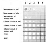
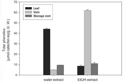
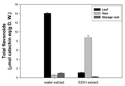
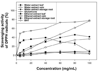
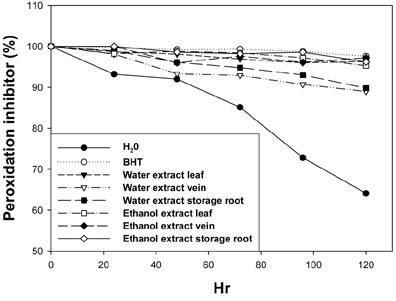
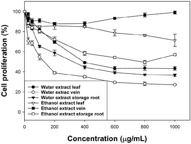
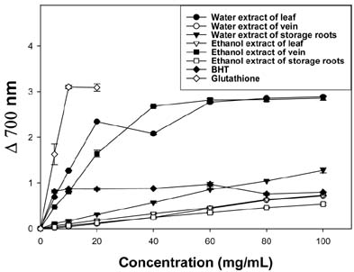
Abstract. The aim of this study is to examine possible antioxidant and antiproliferative activities of the different extracts from sweet potato (Ipomoea batatas [L.] Lam `Tainong 57') organs. DPPH staining, total phenolic compounds and flavonoid content, DPPH radical, reducing power method, FTC method, and cell proliferation were all employed. In the DPPH staining, ethanol extract of vein had the highest radical-scavenging activity when it was diluted to 6.25 mg dry matter/mL. Among all the extracts, the highest amount of total phenolic and flavonoid compounds was found in the ethanol extract of vein. In the DPPH colorimetric method, it was found that ethanol extract of leaf had the highest radical-scavenging activity, followed by water extract of vein. In the reducing power activity assay, it was found that the water extract of leaf had the highest reducing power activity, followed by ethanol extract of vein. Like phenolic compounds, the highest FTC activity was found in the ethanol extract of vein. The antiproliferative activities aof sweet potato were studied in vitro using human lymphoma NB4 cells, and the following results were found: water extract of vein had the highest antiproliferative activity with an EC50 of 449.6 ± 27.73 µg/mL, followed by water extract of storage root, water extract of leaf, ethanol extract of storage root, and ethanol extract of leaf. Although the ethanol extract of vein showed strong antioxidant activity, it had no antiproliferative activity under the experimental conditions tested.

Keywords: Antioxidant; Antiproliferative; Free radical; Sweet potato.

Abbreviations: BHT, butylate hydroxyltoluene; DPPH, 1,1-dipheny-2-picrylhydrazyl; EDTA, ethylenediamine tetraacetic acid; GSH, glutathione; FBS, fetal bovine serum; FTC, ferric thiocyanate; MTT, 3-(4,5-dimethylthiazol-2-yl)-2,5-diphenyl tetrazolium bromide; EC50, dose with 50% efficiency; TI, trypsin inhibitor.

Introduction

It is commonly accepted that under situations of oxidative stress, reactive oxygen species such as superoxide (O2ź-, źOOH), hydroxyl (OHź), and peroxyl (ROOź) radicals are generated. The reactive oxygen species play an important role related to degenerative or pathological processes such as aging (Burns et al., 2001), cancer, coronary heart disease, Alzheimer's disease (Ames, 1983; Gey, 1990; Smith et al., 1996; Diaz et al., 1997), neurodegenerative disorders, atherosclerosis, cataracts, and inflammation (Aruoma, 1998). The use of traditional medicine is widespread, and plants still present a large source of natural antioxidants that might serve as leads for the development of novel drugs. Several anti-inflammatory, digestive, antinecrotic, neuroprotective, and hepatoprotective drugs have recently been shown to have an antioxidant and/or radical scavenging mechanism as part of their activity (Perry et al., 1999; Lin and Huang, 2002; Repetto and Llesuy, 2002). In searching for novel natural antioxidants, some plants have been extensively studied in the past few years for their antioxi

dant and radical scavenging components. These include echinacoside in Echinaceae root (Hu and Kitts, 2000), anthocyanin (Espin et al., 2000), phenolic compounds (Rice-Evans et al., 1997), water extracts of roasted Cassia tora (Yen and Chuang, 2000), and whey proteins (Allen and Wrieden, 1982a, b; Tong et al., 2000).

Sweet potato is a dicotyledonous plant with tubers derived from swollen roots. Its crude protein content has been reported to vary between 1~3% and 10%, but this includes 10~15% non-protein nitrogenous components (Walter et al., 1984). Its major storage protein was reported to account for over 80% of the total protein (Maeshima et al., 1985). Hou and Lin reported that 33 kDa TI had antioxidant activity (Hou et al., 2001; 2002) and that TI also had dehydroascorbate reductase and monodehydroascorbate reductase activities (Hou and Lin, 1997; Hou et al., 1998) associated with intermolecular thiol/disulfide exchange. The biological significance of these observations is not yet clear.

The objectives of this work were to investigate the antioxidant and antiproliferative properties of crude extracts from different tissues of sweet potato and to assay the inhibitory effect on free-radical-related enzymes and the level of inhibition against the growth of human cancer cell lines in vitro.

*Corresponding author. Fax: 886-2-2782-7954; Tel: 886-2- 2789-9590 ext. 320; E-mail: boyhlin@ccvax.sinica.edu.tw


Botanical Bulletin of Academia Sinica, Vol. 45, 2004

Materials and Methods

Materials

BHT, DPPH, EDTA, FTC, MTT, sodium bicarbonate, and Tris (hydroxylmethyl) aminomethane were purchased from Sigma Chemical Co. (St. Louis, MO USA). FBS, L-glutamine, and RPMI medium 1640 were purchased from Gibco BRL Co. (Gaithersburg, MD USA). Hydrogen peroxide was purchased from Showa Chemical Co. (Tokyo, Japan). Ethanol was purchased from Riedel-deHaen Chemical Co. (Germany).

Plant Material

Fresh storage roots of sweet potato (Ipomoea batatas [L.] Lam `Tainong 57') were purchased from a local market. After being cleaned with water, they were placed in a greenhouse and sprayed with water twice a day. After one month, veins and fully expanded green leaves were harvested for experiments.

Sample Preparation

The fully expanded green leaves, veins and storage roots (100 g) were cleaned and air-dried and extracted with 500 mL of 95% ethanol or distilled water by stirring at room temperature for 24 h and then filtered through #1 filter paper (Whatman Inc., Hillsboro, OR, USA). The filtrate was concentrated to a powder by freeze dryer (Christ Alpha, Germany) and stored at -20°C. When the experiment was conducted, each plant extract fraction had acquested by the powder (dry matter) for a suitable concentration.

Rapid Screening of Antioxidant by Dot-Blot and DPPH Staining

An aliquot (3 µL) of a suitable dilution of each plant extract fraction, GSH and BHT was carefully loaded on a 20 cm × 20 cm TLC layer (silica gel 60 F254; Merck) and dried for 3 min. Drops of each sample were loaded in order of decreasing concentration along the row. The staining of the silica plate was based on the procedure of Soler-Rivas et al. (2000). The sheet bearing the dry spots was placed upside down for 10 s in a 0.4 mM DPPH solution. Then, the excess solution was removed with a tissue paper, and the layer was dried with a hair-dryer blowing cold air. The stained silica layer revealed a purple background with white spots at the location of the drops, which showed radical scavenger capacity. The intensity of the white color depends upon the amount and nature of radical scavenger present in the sample.

Determination of Total Phenolic Compounds

Total phenolic compounds were determined using the Folin_Ciocalteu method (Ragazzi and Veronese, 1973). One mL of the extract was added to 10.0 mL distilled water and 2.0 mL of Folin_Ciocalteu phenol reagent (Merck-Schuchardt, Hohenbrun, Germany). The mixture was allowed to stand at room temperature for 5 min, and then 2.0 mL sodium carbonate was added to the mixture. The re

sulting blue complex was then measured at 680 nm. The contents of phenolic compounds were expressed as mg catechin equivalent/g dry weight.

Determination of Total Flavonoid Content

The AlCl3 method (Lamaison and Carnet, 1990) was used for determination of the total flavonoid content of the sample extracts. Aliquots of 1.5 mL of extracts were added to equal volumes of a solution of 2% AlCl3ź6H2O (2 g in 100 mL methanol). The mixture was vigorously shaken, and absorbance at 367 nm was read after 10 min of incubation. Flavonoid contents were expressed as mg catechin equivalent/g dry weight.

Scavenging Activity Against DPPH Radical

The effect of crude extracts on the DPPH radical was estimated according to the method of Yamaguchi et al. (Yamaguchi et al., 1998). An aliquot of crude extract (30 µL) and glutathione (0.04-1.25 mg/mL, 30 µL) was mixed with 100 mM Tris-HCl buffer (120 µL, pH 7.4) and then with 150 µL of the DPPH in ethanol to a final concentration of 250 µM . The mixture was shaken vigorously and left to stand at room temperature for 20 min in the dark. The absorbance at 517 nm of the reaction solution was measured spectrophometrically. The percentage of DPPH decolourization of the sample was calculated according to the equation: % decolourization= [1- ABS sample /ABS control]×100.

Measurement of Reducing Power

The reducing power of the crude extracts and BHT was determined according to the method of Yen and Chen (1995). The crude extract (0, 5, 10, 20, 40, 60, 80, and 100 mg/mL) or BHT was mixed with an equal volume of 0.2 M phosphate buffer, pH 6.6, and 1% potassium ferricyanide. The mixture was incubated at 50°C for 20 min. Then an equal volume of 1% trichloroacetic acid was added to the mixture, which was then centrifuged at 6,000 rpm for 10 min. The upper layer of the solution was mixed with distilled water and 0.1% FeCl3 with a ratio of 1 : 1 : 2, and the absorbance at 700 nm was measured. Increased absorbance of the reaction mixture indicated increased reducing power.

Determination of Antioxidant Activity by the FTC Method

The FTC method was adopted from Osawa and Namiki (1981). Twenty mg/mL of samples dissolved in 4 mL of 95% (w/v) ethanol were mixed with linoleic acid (2.51%, v/v) in 99.5% (w/v) ethanol (4.1 mL), 0.05 M phosphate buffer pH 7.0 (8 mL), and distilled water (3.9 mL) and kept in screw-cap containers at 40°C in the dark. To 0.1 mL of this solution was then added 9.7 mL of 75% (v/v) ethanol and 0.1 mL of 30% (w/v) ammonium thiocyanate. Precisely 3 min after the addition of 0.1 mL of 20 mM ferrous chloride in 3.5% (v/v) hydrochloric acid to the reaction mixture, the absorbance at 500 nm of the resulting red solution was measured, and it was measured again every 24 h until the day when the absorbance of the control reached the


Huang et al. — Antioxidant and antiproliferative activities of sweet potato constituents

maximum value. The percent inhibition of linoleic acid peroxidation was calculated as: (%) inhibition = 100 - [(absorbance increase of the sample/absorbance increase of the control) × 100]. All tests were run in duplicate, and analyses of all samples were run in triplicate and averaged.

Measurement of Cell Proliferation

The NB4 cell line was isolated from long-term cultures of leukemia blast cells on bone-marrow stromal fibroblasts, as reported by Lanotte et al. (1991). NB4 cells were maintained in RPMI 1640 medium supplemented with 10% heat-inactivated FBS, 2 mM L-glutamine, 100 units/mL penicillin, and 100 mg/mL streptomycin in a humidified atmosphere of 5% carbon dioxide. The cells were subcultured every third day, with cell density in the cultures kept below 5 × 10⁵ cells/mL.

MTT-Microculture Tetrazolium Assay

The colorimetric assay for cellular growth and survival was based on Hansen et al. (1989). Suspensions of human histolytic lymphoma NB4 monocytes (2 × 10⁵ cells/mL) were cultured with or without test samples (at various concentrations in 10 µL of suspension) in a 96-well microplate (90 µL suspension/well). After 48 h, 10 µL of MTT solution was added to each well, and the cells were incubated at 37°C for 4 h. Then, 100 µL of lysis buffer were added to each well, and the cells were again incubated at 37°C for 1 h to dissolve the dark blue crystals. Each well was completely pipetted, and then the absorption at 570 nm of formazan solution was measured using a microplate reader. At least three repeats for each sample were used to determine the cell proliferation. The decolourization was plotted against the concentration of the sample extract, and the EC50, which was the amount of sample necessary to decrease 50% of the absorbance of MTT, was calculated.

Statistical Analysis

Means of triplicates were measured. Student's t test was used for comparison between two treatments. A difference was considered to be statistically significant when p<0.05.

Results and Discussion

Rapid Screening of Antioxidant by Dot-Blot and DPPH Staining

To make a semi-quantitative visualization possible, different extract fractions from sweet potato were detected in the TLC plates by the DPPH staining method. For the rapid screening each diluted sample was applied as a dot on a TLC plate that was then stained with DPPH solution (Figure 1). The appearance of white color in the spots has a potential value for the indirect evaluation of the different extracts from sweet potato in the dot plot (Soler-Rivas et al., 2000; Chang et al., 2002). The method is typically based on the inhibition of the accumulation of oxidized products since the generation of free radicals is inhibited by the addition of antioxidants.

Figure 1. Dot blot assay of different extract fractions of sweet potato on a silica sheet stained with a DPPH solution in methanol. For different extract fractions, a 3 µl sample with a concentration of 100, 50, 25, 12.5, and 6.25 mg dry matter/mL, from left to right respectively, was applied to each dot. For GSH or BHT, a 3 µl sample with a concentration of 10, 5, 2.5, 1.25, and 0.625 mg dry matter/mL, from left to right respectively, was applied to each dot.

When different extracts from sweet potato were analyzed, white spots with strong intensity appeared fast up to the dilution of 6.25 mg dry matter/mL for ethanol extract of vein (the final amount in the spot: 18.75 µg dry matter), 12.5 mg dry matter/mL for both water extract and ethanol extract of leaf (final amount: 37.5 µg dry matter), 25 mg dry matter/mL for ethanol extract of storage root (final amount: 75 µg dry matter), 50 mg dry matter/mL for water extract of storage root, and 100 mg dry matter/mL for water extract of vein.

Total Phenolic Compounds and total Flavonoid Content Extracted from Sweet Potato

The total phenolic compounds of six different extract fractions were expressed as µmol of catechin equivalent per gram of dry weight (Figure 2). Ethanol extract of vein had the highest phenolic content (61.79 ± 0.903 µmol/g D.W.), followed by water extract of leaf (44.18 ± 0.647 µmol/g D.W.), ethanol extract of tuber (11.03 ± 0.845 µmol/g D.W.), water extract of tuber (9.66 ± 0.073 µmol/g D.W.), ethanol extract of leaf (8.64 ± 0.757 µmol/g D.W.). Water extract of vein had the lowest phenolic content (4.98 ± 0.045 µmol/g D.W.). There were significant differences (p< 0.05) among the six extract fractions in total phenolic compound content.

The total flavonoid compounds of six different extract fractions were expressed also as µmol of catechin equivalent per gram of dry weight (Figure 3). Water extract of leaf had the highest flavonoid content (14.09 ± 0.168 µmol/g D.W.), followed by ethanol extract of vein (8.79 ± 0.382 µmol/g D.W.), ethanol extract of leaf (1.11 ± 0.092 µmol/g D.W.), water extract of tuber (0.999 ± 0.0666 µmol/g D.W.), water extract of vein (0.554 ± 0.0458 µmol/g D.


Botanical Bulletin of Academia Sinica, Vol. 45, 2004

enging (quercetin and catechin) (Hatano et al., 1989), inhibition of the generation of free radicals and chain-breaking activity, e.g. p-coumaric acids (Laranjinha et al., 1995) and metal chelation (Van-Acker et al., 1998). These compounds are normally phenolic compounds, which are effective hydrogen donors, such as tocopherols, flavonoids, and derivatives of cinnamic acid, phosphatidic and other organic acids.

However, the components responsible for the antioxidative activity of sweet potato are currently unclear. Therefore, further work must be performed to isolate and identify the antioxidative components.

Scavenging Activity Against DPPH Radical

The DPPH radical was widely used in the model system to investigate the scavenging activities of several natural compounds such as phenolic compounds, anthocyanins, or crude mixtures such as ethanol extract of plants. DPPH radical is scavenged by antioxidants through the donation of a hydrogen, forming the reduced DPPH-H. The color changes from purple to yellow after reduction, which can be quantified by its decrease of absorbance at wavelength 517 nm. Figure 4 shows the dose-response curve for the radical-scavenging activity of the different extract fractions of sweet potato, glutathione, and BHT using the DPPH coloring method. It was found in 100 mg dry matter/mL that ethanol extract of leaf had the highest radical-scavenging activity (41.5 ± 0.57%), followed by water extract of vein (36.6 ± 0.90%), ethanol extract storage tuber (8.6 ± 1.04%), water extract of leaf (8.6 ± 2.90%), ethanol extract of vein (4.9 ± 0.61%). Water extract of storage tuber (3.3 ± 0.62%) had the lowest radical-scavenging activity. There were significant differences (p< 0.05) among the six extract fractions in radical-scavenging activity. Water extract of leaf (39.4 ± 1.25%) at 40 mg dry matter/mL had the highest radical-scavenging activity. However the radical-scavenging activity decreased as the concentration increased further. This may be due to the interference substance(s) at critical higher concentrations.

Figure 2. Contents of total phenolic compounds extracted from sweet potato organs (mean ± SD, n =3) (p<0.05).

Figure 3. Contents of total flavonoids extracted from sweet potato organs (mean ± SD, n =3) (p<0.05).

W.). Ethanol extract of tuber had the lowest flavonoid content (0.234 ± 0.0210 µmol/g D.W.). There were significant differences (p< 0.05) among six extract fractions in total flavonoid content.

Both water extract of leaf and ethanol extract of vein had much higher amounts of both phenolic and flavonoid compounds than other tissue extract fractions. Polyphenolic compounds have an important role in stabilizing lipid oxidation and are associated with antioxidant activity (Yen et al., 1993). The phenolic compounds may contribute directly to antioxidative action (Duh et al., 1999). It is suggested that polyphenolic compounds have inhibitory effects on mutagenesis and carcinogenesis in humans, when up to 1.0 g is daily ingested from a diet rich in fruits and vegetables (Tanaka et al., 1998). The antioxidative activities observed can be ascribed to the different mechanisms exerted by different phenolic compounds, and to the synergistic effects of different compounds. The antioxidant assay used in this study measures the oxidation products at the early and final stages of oxidation. The antioxidants present in the six extract fractions may have different functional properties, such as reactive oxygen species scav

Figure 4. DPPH radical scavenging activity of sweet potato extracts (mean ± SD, n =3) (p<0.05).


Huang et al. — Antioxidant and antiproliferative activities of sweet potato constituents

Measurement of Reducing Power

The reducing power of the different extract fractions from sweet potato are shown in Figure 5. Both the reduced glutathione and BHT were used as positive controls. The different extract fractions from sweet potato exhibited a dose-dependent reducing power activity within concentration range of 0, 5, 10, 20, 40, 60, 80, and 100 mg of dry matter/mL. Water extract of leaf had the highest reducing power, followed by ethanol extract of vein, water extract of storage root, ethanol extract of leaf, and water extract of vein. Ethanol extract of storage root had the lowest reducing power. There were significant differences (p< 0.05) among the different extract fractions in reducing power.

FTC Method

Figure 6 shows the time-course plots for the antioxidative activity of the different extract fractions from sweet potato and BHT using the FTC method. Ethanol extract of vein had the highest antioxidative activity, followed by water extract of leaf, ethanol extract of storage root, ethanol extract of leaf, water extract of storage root. Water extract of vein had the lowest antioxidative activity. There were significant differences (p< 0.05) among the different extract fractions in antioxidative activity.

Measurement of Cell Proliferation

Antiproliferative activities of the different extract fractions from sweet potato on the growth of the human lymphoma NB4 cell line in vitro are summarized in Figure 7. Cell proliferation was analyzed at 48 h after NB4 cells had been cultured with an extract fraction of 0, 25, 50, 100, 200, 400, 800, or 1000 µg of dry matter/mL in the media using the MTT assay. NB4 cell proliferation was inhibited in a dose-dependent manner after exposure to the different extract fractions. The antiproliferative activities of each fraction were expressed as the median EC50, with a lower EC50

Figure 6. Inhibition of linoleic peroxidation by ethanol or water extract of leaf, vein or storage root of sweet potato as measured by the FTC method (mean ± SD, n =3) (p<0.05).

Figure 7. Percent inhibition of NB4 cell proliferation by different extracts from sweet potato organs (mean ± SD, n =3) (p<0.05).

value indicating a higher antiproliferative activity. Water extract of vein had the highest antiporliferative activity with the lowest EC50 of 449.6 ± 27.73 µg dry matter/mL, followed by water extract of storage root (594.6 ± 12.49 µg/mL), water extract of leaf (697.8 ± 37.70 µg/mL), ethanol extract of tuber (791.9 ± 28.84 µg/mL), and ethanol extract of leaf (1221.1 ± 16.80 µg/mL). The ethanol extract of vein had no antiproliferative activities under the experimental conditions. The antioxidant activity of the different extract fractions was directly correlated to the total amount of phenolics and flavonoids found in the fraction, but there was no relationship between antioxidant activity and antiproliferative activity. This experiment suggests that the inhibition of tumor cell proliferation in vitro by the water extract of vein can not be solely explained by the concentration of phenolic/flavonoid compounds. The inhibition of cancer cell proliferation is also attributed to some unknown compound(s) present in the sweet potato extracts. Other phytochemicals may play a major role in the antiproliferative activity.

Figure 5. Antioxidative activities of ethanol or water extract of leaf, vein or storage root of sweet potato as measured by the reducing power method. Absorbance values represent means of triplicates (p<0.05).


Botanical Bulletin of Academia Sinica, Vol. 45, 2004

Hatano, T., R. Edamatsu, M. Hiramatsu, A. Moti, Y. Fujita, T. Yasuhara, T. Yoshida, and T. Okuda. 1989. Effects of tannins and related polyphenols on superoxide anion radical, and on 1,1-diphenyl-2-picrylhydrazyl radical. Chem. Pharm. Bull. 37: 2016-2021.

Hou, W.C. and Y.H. Lin. 1997. Dehydroascorbate reductase and monodehydro- ascorbate reductase activities of trypsin inhibitors, the major sweet potato (Ipomoea batatas [L.] Lam) root storage protein. Plant Sci. 128: 151-158.

Hou, W.C., Y.C. Chen, and Y.H. Lin. 1998. Chitinase activity of sweet potato (Ipomoea batatas [L.] Lam var. Tainong 57). Bot. Bull. Acad. Sin. 39: 93-97.

Hou, W.C., Y.C. Chen, H.J. Chen, Y.H. Liu, L.L. Yang, and M.H. Lee. 2001. Antioxidant activities of a 33 KDa root storage protein of sweet potato (Ipomoea batatas (L.) Lam cv. Tainong 57). J. Agric. Food Chem. 49: 2978-2981.

Hou, W.C., D.J. Huang, and Y.H. Lin. 2002. An aspartic type protease degrades trypsin inhibitors, the major root storage proteins of sweet potato (Ipomoea batatas (L.) Lam cv. Tainong 57). Bot. Bull. Acad. Sin. 43: 271-276.

Hu, C. and D.D. Kitts. 2000. Studies on the antioxidant activity of Echinaceae root extract. J. Agric. Food Chem. 48: 1466-1472.

Lamaison, J.L.C. and A. Carnet. 1990. Teneurs en principaux flavonoids des fleurs de Crataegeus monogyna Jacq et de Crataegeus laevigata (Poiret D. C) en fonction de la vegetation. Pharm. Acta. Helv. 65: 315-320.

Lanotte, M., V. Martin-Thouvenin, S. Najman, P. Balerini, F. Valensi, and R. Berger. 1991. NB4, a maturation inducible cell line with t (15;17) marker isolated from a human acute promyelocytic leukemia (M3). Blood. 77: 1080-1086.

Laranjinha, J., O. Vieira, V. Madeira, and L. Almeida. 1995. Two related phenolic antioxidants with opposite effects on vitamin E content in low density lipoproteins oxidized by ferrylmyoglobin: consumption versus regeneration. Arch. Biochem. Biophys. 323(2): 373-381.

Lin, C.C. and P.C. Huang. 2002. Antioxidant and hepatoprotective effects of Acathopanax senticosus. Phytother. Res. 14: 489-494.

Maeshima, M., T. Sasaki, and T. Asahi. 1985. Characterization of major proteins in sweet potato tuberous roots. Phytochemistry 24: 1899-1902.

Osawa, T. and M. Namiki. 1981. A novel type of antioxidant isolated from leaf wax of Eucalyptus leaves. Agric. Biol. Chem. 45(3): 735-739.

Perry, E.K., A.T. Pickering, W.W. Wang, P.J. Houghton, and N.S. Perru. 1999. Medicinal plants and Alzheimer's disease: from ethnobotany to phytotherapy. J. Pharm. Pharmacol. 51: 527-534.

Ragazzi, E. and G. Veronese. 1973. Quantitative analysis of phenolics compounds after thin-layer chromatographic separation. J. Chromatogr. 77: 369-375.

Repetto, M.G. and S.F. Llesuy. 2002. Antioxidant properties of natural compounds used in popular medicine for gastric ulcers. Braz. J. Med. Biol. Res. 35: 523-534.

Rice-Evans, C.A., N.J. Miller, and G. Paganga. 1997. Antioxidant properties of phenolic compounds. Trends Plant Sci. 2: 152-159.

Smith, M.A., G. Perry, P. L. Richey, L.M. Sayre, V.E. Anderson, M.F. Beal, and N. Kowal. 1996. Oxidative dam

In conclusion, the results from in vitro experiments—including DPPH staining (Figure 1), total phenolic compounds and flavonoid content (Figures 2, 3), DPPH radical (Figure 4), reducing power method (Figure 5), FTC method (Figure 6), and cell proliferation (Figure 7)—demonstrated that the phytochemicals in sweet potato may have a significant effect on antioxidant and anticancer activities. Additionally, the antioxidant activity was directly related to the total amount of phenolics and flavonoids found in the sweet potato extracts. The additive roles of phytochemicals may contribute significantly to the potent antioxidant activity and the ability to inhibit tumor cell proliferation in vitro. Hence, sweet potato can be used as an easy accessible source of natural antioxidants, as a food supplement, or in the pharmaceutical and medical industries. Further work should be performed to isolate and identify the antioxidative or antiproliferative components.

Literature Cited

Allen, J.C. and W.L. Wrieden. 1982a. Influence of milk proteins on lipid oxidation in aqueous emulsion I. Casein, whey protein and R-lactalbumin. J. Dairy Res. 49: 239-248.

Allen, J.C. and W.L. Wrieden. 1982b. Influence of milk proteins on lipid oxidation in aqueous emulsion II. Lactoperoxidase, lactoferrin, superoxide dismutase and xanthine oxidase. J. Dairy Res. 49: 249-263.

Ames, B.N. 1983. Dietary carcinogens and anticarcinogens: oxygen radicals and degenerative diseases. Science 221: 1256-1264.

Aruoma, O.I. 1998. Free radicals, oxidative stress, and antioxidants in human health and disease. JAOCS 75: 199-212.

Burns, J., P.T.Gardner, D. Matthews, G.G. Duthie, M.E. Lean, and A. Crozier. 2001. Extraction of phenolics and changes in antioxidant activity of red wines during vinification. J. Agric. Food Chem. 49: 5797-5808.

Chang, W.C., S.C. Kim, S.S. Hwang, B.K. Choi, H.J. Ahn, M.Y. Lee, S.H. Park, and S.K. Kim. 2002. Antioxidant activity and free radical scavenging capacity between Korean medicinal plants and flavonoids by assay-guided comparison. Plant Sci. 163: 1161-1168.

Diaz, M.N., B. Frei, J.A. Vita, and J.F. Keaney. 1997. Antioxidants and atherosclerotic heart disease. N. Engl. J. Med. 337: 408-416.

Duh, P.D., Y.Y. Tu, and G.C. Yen. 1999. Antioxidant activity of water extract of Harng Jyur (Chyrsanthemum morifolium Ramat). Lebensmittel- Wissenschaft und Technologie. 32: 269-277.

Espin, J.C., C. Soler-Rivas, H.J. Wichers, and C. Viguera-Garcia. 2000. Anthocyanin-based natural colorants: a new source of antiradical activity for foodstuff. J. Agric. Food Chem. 48: 1588-1592.

Gey, K.F. 1990. The antioxidant hypothesis of cardiovascular disease: epidemiology and mechanisms. Biochem. Soc. Trans. 18: 1041-1045.

Hansen, M.B., S.E. Nielsen, and K. Berg. 1989. Reexamination and further development of a precise and rapid dye method for measuring cell growth/cell kill. J. Immunol. Methods. 119: 203-210.


Huang et al. — Antioxidant and antiproliferative activities of sweet potato constituents

Walter, A.M., W.W. Collins, and A.E. Purcell. 1984. Sweet potato protein: a review. J. Agric. Food Chem. 32: 695-699.

Yamaguchi, T., H. Takamura, T. Matoba, and J. Terao. 1998. HPLC method for evaluation of the free radical-scavenging activity of foods by using 1,1,-diphenyl-2-picrylhydrazyl. Biosci. Biotechnol. Biochem. 62: 1201-1204.

Yen, G.C., P. D. Duh, and C.L. Tsai. 1993. Relationship between antioxidant activity and maturity of peanut hulls. J. Argic. Food Chem. 41: 67-70.

Yen, G.C. and H.Y. Chen. 1995. Antioxidant activity of various tea extracts in relation to their antimutagenicity. J. Argic. Food Chem. 46: 849-854.

Yen, G.C. and D.Y. Chuang. 2000. Antioxidant properties of water extracts from Cassia tora L. in relation to the degree of roasting. J. Agric. Food Chem. 48: 2760-2765.

age in Alzheimer's. Nature 382: 120-121.

Soler-Rivas, C., J.C. Espín, and H.J. Wichers. 2000. An easy and fast test to compare total free radical scavenger capacity of foodstuffs. Phytochem. Anal. 11: 330-338.

Tanaka, M., C.W. Kuei, Y. Nagashima, and T. Taguchi. 1998. Application of antioxidative maillrad reaction products from histidine and glucose to sardine products. Nippon Suisan Gakkaishil. 54: 1409-1414.

Tong, L.M., S. Sasaki, D.J. McClements, and E.A. Decker. 2000. Mechanisms of the antioxidant activity of a high molecular weight fraction of whey. J. Agric. Food Chem. 48: 1473-1478.

Van-Acker, S.A.B.E., G.P. van-Balen, D.J. van-den-Berg, A. Bast, and S.A.B. E.van-der-Vijgh. 1998. Influence of iron chelation on the antioxidant activity of flavonoids. Biochem. Pharmacol. 56(8): 935-943.


Botanical Bulletin of Academia Sinica, Vol. 45, 2004