Bot. Bull. Acad. Sin. (2004) 45: 187-196

Hsu et al. — Rice starch phosphorylase purification and characterization

Purification and characterization of a cytosolic starch phosphorylase from etiolated rice seedlings

Jen-Hung HSU, Chien-Chih YANG, Jong-Ching SU, and Ping-Du LEE*

Biochemistry Laboratory, Department of Agricultural Chemistry, and Department of Biochemical Science and Technology, National Taiwan University, No.1 Roosevelt Rd., Sec 4, Taipei 106, Taiwan

(Received April 28, 2003; Accepted May 18, 2004)

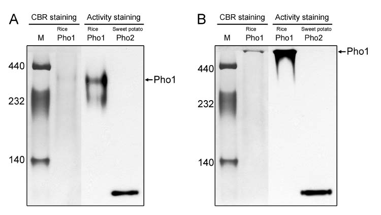
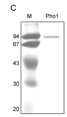
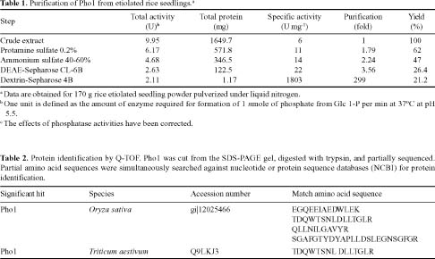
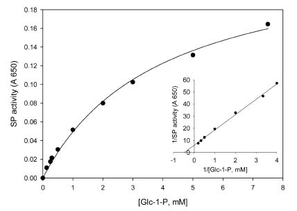
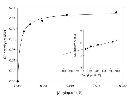
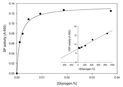
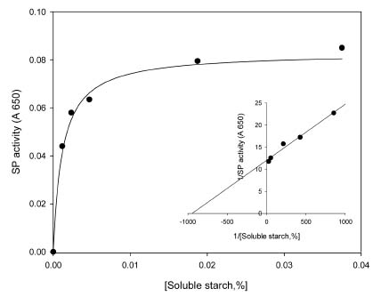
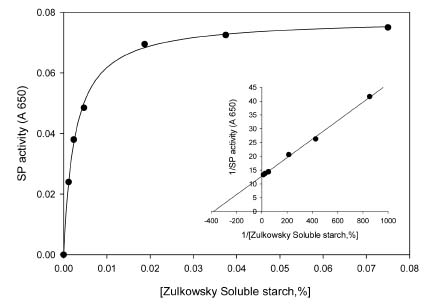
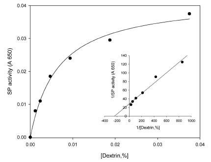
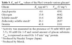
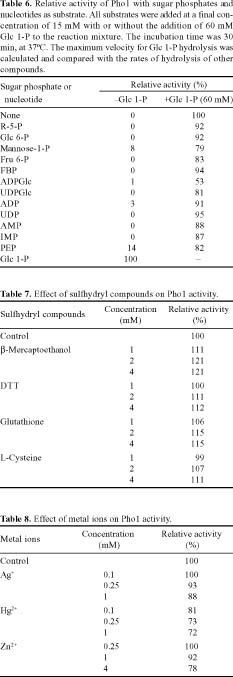
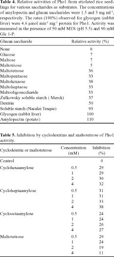
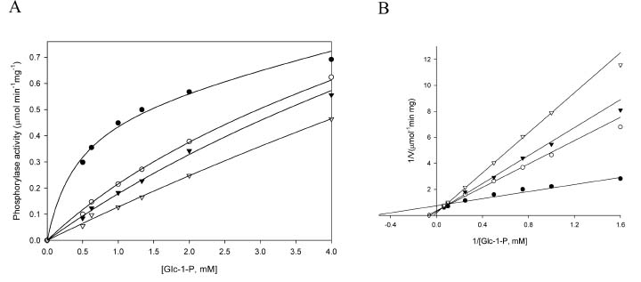
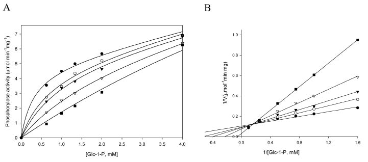
Abstract. Starch phosphorylase1 (Pho1) from etiolated rice (Oryza sativa L. cv. Tainong 67) seedlings was purified by ammonium sulfate fractionation, DEAE-Sepharose CL-6B anion exchange chromatography, and dextrin-Sepharose 4B affinity chromatography. The purification fold was 299, and the enzyme activity recovery was about 21%. The molecular mass of the native Pho1 on Superose 12 gel filtration was 145 kDa. The subunit molecular weight as determined by SDS-PAGE was 85 kDa. The enzyme has an optimum pH of 5 and an optimum reaction temperature of about 45oC~50 oC. In the synthetic reaction for Glc 1-P, the Km value was 2.1 mM, and the Vmax value was 5.85 U mg-1. In the phosphorolytic direction for orthophosphate, the Km value was 3.8 mM. Pho1 has a higher affinity for amylopectin, glycogen, soluble starch and dextrin than for maltooligosaccharide (6 to 10 glucose units). In addition, the Km value for amylopectin was ninefold lower than for dextrin. Cyclohexaamylose, cycloheptaamylose, cyclooctaamylose, and maltotetrose were inhibitors of Pho1. Mannose 1-P, Fru 6-P, ADPGlc, UDPGlc, AMP, IMP and PEP also inhibit Pho1. The metal ions Ag+, Hg2+ and Zn2+ also reduce the enzyme activity. However, thiol reagents activate Pho1 activity, suggesting that sulfhydryl-group(s) may be required for enzyme stability.

Keywords: Affinity chromatography; Dextrin; Glucan; Rice; Starch phosphorylase.

Abbreviations: Pho1, H type starch phosphorylase; Pho2, L type starch phosphorylase; Glc 1-P, glucose 1-phosphate.

Introduction

The plant starch phosphorylases (Pho, SP, a-glucan phosphorylase, EC 2.4.1.1) catalyze the reversible phosphorolysis of a-1,4-glucans and have been reported in many higher plants (Fukui, 1983; Steup and Schachtele, 1986). The reaction can be described essentially by the following equation:

(a-1,4-D-glucose)n + Glc 1-P (a-1,4-D-glucose)n+1 + Pi

A glucosyl unit is transferred from Glc 1-P to starch of increasing chain length, and this is termed as "the synthetic direction." In the other direction, the addition of inorganic phosphate generates Glc 1-P from starch as the chain length decreases, and this is called "the phosphorolytic direction."

Pho is generally regarded as a starch-degrading enzyme, but some evidence suggests it might also have an important synthetic function. In most plants, Pho has been found in both cytosolic and amyloplastic forms in pea seeds and leaves (Hanes, 1940), broad beans (Suda et al., 1987), potato tubers (Hanes, 1940), maize (Tsai and Nelson, 1968; Mu et al., 2001), barley (Baxter and Duffus, 1973), rice seeds (Richardson and Matheson, 1977), spinach (Steup and Latzko, 1979), sweet potato (Chang et al., 1987; Lu et al., 1995), bananas (Richardson and Matheson, 1977; Da Mota

et al., 2002), and seaweed (Fredrick, 1973; Yu and Pedersen, 1991). Specifically, the Pho1 or H (high affinity for glycogen) type of starch phosphorylase that occurs in the cytosol of plant cells has a molecular mass of about 90 kDa. Pho2 or L (low affinity for glycogen) form, is located in the amyloplast and has a molecular mass of about 100 kDa (Steup and Latzko, 1979; Conrads et al., 1986; Mu et al., 2001; Da Mota et al., 2002).

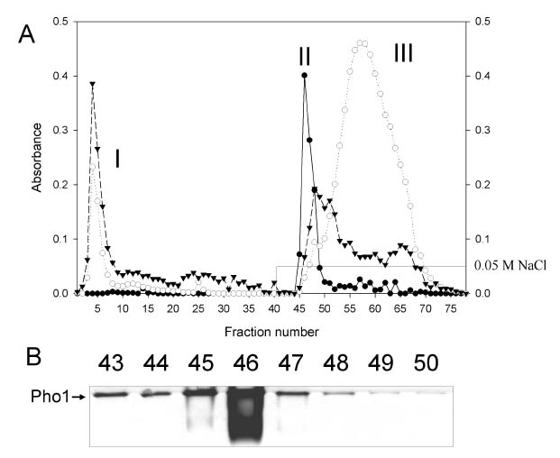
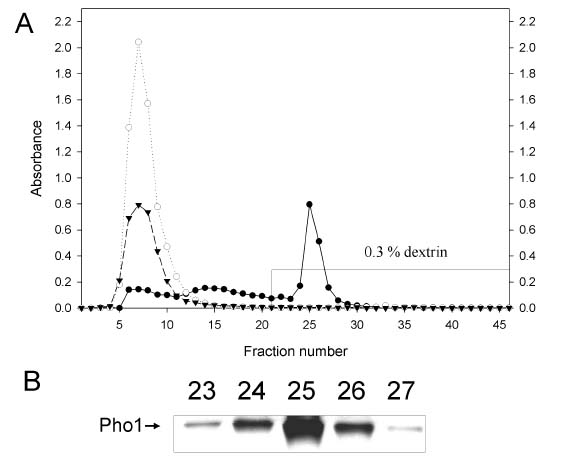
Here we report the purification and enzymatic characterization of a rice starch phosphorylase, Pho1. Since the level of Pho1 protein in etiolated rice seedlings is very low, we developed a procedure that greatly increases the yield and results in a Pho1 enzyme of high purity. Pho1 purified by this procedure was analyzed, and its molecular mass and kinetic properties was determined.

Material and Methods

Materials

Rice seeds (Oryza sativa L. cv. Tainong 67) were grown in the dark for 14 days at 30oC in a growth chamber. These fresh etiolated rice seedlings were used for enzyme purification. Imidazole, protease inhibitors, protamine, MES, glucan saccharides, cyclodextrins, sugar phosphate, nucleotides, thiol reagents, and ammonium molybdate were obtained from Sigma (USA). Soluble starch was purchased from Nacalai Tesque Inc. (Kyoto, Japan). Zulkowsky soluble starch, AgNO3, ammonium sulfate, Tris-Cl were supplied

*Corresponding author. Tel:+886-2-2363-3610; Fax: +886-2- 2363-3610; E-mail: pingdu@ntu.edu.tw


Botanical Bulletin of Academia Sinica, Vol. 45, 2004

by Merck (USA). All of the chromatographic gel materials were purchased from Amersham Pharmacia Biotech (UK).

Purification of Pho1

All purification steps were carried out at 0°C to 4°C (except where noted). The etiolated rice seedlings were cut to pieces with scissors and pulverized under liquid nitrogen using a mortar-pestle. Approximately 170 g of etiolated rice seedlings powder was added to 400 ml of buffer A [50 mM imidazole-HCl (pH 7), 1 mM PMSF, 1 mM benzamidine, 1 mM EDTA, 1 mM DTT] and filtered through four layers of cloth. Protamine sulfate was added to the filtrate to a concentration of 0.2%, and the solution was then kept 4°C for 30 min. At that point, the solution was centrifuged at 10,000 g for 20 min. The supernatant was made up to 40% saturation with solid ammonium sulfate, chilled and centrifuged at 10,000 g for 30 min. The pellet was discarded and the supernatant fraction was raised to 60% saturation with solid ammonium sulfate, chilled, and centrifuged at 10,000 g for 30 min. The pellet was resuspended in a minimal volume of buffer B [50 mM imidazole-HCl (pH 7), 1 mM EDTA, 1 mM DTT] and dialyzed overnight against buffer B. The solution containing the enzyme was then applied to a DEAE-Sepharose CL-6B column (2 cm2×15 cm) pre-equilibrated with buffer B at a flow rate of 30 ml h-1. The column was then washed with 10 volume of the same buffer to wash away the bulk of the unwanted proteins. The enzyme fractions were sequentially eluted with buffer B containing 0.05 M NaCl. Active fractions were applied to a dextrin-Sepharose 4B column (2 cm2×11 cm) pre-equilibrated with buffer B containing 0.05 M NaCl at a flow rate of 30 ml h-1. Phosphorylase was eluted with buffer B containing 0.3% dextrin. Fractions with phosphorylase activity were pooled and dialyzed against buffer B overnight.

Preparation of dextrin-Sepharose

Freeze-dried CNBr-activated Sepharose 4B powder (Amersham Pharmacia Biotech) was washed with 200 ml of 1 mM HCl per gram and then reswelled on a sintered glass filter (G3). A solution of 30 mg dextrin per ml of gel was made up in coupling buffer (0.1 M NaHCO3, pH 8.3, containing 0.5 M NaCl). The dextrin solution was mixed with the gel suspension in an end-over-end mixer for 2 h at room temperature or overnight at 4°C. It is important not to use a magnetic stirrer. The gel was transferred to buffer containing the blocking agent (1 M ethanolamine or 0.2 M glycine, pH 8.0) for 16 h at 4°C or 2 h at room temperature. The excess adsorbed dextrin was washed away with coupling buffer followed by 0.1 M acetate buffer (pH 4) containing 0.5 M NaCl. This was followed by a second wash with coupling buffer to further remove excess blocking agent. The dextrin-Sepharose gel was stored in experimental buffer at 4-8°C.

Assay of Pho1

Glucan synthesis activity: One unit of specific (SP) activity was defined as the amount of enzyme that catalyzed the formation of 1 nmole of product per min. Specific ac

tivity was defined as units per mg of protein. SP activity was measured in the synthetic reaction by following the formation of Pi using a molybdate-based assay (Fiske and Subbarow, 1925). The reaction mixture contained 50 mM MES, pH 5.5, 0.3% glycogen, 90 mM Glc 1-P (except where noted), and the enzyme sample and had a final volume of 100 µl. After incubation for 30 min at 37°C, the inorganic phosphate released was determined by adding 200 µl of ferrous sulphate molybdate solution followed by incubation at room temperature for 10 min. The absorbance was measured using an ELISA reader at 650 nm (Chen et al., 2002).

The Pho1 activity was determined by assaying the total phosphate production minus the amount of phosphate produced by the phosphatase activity. The resulting phosphate amount was then used to calculate the real phosphorylase synthetic activity.

Phosphatase activity: The reaction was conducted under the same incubation conditions as described for the assay of glucan synthesizing activity of phosphorylase in the absence of glycogen (Suda et al., 1987).

Phosphorolytic activity: The reaction mixture contains 50 mM MES buffer (pH 6), 3% glycogen, and 50 mM Pi in a total volume of 100 µl. The reaction was stopped by heating the mixture for one min at 100°C. Glc 1-P produced was determined in a coupled reaction via phosphoglucomutase (EC 5.4.2.2) and glucose-6-phosphate dehydrogenase (EC 1.1.1.49) (Michal, 1986; Toroser et al., 2000; Da Mota and Cordenunsi, 2002).

Protein determination

Protein concentration was determined by the dye-binding method (Bradford, 1976) using bovine serum albumin as a standard.

Activity staining of Polyacrylamide Gel Electrophoresis

All analytical gels used were 0.75 mm in thickness. After native-PAGE, activity staining was done by incubating the gel with shaking in 50 mM MES buffer (pH 5.5) containing 20 mM Glc 1-P and 0.3% glycogen at 37°C for 6 h, followed by iodine staining.

Kinetic assay of Pho1

Km of Pho1 for Glc 1-P: Pho1 activity was measured in the synthetic direction as a function of the concentration of glycogen at the indicated set concentrations of Glc 1-P. Km of Pho1 for glycogen: Pho1 activity was measured in the synthetic direction as a function of the concentration of Glc 1-P at the indicated set concentrations of glycogen. Data given in Table 3 were the average of three replicates of separated experiments.

Mass spectrometry analysis

After CBR staining on a SDS-PAGE gel, the major band was cut from the gel, digested with trypsin (Jimenez et al.,


Hsu et al. — Rice starch phosphorylase purification and characterization

1998), and partially sequenced by Q-TOF (Micromass) analysis.

Results

Purification of the Pho1

Pho1 was stable in 50 mM imidazole, pH 7.0, 1 mM EDTA, 1 mM DTT at 4°C for at least two weeks. Fractionation with protamine sulfate and ammonium sulfate of the initial extract, followed by DEAE-Sepharose CL-6B chromatography and dextrin-Sepharose 4B affinity chromatography resulted in a 299-fold purification of SP with a 21%

recovery from the initial extract. Elution profiles of the enzyme on DEAE-Sepharose CL-6B chromatography and dextrin-Sepharose chromatography are shown in Figures 1 and 2. The specific activity of the enzyme was 1.8 U mg-1 protein. On DEAE-Sepharose CL-6B chromatography, the enzyme was eluted at 0.05 M NaCl. Only peak II represented Pho1 activity when tested on a 5% native-PAGE activity gel (Figure 1).

The final step of dextrin-Sepharose chromatography was an efficient step. A large amount protein was not retained on the dextrin matrix while Pho1 was eluted at 0.3% dextrin (Figure 2). Its native-PAGE and SDS-PAGE patterns are shown in Figure 3. Pho1 has a high affinity toward

Figure 1. Elution profiles and activity staining of the phosphorylase activity from etiolated rice seedlings on DEAE-Sepharose CL-6B. Panel A: Phosphorylase activity (l); phosphatase activity (m); protein (q); NaCl concentration (_) are shown. Peak II represents phosphorylase activity. Peak I and III were ascribed to phosphatase activity. Panel B: Native-PAGE adding 0.05% glycogen in separation gel.

Figure 2. Affinity chromatography of phosphorylase activity from etiolated rice seedlings on Dextrin-Sepharose 4B. Panel A: Phosphorylase activity (l); phosphatase activity (m); protein (q) are shown. The full line represents the elution concentration of dextrin. Panel B: Activity staining of Native-PAGE adding 0.05% glycogen in separation gel.

Figure 3. Native-PAGE and SDS-PAGE of Pho1 fraction separated after dextrin-Sepharose 4B chromatography. Approximately 4 ug of total protein were loaded in each lane. Proteins were stained with Coomassie blue. Panel A: Native-PAGE. M, molecular mass markers in kDa. Panel B: Native-PAGE adding 0.05% glycogen in separation gel. Panel C: SDS-PAGE.


Botanical Bulletin of Academia Sinica, Vol. 45, 2004

branched glucan saccharides, and therefore it was retarded on a 5% native-PAGE gel when 0.05% glycogen was added (Figure 3B). The results of the stepwise purification of the Pho1 are summarized in Table 1.

Molecular Mass and Q-TOF Analysis

The molecular weight of native Pho1 is 145 kDa, as measured by fast protein liquid chromatograph (Superose 12) (data not shown). Figure 3A shows that the native enzyme has an apparent size of about 300 kDa. When analyzed by denaturing SDS-PAGE, the enzyme produced a single major band with a subunit size of 85 kDa (Figure 3C). It was cut from the SDS-PAGE gel, digested with trypsin overnight, and partially sequenced by Q-TOF. The enzyme obtained from the etiolated rice seedlings matched with four peptide sequences of rice Pho1 and one of wheat Pho1 (Table 2).

Enzyme Kinetics Assay

The Pho1 kinetics assay was carried out in the direction of polysaccharide synthesis. Under these conditions, the reaction was linear with time for at least 50 min. For Glc 1-P, the apparent Km value was measured as 2.1 mM, and the Vmax value was 5.85 U mg-1 (Figure 4). For the various saccharides as the acceptor substrate, the different Km and Vmax values of the purified Pho1 from etiolated rice seedlings are listed in Table 3. Amylopectin was the best substrate, and dextrin was the poorest with the Km value for amylopectin being ninefold lower than that for dextrin.

In the phosphorolytic direction, the Km for glycogen and soluble starch of Pho1 were 1.19 and 0.76 mg ml-1,

respectively. For orthophosphate, the Km value was 3.8 mM.

Optimum pH and Temperature

The purified enzyme exhibited an optimal activity at pH 5.0 in the polysaccharide synthesis direction with the highest stability at pH 7.0. The maximum enzyme activity obtained at 45~50°C over an incubation time of 30 min.

Effect of Glucan Saccharides

The different synthetic activities of Pho1 with various sized glucan substrates are displayed in Table 4. Glucose, maltose, and maltotriose showed almost no activity with Pho1. Maltotetrose and maltohexaose showed higher activity than maltopentaose and maltoheptaose. Maltooligosaccharide, containing 6 to 10 glucose subunits, showed an apparently low relative activity. Amylopectin showed the highest relative activity.

Inhibition by Cyclodextrins and Maltotetrose

Table 5 shows that cyclodextrin and maltotetrose both inhibit Pho1. At a range of different concentrations, the cyclodextrins showed a higher level of inhibition than maltotetrose, especially cycloheptaamylose.

Effect of Sugar Phosphates, Nucleotides, and Nucleotide Sugars

In the absence of Glc 1-P as reaction substrate, sugar phosphates had no effect on Pho1 activity. This indicates that Pho1 has a high substrate specificity for Glc 1-P (Table


Hsu et al. — Rice starch phosphorylase purification and characterization

(a)

(b)

(c)

(d)

(e)

(f)

Figure 4. Effect of glucose-1-phosphate (a), amylopectin (b), glycogen (c), soluble starch (produced by Nacalai tesque) (d), Zulkowsky soluble starch (produced by Merck) (e) and dextrin (f) concentrations on activities of Pho1 and respective double reciprocal plot.

6). Mannose 1-P, Fru 6-P, ADPG, UDPG and PEP showed a high level of inhibition of Pho1.

Effect of Thiol Reagents and Metal Ions

Table 7 shows that thiol reagents increased enzyme activity by 10~20% at 1~4 mM, especially b-mercaptoethanol. Thiol reagents at a higher concentration activated the enzyme. The effect of various metal ions on enzyme activity was shown in Table 8. Zn2+, Hg+2 and Ag+ reduced the enzyme activity observably. However, other metal ions like Na+, K+, Mg2+, Ca2+ had no significant effect on it (data not shown).


Botanical Bulletin of Academia Sinica, Vol. 45, 2004

Discussion

This is the first detailed study of Pho1 in rice. Maximal activity for Pho1 was detected in 14-d-old dark-grown rice seedlings. Pho1 is the predominant form. During the extraction treatment, Pho2 could only be detected on the first day. Under DEAE-Sepharose CL-6B chromatography (Figure 1), Pho1 showed a low negative charge binding. It could easily be eluted with 50 mM NaCl. On DEAE-Sepharose CL-6B chromatography, peak II presented real phosphorylase activity, while peak I and III were false phosphatase activity (Figure 1).


Hsu et al. — Rice starch phosphorylase purification and characterization

The purification key step was the dextrin-Sepharose affinity chromatography (Figure 2), during which all unwanted proteins were washed away. This used the principle that Pho1 has highly binding affinity for glucan saccharides, especially branching glucan saccharides. We modified the manufacturer's method of dextrin-Sepharose preparation (Steup et al., 1980) by using commercial CNBr-activated Sepharose (Amersham Pharmacia Biotech). This protocol avoided the use of toxic CNBr during the preparation of dextrin-Sepharose. The Pho1 was purified 299-fold with a yield of about 21% (Table 1).

The high affinity of Pho1 for glycogen was demonstrated by the native-PAGE (Figure 3A, B). It was obviously retarded on native-PAGE when 0.05% glycogen was added to the separation gel (Figure 3B). However, Pho2 of sweet potato was not retarded by glycogen on native-PAGE. Thus, affinity-gel electrophoresis indicated a difference in the affinity of Pho1 and Pho2 for glycogen. Figure 3 shows that activity staining of Pho1 was much more sensitive than CBR staining. The enzyme apparently undergoes diffusion in the native PAGE gel and appears as a smear band (Figure 3A).

After fast protein liquid chromatography Superose 12 gel filtration, the Pho1 was measured as having an apparent molecular weight of 145 kDa (data not shown). However, the subunit of the enzyme was 85 kDa on SDS-PAGE (Figure 3C). The result is quite similar to that observed for the spinach Pho1 (native: 150 kDa; subunit: 89 kDa) (Steup et al., 1980) and banana Pho1 (native: 155 kDa; subunit: 90 kDa) (Da Mota et al., 2002). The enzyme was partially sequenced by Q-TOF, and this matched the rice Pho1 from the genome database and the wheat Pho1 from the protein database (Table 2). It also showed a match to animal glycogen phosphorylase (data not shown). This result confirms that the purified enzyme was Pho1 from etiolated rice seedlings.

The even numbers chain length of the short chain maltosaccharides, like maltotetrose and maltohexaose, had a higher relative activity than the odd numbers chains (Table 4). On the other hand, glucose, maltose, and maltotriose were ineffective primers while maltodextrins that contained more than 4 glucose units at a concentration 3 mg ml-1 showed greater than 30% activity. In vivo Pho2 from maize utilizes maltooligosaccharide substrates ranging in size from 4 to 7 glucose units (Mu et al., 2001). Kinetic studies of maize Pho2 show that the phosphorolytic reaction was favored over the synthetic reaction and that the enzyme preferred longer chain substrates (Mu et al., 2001). Nevertheless, the Pho1 of red seaweeds and etiolated soybean seedlings show a higher activity with maltodextrins that contain more than 5 glucose units (Suda et al., 1987; Yu and Pedersen., 1991).

Cyclodextrins isolated from potato and spinach are efficient inhibitors for Pho1 (Table 5). They competed with the glucan substrate on Pho1, but had no effect on the rabbit muscle glycogen phosphorylase. The degree of inhibition by cyclodextrins varies with their degree of polymerization and the particular phosphorylase (Shimomura et al., 1982; Yu and Pedersen., 1991). In the case of etiolated rice seedlings, cycloheptaamylose was more inhibitory than cyclohexaamylose or cyclooctaamylose (Table 5).

Table 6 shows ADPGlc and UDPGlc are potent inhibitors of the synthetic reaction. Both ADPGlc and UDPGlc were found to be non-Michaelian inhibitors with respect to Glc 1-P in the synthetic reaction (Figure 5, 6). The phenomenon was partially similar to the study in maize, where only ADPGlc was found to be a non-Michaelian inhibitor (Burr and Nelson, 1975; Mu et al., 2001). Since ADPGlc and UDPGlc can both serve as precursors for starch synthesis, directly or indirectly, it is reasonable to believe they possess inhibitory properties to Pho1.

Figure 5. Effects of ADPGlc concentrations on phosphorylase activity (A) and respective double reciprocal plot (B). No inhibitor (l); 0.675 mM ADPGlc (m); 1.25 mM ADPGlc (q) or 2.5 mM ADPGlc (s).


Botanical Bulletin of Academia Sinica, Vol. 45, 2004

Figure 6. Effects of UDPGlc concentrations on phosphorylase activity (A) and respective double reciprocal plot (B). No inhibitor (l); 0.675 mM UDPGlc (m); 1.25 mM UDPGlc (q) or 2.5 mM UDPGlc (s) or 5 mM UDPGlc (n).

The rice Pho1 showed a requirement for free -SH groups to ensure stability (Table 7). The -SH group might be directly involved in catalysis or be required to maintain an appropriate enzyme conformation. Thus, it would seem that the presence of essential sulfhydryl groups is required for enzyme activity. In addition, Tsai and Nelson (Tsai and Nelson, 1968) reported that Pho2 activity was stimulated threefold by 10 mM MgCl2 or 2 mM EDTA. This does not occur with Pho1 from etiolated rice seedlings (Table 8).

In the synthetic reaction for Glc 1-P, the Km value was 2.1 mM. The Km for orthophosphate of Pho1 in the physiological direction of Glc 1-P production was 3.83 mM. The Km value for Glc 1-P was lower than for orthophosphate. In phosphorolytic direction, Pho1 favored soluble starch more than glycogen. The Km values were 0.76 and 1.19 mg ml-1, respectively, which is in contrast with the synthetic reaction (Table 3). Based on our results, the kinetics data indicates that Pho1 favors synthetic direction over phosphorolytic direction. Therefore, in the etiolated rice seedlings, Pho1 may play a role in starch synthesis.

In higher plants, the flow of carbon from Glc 1-P to starch was focused on ADPGlc pyrophosphorylase, starch synthase, starch branching enzyme, and starch debranching enzyme (Smith et al., 1997). However, the expression of Pho2 cDNA in potato (Brisson et al., 1989; St-Pierre and Brisson, 1995), spinach (Euwenig et al., 1997), and pea (Van Berkel et al., 1991) suggests that the enzyme is involved in starch biosynthesis. Thus, it is reasonable to speculate that starch phosphorylase might play some role in starch metabolism. The glucan-trimming model (Ball et al., 1996; Myers et al., 2000) suggests that starch synthase elongates glucan chains and branching enzyme inserted branches to form a highly branched structure known as preamylopectin. Preamylopectin molecules are then trimmed by the starch debranching enzyme. The short

chain maltooligosaccharides liberated in the trimming reaction are converted to longer chain glucan molecules by the action of glucan-degrading enzymes such as isoamylase, starch phosphorylase, and debranching-enzyme (Takaha et al., 1998; Mu et al., 2001). It has been proposed that debranching-enzyme and starch phosphorylase might participate in the recycling of maltooligosaccharides for use in starch synthesis (Myers et al., 2000; Takaha et al., 1998; Mu et al., 2001; Yu et al., 2001). The longer-chain glucan molecules can then be utilized by starch phosphorylase via the phosphorolytic reaction to generate Glc 1-P. Glc 1-P could be utilized by ADPGlc pyrophosphorylase for the synthesis of starch (Mu et al., 2001; Yu et al., 2001).

Pho1 has high similarity to the animal glycogen phosphorylase in structure (Nakano and Fukui, 1986; Mori et al., 1993). When compared to Pho2 from sweet potato, Pho1 lacks the loop 78 made up of inserted 78 amino acids. Loop 78 was hypothesized to be inserted in the starch-binding site of the ancestor phosphorylase gene during evolution (Chen et al., 2002). Loop 78 might serve as a switch that regulates Pho2 by initiating the synthesis of amylose to act as a primer in starch biosynthesis or, perhaps, catalyzing the phosphorolytic reaction to degrade starch in sweet potato (Chen et al., 2002).

The Pho1 content in rice is low. Nevertheless, it is still much higher than Pho2. Pho2 in rice was only present during the first day of enzyme extraction. We therefore focus on the analysis of Pho1 in this study. The identification and characterization of Pho1 from etiolated rice seedlings presented here provides a foundation for future studies of this rice enzyme.

Acknowledgements. This work was supported by grants from the National Science Council of the Republic of China (NSC90-2313-B-002-282).


Hsu et al. — Rice starch phosphorylase purification and characterization

Literature Cited

Ball, S., H.P. Guan, M. James, A. Myers, P. Keeling, G. Mouille, A. Buleon, P. Colonna, and J. Preiss. 1996. From glycogen to amylopectin: a model for the biogenesis of the plant starch granule. Cell 86: 349-52.

Baxter, E.D. and C.M. Duffus. 1973. Phosphorylase activity in relation to starch synthesis in developing Hordeum distichum grain. Phytochemistry 12: 2321-2330.

Bradford, M.M. 1976. A rapid and sensitive method of the quantitation of microgram quantities of protein utilizing the principle of protein-dye binding. Anal. Biochem. 72: 248-254.

Brisson, N., H. Giroux, M. Zollinger, A. Camirand, and C. Simard. 1989. Maturation and subcellular compartmentation of potato starch phosphorylase. Plant Cell. 1: 559-566.

Burr, B. and O.E. Nelson. 1975. Maize a-Glucan phosphorylase. Eur. J. Biochem. 56: 539-546.

Chang, T.C., S.C. Lee, and J.C. Su. 1987. Sweet potato starch phosphorylase-purification and characterization. Agric. Biol Chem. 51: 187-195.

Chen, H.M., S.C. Chang, C.C. Wu, T.S. Cuo, J.S. Wu, and R.H. Juang. 2002. Regulation of the catalytic behaviour of L-form starch phosphorylase from sweet potato roots by proteolysis. Physiol. Plant. 114: 506-515.

Conrads, J., J. van Berkel, C. Schachtele, and M. Steup. 1986. Non-chloroplast a-1,4-glucan phosphorylase from pea leaves: characterization and in situ localization by indirect immunofluorescence. Biochim. Biophys. Acta. 882: 452-463.

Da Mota, R.V., B.R. Cordenunsi, J.R. do Nascimento, E. Purgatto, M.R. Rosseto, and F.M. Lajolo. 2002. Activity and expression of banana starch phosphorylases during fruit development and ripening. Planta 216: 325-333.

Fiske, C.H. and Y. Subbarow. 1925. The colorimetric determination of phosphorous. J. Biol. Chem. 66: 375-400.

Fredrick, J.F. 1973. Further studies of primer-independent phosphorylase isozymes in the algae. Phytochemistry 12: 1933-1936.

Fukui, T. 1983. Plant Phosphorylase. In T. Akazawa, T. Asahi, and H. Imase (eds.), The New Frontiers in Plant Biochemistry. Japan Scientific Societies Press, Tokyo, pp. 71-82.

Hanes, C.S. 1940. The breakdown and synthesis of starch by enzyme system from pea seeds. Proc. Roy. Soc. (London) B128: 421-450.

Hanes, C.S. 1940. The reversible formation of starch from Glucose-1-Phosphate catalyzed by potato phosphorylase. Proc. Roy. Soc. (London) B129: 174-208.

Jimenez, C.R., L. Huang, Y. Qiu, and A.L. Burlingame. 1998. Current Protocols in Protein Science: In-gel digestion of proteins for MALDI-MS fingerprint mapping, pp. 16.4.1-16.4.5.

Lu, C.H., P.D. Lee, and J.C. Su. 1995. Preparation of amyloplasts from sweet potato callus culture. Bot. Bull. Acad. Sin. 36: 223-228.

Michal, G. 1986. Methods of Enzymatic Analysis. In H.U. Bergmeyer, J. Bergmeyer, and M. Grabl (eds.), Verlag Chemie, Weinheim, Basel/Deerfield Beach, FL, pp. 185-191.

Mori, H., K. Tanizawa, and T. Fukui. 1993. A chimeric a-glucan phosphorylase of plant type L and H isozymes. J. Biol. Chem. 268: 5574-5581.

Mu, H.H., Y. Yu, B.P. Wasserman, and G.M. Carman. 2001. Purification and characterization of the maize amyloplast stromal 112-kDa starch phosphorylase. Arch. Biochem. Biophys. 388: 155-164.

Myers, A.M., M.K. Morell, M.G. James, and S.G. Ball. 2000. Recent progress toward understanding biosynthesis of the amylopectin crystal. Plant Physiol. 122: 989-998.

Nakano, R. and T. Fukui. 1986. The complete amino acid sequence of potato a-glucan phosphorylase. J. Biol. Chem. 261: 8230-8236.

Richardson, R.H. and N.K. Matheson. 1977. Some molecular properties of plant starch phosphorylase and the levels of activity of the multiple forms. Phytochemistry 66: 1875-1879.

Shimomura, S., M. Nagai, and T. Fukui. 1982. Comparative glucan specificities of two types of spinach leaf phosphorylase. J. Biochem. 91: 703-717.

Smith, A.M., K. Denyer, and C. Martin. 1997. The synthesis of the starch granule. Annu. Rev. Plant. Physiol. Mol. Biol. 48: 65-87.

Steup, M. and C. Schachtele. 1986. a-1,4-Glucan phosphorylase from leaves of spinach II. Peptide patterns and immunological properties. A comparison with other phosphorylase forms. Planta 168: 222-231.

Steup, M., C. Schachtele, and E. Latzko. 1980. Purification of a non-chloroplastic a-glucan phosphorylase from spinach leaves. Planta 148: 168-173.

Steup, M. and E. Latzko. 1979. Intracellular localization of phosphorylase in spinach and pea leaves. Planta 145: 69-75.

St-Pierre, B. and N. Brisson. 1995. Induction of the plastidic starch-phosphorylase gene in potato storage sink tissue. Planta 195: 339-344.

Suda, M., T. Watanabe, M. Kobayashi, and K. Matsuda. 1987. Two types of phosphorylases from etiolated soybean cotyledons. J. Biochem. 102: 471-479.

Takaha, T., J. Critchley, S. Okada, and S.M. Smith. 1998. Normal starch content and composition in tubers of antisense potato plants lacking D-enzyme (4-a-glucanotransferase). Planta 205: 445-451.

Toroser, D., Z. Plaut, and S.C. Huber. 2000. Regulation of a plant SNF-related protein kinase by glucose-6-phosphate. Plant Physiol. 123: 403-412.

Tsai, C.Y. and O.E. Nelson. 1968. Phosphorylase I and II of Maize endosperm. Plant Physiol. 43: 103-112.

Van Berkel, J., J. Conrads-Stauch, and M. Steup. 1991. Glucan phosphorylase forms in cotyledons of Pisum sativum L.: location, developmental change, in vitro translocation, and processing. Planta 185: 432-439.

Yu, S. and M. Pedersen. 1991. One-step purification to homogeneity and isoforms of a-1,4-glucan phosphorylase of red seaweed. Plant Physiol. Biochem. 29: 341-347.

Yu, Y., H.H. Mu, B.P. Wasserman, and G.M. Carman. 2001. Identification of the maize amyloplast stromal 112-kD protein as a plastidic starch phosphorylase. Plant Physiol. 125: 351-359.


Botanical Bulletin of Academia Sinica, Vol. 45, 2004