Bot. Bull. Acad. Sin. (2004) 45: 237-245

So — The occurrence of extrafloral nectaries in Hong Kong plants

The occurrence of extrafloral nectaries in Hong Kong plants

May Ling SO*

Biology Department, Hong Kong Baptist University, 224 Waterloo Road, Hong Kong

(Received September 3, 2003; Accepted March 2, 2004)

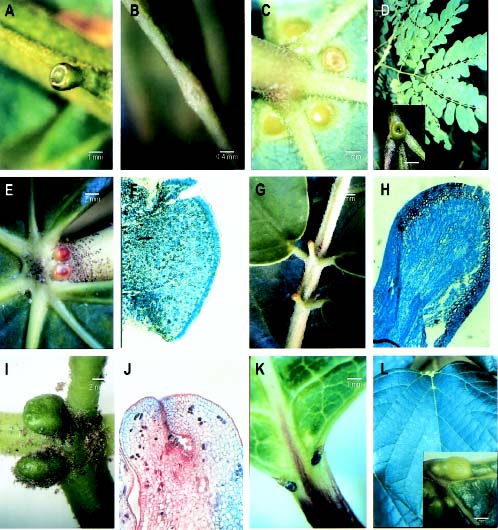
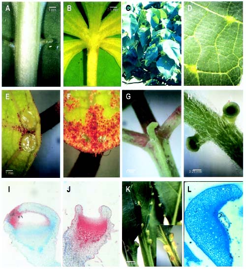
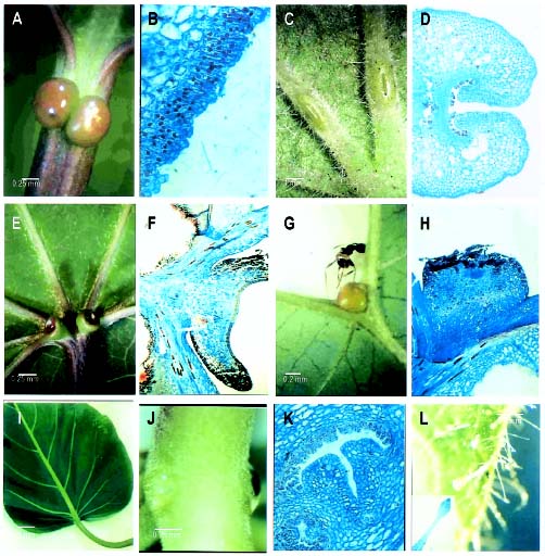
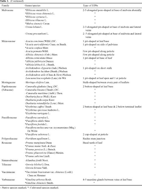
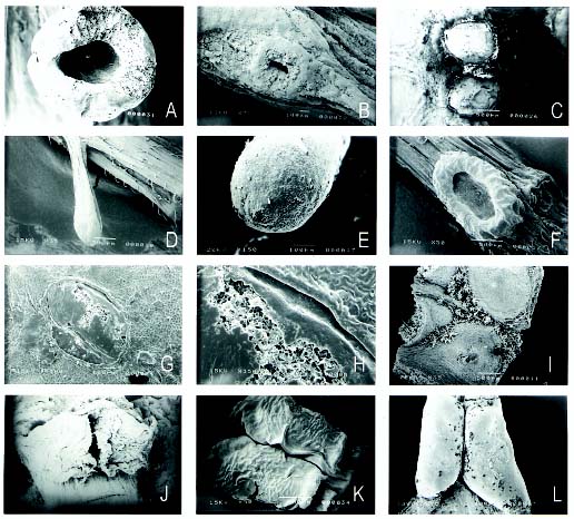
Abstract. This is a study of the extrafloral nectaries in Hong Kong plants. Five major types can be discerned: button-shaped, cup-shaped, stalk-shaped, pit-shaped, and pore-shaped. Euphorbiaceae is the largest family with extrafloral nectaries which are always visible structures, attracting ants. SEM micrographs of extrafloral nectaries are included.

Keywords: Extrafloral nectaries; Hong Kong plants.

Introduction

Nectaries not involved in pollination are called extrafloral nectaries (EFNs), sugar producing glands found outside the flower. They occur in at least 66 families (Elias, 1983), many in the tropics. Hong Kong, situated in a subtropical region, is rich in flowering plants, many of them either cultivated in parks and gardens or grow in the wild in many countryparks. EFNs have been shown to play an important part in the defense mechanism of plants against herbivores (Nees, 2003). Even though the debate over the role of extrafloral nectaries has lasted over a century, numerous scientists are still attracted by the role these small, fascinating structures play in the life of a flowering plant (Bentley, 1977; Koptur, 1992). The animals usually associated with EFNs are ants, and it is not uncommon to see ants crawling over leaves and petioles, looking for nectar. This ants/plant facultative mutualistic association has been described as beneficial to both parties (Pemberton, 1998). The ants, while obtaining food for their colony, can deter other herbivores from attacking the plants. Heil et al. (2001) even showed that the production of extrafloral nectar in Macaranga tanarius is an induced, indirect defensive reponse that strongly reduces herbivory. In a series of experiments to investigate the various factors affecting nectar production in Macaranga tanarius (Heil et al., 2000), nectar production is highest in unfolded young leaves, and the rate of secretion remains relatively constant throughout the day, peaking at dusk. The composition of the secretion is mostly fructose, glucose, sucrose (Heil et al., 2000), with traces of various amino acids (Ness, 2003). The ontogeny of EFNs in Cappris retusa has been studied by Di Sapio et al. (2001), who confirmed that nectar secretion appears early in the development process. Studies by Farji

Brenar et al. (1992) also indicate that secretion is apparently related to ant patrolling activities. Ness (2003) also showed that production of nectar increases two- to three-fold when leaves are attacked by caterpillars, together with further attraction of ant bodyguards. The presence of EFNs may further add an ecological advantage to these plants in self-protection, reduce vegetative damage, and help to prevent heavy foraging by other animals (Bentley, 1976; Pemberton, 1998). An experiment conducted on Sapium sebiferum by Rogers et al. (2003) indicated that simulated leaf herbivory significantly stimulated effluent production on EFN glands on seedlings. The beneficial effect of ants on the reproductive success of Dyckia floribunda (Bromeliaceae), an extrafloral nectary plant, is shown by Vesprini et al. (2003) that total seed production per plant was strongly affected by ant exclusion. However, other studies by Freitas et al. (2000) indicated no significant differences in either the degree of herbivory or in the reproductive output between stems of Croton sarcopetulus with ants and those without. Another hypothesis on the secretion of extrafloral nectar is that it is an attempt to distract insects from flowers (Rosenzweig, 2002). Since the secretion of nectar is an energy intensive process, Rosenzweig (2002) theorized that the cost of each extrafloral nectary divided by the cost of each flower must be less than the proportion of reproduction threatened by insect visits.

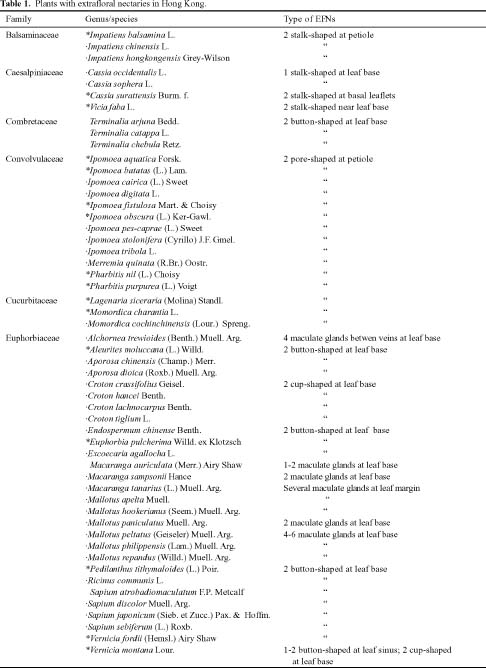
The purpose of this paper is to study the EFNs in Hong Kong plants and examine the type, shape, number, and position on the leaves. Extrafloral nectaries differ considerably in gross morphology. Since many of the EFNs are minute and inconspicuous, e.g. Acacia sp., they are easily overlooked.

Materials and Methods

A number of field trips to numerous localities in various parts of Hong Kong were conducted to look for plants

*Corresponding author. E-mail: mlso@hkbu.edu.hk


Botanical Bulletin of Academia Sinica, Vol. 45, 2004

with extrafloral nectaries. Approximately 200 collections were examined in the course of this study. Scanning electron micrographs were made with a JOEL 5SM U-3 electronmicroscope, and all specimens were air dried or freeze-dried before gold coating. For the study of EFN anatomy, leaves were fixed in FAA, dehydrated in an ascending ethanol series (50%, 70%, 90%, 95%, and 100%), then embedded in paraffin. Transverse sections 10 µm in thickness were cut on a Leica RM 2125 RT rotary microtome and stained with fast green, safranin, or methylene blue. Photomicrographs were made with an Olympus microscope CX-40 mounted with a camera DP-10. The presence of sugar was tested for by using the Clinistix glucose reagent strips (Pemberton, 1990).

Results and Discussion

Extrafloral nectaries are especially common in the family Euphorbiaceae, which consists of at least 30 genera, among which 27 species belonging to 13 genera possess these structures. A few genera in the families Caesalpiniaceae, Mimosaceae, Convolvulaceae, Papilionaceae, Passifloraceae and Balsamaceae also have these nectaries. Most of these species are trees or shrubs. Herbaceous plants with EFNs are not common. EFNs vary considerably in size, shape, and position on the leaves. They are usually found at the base of lamina as small outgrowths, probably from modified stipules, rarely from marginal teeth. Some of the EFNs are found near the apical margin of the lamina, as seen in Macaranga tanarius.

In the present study of plants with extrafloral nectaries, these structures are best seen in young and developing leaves, secreting copious amount of sugars and attracting numerous ants. In mature leaves, these structures seem to have passed their prime stage, appearing brownish, becoming dark when the leaf is fully mature (Figure 2K). Vertical sections of the EFNs show glandular cells at the apex of the structure (Figure 1F, H). These cells have densely stained cytoplasm due to a very high concentration of ribosomes and mitochondria, and the nuclei are large (Figure 3B). Basically, five types of EFNs can be distinguished locally, and Table 1 shows the species with EFNs in Hong Kong plants. From the types of EFNs observed, the species specificity seems rather weak. For example, members of the family Caesalpinaceae have stalk-shaped EFNs between the leaflets (Figure 1G) while members of the Convolvulaceae have pore-shaped EFNs on the petiole just below the lamina (Figure 2B). Members of Mimosaceae have pit-shaped EFNs (Figure 1B). However, members of Euphorbiaceae have varied EFNs, from the cup-shaped in Vernicia montana (Figure 3E) to four maculate glands in Alchornea trewioides (Figure 1C).

Types of EFNs

A. Button-shaped EFNs

Button-shaped EFNs seem to be the most common type locally. They occur as a pair of round structures at the

base of the lamina, alongside the midvein or slightly below. When the leaf is expanding these EFNs are green and often covered with a weft of light brown scales during their development (Figure 2F). When the leaf is fully expanded, the scales fall off, and the EFNs are green, dark green, or brown (Figures 1I, 2E). These button-shaped EFNs may be round, as in Aleurites moluccana (Figure 1E), or oval, as in Sapium discolor (Figure 3A). A vertical section of the gland reveals a layer of glandular cells at the periphery of the gland (Figure 1F, 3B). When the leaf ages, these EFNs turn to dark brown, leaving a depression-like brown scar, seen here in Euphorbia pulcherrima (Figure 1K). The most peculiar position of button-shaped EFNs is found in Prunus persica. The fine marginal teeth of the leaves are light brown, however, the 2-4 (6) basal teeth near the petiole often become EFNs, arising as yellowish green knobs, gradually enlarged in size and finally becoming secretory. As the EFNs age, pit-like brown scars remain (Figure 2K). The button-shaped EFNs in Ricinis communis have a depressed centre as they age (Figure 2L). In addition to the two small button-shaped EFNs, glandular hairs are found in species of Passiflora, and those in Passiflora foetida are shown here (Figure 3L). In some species, button-shaped EFNs are rather thin and are called maculate glands, usually found at the base of lamina. Macaranga tanarius with its very large peltate leaves has several such minute glands bordering the apical margin. They number from 5 to 7 in total (Figures 2C, D, 4G, H) and are oval in shape with a flat or concave surface. Maculate glands are more conspicuous in Alchornea trewioides (Figures 1C).

B. Cup-shaped EFNs

Cup-shaped EFNs are uncommon locally, occurring in six species only. One example is found in Passiflora suberosa, a herbaceous climber. These EFNs arise on both sides of the petiole as two small pubescent knobs. Sections of these glands during development show that these knobs gradually become cup-shaped, with a stalk at the base (Figure 2I, J). The rim of the cup is thin and secretory cells occur at the centre of the cup (Figure 2H). In contrast, Vernicia montana also has somewhat cup-shaped EFNs which occur at the base of the lamina (Figure 3E). A longitudinal section of the gland shows the sessile cup arising directly from the leaf (Figure 3F).

C. Stalk-shaped EFNs

Stalk-shaped EFNs are also common locally, found in several genera belonging to different families. Among the 13 species of Cassia in Hong Kong, C. surattensis is the most common roadside tree. Stalk-shaped EFNs occur as thin stalks on the leaf axis between the lowest 2nd to 4th pairs of leaflets (Figures 1G, 4D, E). The apex of the EFN is round and remains so as long as it is secretory (Figure 1H). Similar stalk-shaped EFNs are also seen in Moringa oleifera, but they are found between every pair of leaflets (Figure 2G). Species of Impatiens have slender, fleshy, inconspicuous, and almost ephemeral EFNs at a distance from the base of the lamina (Figure 2A).


So — The occurrence of extrafloral nectaries in Hong Kong plants

D. Pit-shaped EFNs

Pit-shaped EFNs are not obvious externally and can only be found by careful examination. A typical pit is often seen in Leucaena leucocephala and species of Archidendron where a raised circular structure occurs at the junction between two leaflets. At the sunken centre of this structure

are secretory cells (Figure 4A, F). These pits vary in their number and position in different species. An even more obscure pit is seen in species of Acacia. Acacia confusa, a very widespread introduced species has very small phyllodes (up to 9 cm long, 1 cm wide), and a small inconspicuous pit occurs along the ridge adjacent to the stem (Figures

Figure 1. Extrafloral nectaries in Hong Kong plants. A, Archidendron clypearia showing a pit-shaped EFN on short stalk; B, Acacia confusa with a pit-shaped EFN; C, Alchornea trewioides with 4 maculate glands; D, Albizia lebbeck (insert: close up view of a pit-shaped EFN). Aleurites moluccana; E, Surface view of 2 button-shaped EFNs; F, Vertical section of a gland. Cassia surattensis; G, Stalk-shaped EFNs; H, Vertical section of a gland. Erythrina speciosa. I, Surface view of the button-shaped glands; J, Vertical section of a gland; I, Euphorbia pulcherima; J, Gmelina chinensis (insert: close-up view of EFNs).


Botanical Bulletin of Academia Sinica, Vol. 45, 2004

1B, 4B). Acacia auriciliformis, an introduced species from New Zealand, has a much larger phyllode (up to 15 cm long, 5 cm wide), but the pit-shaped EFNs are similar in position and almost in size to A. confusa. The elongated pits in Urena lobata are barely visible, occurring as slits

1.5 mm long near the base on the abaxial surface of the midrib and 2-3 lateral veins (Figure 3C). A vertical section of such an EFN shows the longitudinal slit at the centre of the gland, where secretory cells occur (Figure 3D).

Figure 2. Extrafloral nectaries in Hong Kong plants. A, Impatiens balsamina showing 2 stalk-shaped EFNs at petiole; B, Ipomoea cairica, showing 2 pore-shaped EFNs below leaf base. C & D, Macaranga tanarius; C, Habit view; D, Close-up view of 2 maculate glands. E & F, Mallotus paniculatus. E, Surface view of button-shaped EFNs; F, Close-up view of young glands covered with brown scales; G, Moringa oleifera showing a stalk-shaped EFN. H, I, J, Passiflora suberosa; H, Petiole showing two cup-shaped EFNs; I, Vertical section of an immature gland; J, Vertical section of a mature gland; K, Prunus persica (insert: close-up view of several button-shaped EFNs); L, Vertical section of an EFN in Ricinus communis.


So — The occurrence of extrafloral nectaries in Hong Kong plants

E. Pore-shaped/embedded-type EFN

These EFNs are closely appressed to the surface and barely visible, and these cryptic structures often elude the naked eye. Species in Ipomoea and Pharbitis have such obscure pore-shaped embedded-type EFNs at the base of

lamina. They are so minute that they are almost invisible externally (Figures 2B, 3I) and seem to secrete sugars simply from the epidermal cells (Figure 3J). A section of this gland shows the pore with dense concentration of secretory cells (Figure 3K).

Figure 3. Extrafloral nectaries in Hong Kong plants. A & B, Sapium discolor. A, Surface view of 2 button-shaped EFNs; B, Portion of the vertical section of one gland. C &D, Urena lobata; C, Surface view of 2 slit-shaped EFNs; D, Vertical section of a gland showing the slit. E & F, Vernicia fordei. E, Surface view of 2 cup-shaped EFNs; F, Vertical section of a gland. G & H, Vernicia Montana; G, Surface view of a button-shaped EFN at leaf sinus with a visiting ant; H, Vertical section of the gland. I, J, K, Ipomoea fistulosa; I, Leaf; J, Close-up view of 2 pore-shaped glands at petiole; K, Section of one gland; L, Petiole of Passiflora foetica showing hairs (insert: a glandular hair).


Botanical Bulletin of Academia Sinica, Vol. 45, 2004


So — The occurrence of extrafloral nectaries in Hong Kong plants


Botanical Bulletin of Academia Sinica, Vol. 45, 2004

Figure 4. SEM micrographs of extrafloral nectaries. A, Archidendron clypearia; B, Acacia confuse; C, Aleurites moluccana; D & E, Cassia surattensis; F, Leucaena leucocephala; G & H, Macaranga tanarius; I, Mallotus paniculatus; J, Ricinus communis; K, Sapium discolor; L, Sapium sebiferum.

Acknowledgements. The author thanks K. Y. Tam and Y. W. Lam of HK for identification of some specimens; thanks go to Y. Cheng, W. C. Chu, and L. Y. Man for technical assistance; thanks also go to the two anonymous reviewers for helpful comments and suggestions.

Literature Cited

Bentley, B.L. 1976. Plants bearing extrafloral nectaries and the associated ant community: Interhabitat differences in the reduction of herbivore damage. Ecology 57: 815-820.

Bentley, B.L. 1977. Extrafloral nectaries and protection by pugnacious bodyguards. Ann. Rev. Ecol. Syst. 8: 407-427.

Di Sapio, O.A., M.A. Gattuso, and D.E. Prado. 2001. Structure and development of the axillary complex and extrafloral nec

taries in Capparis retusa Griseb. Plant Biol. 3: 598-606.

Elias, T.S. 1983. Extrafloral Nectaries. In B.L. Bentley and T.S.Elias (eds.), The Biology of Nectaries. Columbia Univ. Press. New York, pp. 174-203.

Farji Brener, A., G.P. Fogarait, and J. Protamastro. 1992. Asociación entre el arbusto Cappris retusa (Capparidaceae) y las hormigas Camponotus blandus y Acromyrmex striatus (Hymennoptera: formidicae). Rev. Biol. Trop. 40: 341-344.

Freitas, L., L. Galetto, G. Bernardello, and A.A.S. Paoli. 2000. Ant exlusion and reproduction of Croton sarcopetulus (Euphorbiaceae). Flora 195: 398-402.

Heil, M., B. Fiala, B. Baumann, and K.E. Linsenmair. 2000. Temporal, spatial and biotic variations in extrafloral nectar secretion by Macaranga tanarius. Funct. Eco. 14: 749-757.

Heil, M., T. Koch, A. Hilpert, B. Fiala, W. Boland, and K.E.


So — The occurrence of extrafloral nectaries in Hong Kong plants

sive mutualism, along a latitudinal gradient in east Asia. J. Biogeog. 25: 661-668.

Rogers, W.E., E. Siemann, and R.A. Lankau. 2003. Damage induced production of extrafloral nectaries in native and invasive seedlings of Chinese tallow tree (Sapium sebiferum). Amer. Midl. Nat. 149: 413-417.

Rosenzweig, M.L. 2002. The distraction hypothesis depends on relatively cheap extrafloral nectaries. Evol. Eco. Res. 4: 307-311.

Vesprini, J.L., L. Galetto and G. Bernardello. 2003. The beneficial effect of ants on the reproductive success of Dyckia floribunda (Bromeliaceae), an extrafloral nectary plant. Canadian J. Bot. 81: 24-27.

Linsenmair. 2001. Extrafloral nectar production of the ant-associated plant, Macaranga tanarius, is an induced, indirect, defensive response elicited by Jasmonic acid. Proc. Natl. Acad. Sci. 98: 1083-1088.

Koptur, S. 1992. Extrafloral nectary-mediated interactions between insects and plants. In E. Bernays (ed.), Insect-plant interactions, CRC Press. Boca Raton, Fl., Vol. IV, 81-129.

Ness, J.H. 2003. Catalpa bignonioides alters extrafloral nectar production after herbivory and attracts ant bodyguards. Oecologia 134: 210-218.

Pemberton, R.W. 1990. The occurrence of Extrafloral Nectaries in Korean Plants. Korean J. Ecol. 13: 251-266.

Pemberton, R.W. 1998. The occurrence and abundance of plants with extrafloral nectaries, the basis for antiherbivore defen