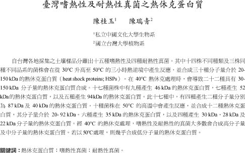
Bot. Bull. Acad. Sin. (2004) 45: 247-257

Chen and Chen — HSPs of thermophilic and thermotolerant fungi from Taiwan

Heat shock proteins of thermophilic and thermotolerant fungi from Taiwan

Kuei-Yu CHEN1,* and Zuei-Ching CHEN2

1Department of Biology, Chinese Culture University, Taipei 111, Taiwan

2Department of Botany, National Taiwan University, Taipei 106, Taiwan

(Received December 18, 2003; Accepted April 22, 2004)

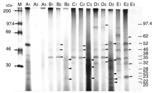
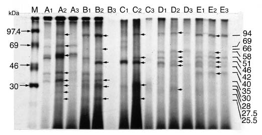
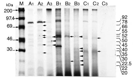
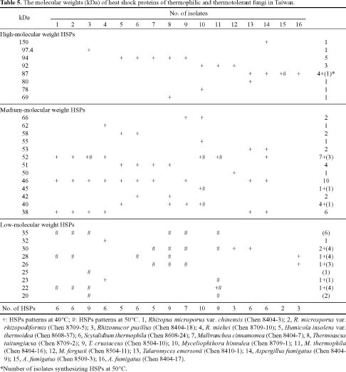
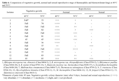
Abstract. Fifteen species of thermophilic and four species of thermotolerant fungi were isolated from soil samples collected at various localities in Taiwan. Fourteen species and three strains responded to three h of thermal stress at elevated temperature from 30°C to 50°C by synthesizing 30 heat shock proteins (HSPs) with molecular weights ranging between 20-150 kDa. Heat shock treatments at 40°C, resulted in the synthesis of 22 HSPs with molecular weights of 30-150 kDa. Nine of the seventeen fungal species produced a 46 kDa HSP, seven species a 52 kDa HSP, and five species a 94 kDa HSP. Four of the seventeen species produced two kinds of HSPs with molecular weights of 87 kDa and 40 kDa. Ten fungal strains responded to an elevated temperature of 50°C and synthesized 12 HSPs of molecular weights ranging from 20 to 92 kDa. Six species produced a 35 kDa HSP, and four species produced HSPs with molecular weights of 30 kDa, 28 kDa and 22 kDa. When subjected to heat shock treatments at 40°C, thermophilic and thermotolerant fungi synthesized mostly high and medium molecular weight HSPs while at 50°C, they synthesized mostly low molecular weight HSPs.

Keywords: Heat shock proteins; Thermophilic fungi; Thermotolerant fungi.

Introduction

Fungi are heterotrophic eukaryotes which exhibit a great diversity in morphology and distribution. Some species can survive at the elevated temperatures of hot springs, in the high salinity of sea-water, or in various other adverse environments. Environmental stresses or stimuli will change the structure or metabolism of an organism by acting as elicitors which affect gene expression and result in the synthesis of stress-specific compounds to protect the organism.

Adverse factors can be either biotic or abiotic. Biotic factors include bacteria, fungi, insects, or disease-causing organisms. They elicit changes in host genetic expression so that stress-specific compounds are synthesized to enhance host resistance to the foreign organism. Abiotic factors include temperature, excess water (Ben-Zioni et al., 1967; Hsiao, 1970), salinity (Ben-Zioni et al., 1967; LaRosa et al., 1989; Burk and Jennings, 1990), anaerobic conditions (Frelling and Bennett, 1985), heavy metals (Jackson et al., 1984; Curle and Kapoor, 1988; Gruhn and Miller, 1991), growth regulator (Heikkila et al., 1984), ultraviolet irradiation (Chappell and Hahlbrock, 1984), metabolic repressors, oxidizers (Curle and Kapoor, 1988), famine (Curle and Kapoor, 1988), and pH (LeJohn and Braithwaite, 1984). Among the environmental stressors listed above, thermal stress has been most widely studied. Both heat shock and cold shock can induce the synthesis or storage of a group

of proteins which increase resistance to thermal stress (Ketola-Pirie and Atkinson, 1983; Yacoob and Filion, 1987).

When thermal stress is applied, the most prominent physiological reactions are the production of a set of novel proteins or an increase in the quantity of certain types of existing proteins. These proteins are known as heat shock proteins (HSPs) (Ketola-Pirie and Atkinson, 1983; Neidhardt et al., 1984; Lindquist, 1986; Lindquist and Craig, 1988; Freeman et al., 1989). This phenomenon was first discovered in 1962 by Ritossa in fruit fly larvae (Drosophila buskii) and has since been manifested in other living organisms (Lindquist, 1986; Lindquist and Craig, 1988).

The optimum temperature for the production of HSPs varies from organism to organism. The heat shock temperature range for Escherichia coli is 43- 47°C (Neidhart et al., 1984), for the yeast Saccharomyces cerevisiae 36°C (McAlister and Finkelstein, 1980), and for the sickle fungus Fusarium oxysporum 40°C or 43°C (Freeman et al., 1989). In general, a rise of 5°C above the normal physiological temperature will induce the synthesis of HSPs.

Heat shock proteins can be classified into three categories according to their molecular size: (I) high-molecular size, with molecular weight between 69 and 120 kDa, (II) medium-molecular size, with molecular weight between 39 and 68 kDa, and (III) low-molecular size, with molecular weight below 38 kDa.

In fungi, the synthesis of HSPs is a rapid process. For example, 10 min after a heat treatment, F. oxysporum began to synthesize HSPs (Freeman et al., 1989) while for S. cerevisiae, a period of 20 to 30 min was required. The synthesis of HSPs peaked at 60 min after heat treatment of

*Corresponding author. Tel: 886-2-28610511-652; Fax: 886-2-28623724; E-mail: thermophilic@ms52.url.com.tw


Botanical Bulletin of Academia Sinica, Vol. 45, 2004

Materials and Methods

Test Fungi

Fourteen species and three strains of thermophilic and thermotolerant fungi, all isolated from Taiwan soils, were used to study the effects of heat on protein synthesis. These were:

1. Rhizopus stolonifer (Ehrenb.: Fr.) Vuill.

2. R. microsporus v. Tiegh. var. chinensis (Saito) Schipper and Stalper (Chen 8404-3).

3. R. microsporus v. Tiegh. var. rhizopodiformis (Cohn) Schipper and Stalper (Chen 8709-5).

4. Rhizomucor pusillus (Lindt.) Schipper (Chen 8404-18).

5. Rhizomucor miehei (Cooney and Emerson) Schipper (Chen 8709-10).

6. Thermoascus taitungiacus K. Y. Chen and Z. C. Chen (Chen 8709-2).

7. T. crustaceus (Apinis and Chesters) Stolk (Chen 8504-10).

8. Malbranchea cinnamomea (Lib.) Van Oorschot and de Hoog (Chen 8404-7).

9. Scytalidium thermophila (Cooney and Emerson) Austwick (Chen 8608-24).

10. Humicola insolens var. thermoidea (Cooney and Emerson) Ellis (Chen 8608-37).

11. Aspergillus fumigatus Fres. (Chen 8404-9).

12. A. fumigatus (Chen 8509-3).

13. A. fumigatus (Chen 8404-17).

14. Talaromyces emersonii Stolk (Chen 8410-1).

15. Myceliophthora hinnulea Awao and Udagawa (Chen 8709-1).

16. M. thermophila (Apinis) van Oorschot (Chen 8404-16).

17. M. fergusii (Klopotek) van Oorschot (Chen 8504-11).

Heat Shock Treatment

Fungi for inoculum were first cultured on Yeast starch agar medium (YpSs: Difco-Yeast extract 4 g, Soluble starch 15 g, K2HPO4 1 g, MgSO4·7H2O 0.5 g, Agar 20 g, Distilled H2O 1,000 mL) at 30°C for 3 to 7 days. An inoculum consisting of 5-10 pieces of 3 mm diameter of mycelial mats was then transferred to a 250-mL Erlenmeyer flask containing 150 mL YpSs broth and incubated at 30°C for 3 or 7 days with agitation.

Aliquots (5 mL) of mycelial suspension prepared by YpSs broth were added to an Erlenmeyer flask containing 50 mL of YpSs broth. The cultures were then placed in water baths set at 30°C, 40°C or 50°C for 15 min as the pre-treatment of heat shock, and then 5 µl of 35S-Methionine was added to each flask. The cultures were maintained at the different treatment temperatures for three h and then, after the heat shock treatment, all cultures were incubated at 30°C for two more h. Flask contents were filtered and then washed 2-3 times with phosphate buffer solution (PBS buffer: KCl 0.2 g; NaCl 0.8 g; KH2PO4 0.2 g; 1,000 mL

Achlya (Silver et al., 1983) and Neurospora crassa (Plesofsky-Vig and Brambl, 1985b). However, under high temperature treatments, fungi differ from plants and animals in needing a long recovery time before synthesizing normal proteins (Plesofsky-Vig and Brambl, 1985a).

Although the production of heat shock proteins is under genetic control, the control mechanisms in fungi differ from those of fruit flies and soybeans. In S. cerevisiae and N. crassa, the production of normal proteins is inhibited at high temperatures, and mRNA slowly disintegrates. In the case of the fruit fly, even though protein production is inhibited by heat stress, the original mRNA is not destroyed. Thus when the normal temperature is restored, normal protein synthesis begins immediately. In fungi, the resumption of normal protein production takes longer (Plesofsky-Vig and Brambl, 1985a).

Fungi such as Achlya klebsiana (LeJohn and Braithwaite, 1984), A. ambisexualis (Gwynne and Brandhorst, 1982; Silver et al., 1983), Dictyostelium sp. (Loomis and Wheeler, 1982), N. crassa (Plesofsky-Vig and Brambl, 1985b), F. oxysporum f. sp. niveum (Freeman et al., 1989), Uromyces sp. (Staples et al., 1989) and yeasts can synthesize three different groups of HSPs: (1) high-molecular weight: 70 kDa - 98 kDa HSPs, (2) medium-molecular weight: 40 kDa - 69 kDa HSPs, and (3) low-molecular weight: 23 kDa - 38 kDa HSPs. The high-molecular weight HSPs are similar to those of other organisms. For example, during conidia exposure to a 45°C heat treatment, N. crassa will produce 98 kDa, 80 kDa, and 70 kDa HSPs similar to those synthesized by fruit flies, chickens, and yeast (Plesofsky-Vig and Brambl, 1985b). Some fungi have HSPs of even higher molecular weights, such as the 105 kDa HSP of Physarium polycephalium (Silver et al., 1983). High-molecular weight HSPs, such as 83 kDa and 70 kDa, in fungi exist prior to a heat shock treatment, as in other organisms. Upon the heat shock, they only increase in quantity (Plesofsky-Vig and Brambl, 1985a). The low-molecular weight HSPs are rather common in fungi, i.e., in A. ambisexualis 23 and 28 kDa (Silver et al., 1983), in Dictyostelium discoideum, 26 - 32 kDa (Loomis and Wheeler, 1982), in F. oxysporum f. sp. niveum, 18 kDa (Freeman et al., 1989), and in Uromyces sp. 14 kDa, 17 kDa and 18 kDa, (Staples et al., 1989).

In fungi HSPs carry out physiological functions as in other organisms: i.e. they confer increased heat resistance. They can bind to organelles. According to Neuman et al. (1987), high-molecular weight HSPs are assembled near the nucleus but are restricted to the cytoplasm. In contrast, low-molecular weight HSPs are bound to the chromatin, as in Dictyostelium (Loomis and Wheeler, 1982), present inside the nucleus, as in A. ambisexua, or near the mitochondria, as in N. crassa. HSPs produced in Physarium can extend the time of interface during the G1 phase (Plesofsky-Vig and Brambl, 1985a).

In this paper, the responses of several thermophilic and thermotolerant fungi to heat shock stress are investigated, and the chemotaxonomic implications of these responses are discussed.


Chen and Chen — HSPs of thermophilic and thermotolerant fungi from Taiwan

deionized water) prior to extraction of heat shock proteins. If a culture was not used immediately, it was frozen in liquid nitrogen and stored at -70°C.

Protein Extraction

Samples were ground in a grinding tube containing 1 mL of buffer consisting of Tris-HCl (pH 8.5) 50 mM; Sodium dodecyl sulfate (SDS) 2%; b-mercaptoethanol 2%; Phenylmethylsulfonyl fluoride (PMSF in 95% alcohol) 1 mM. The homogenate was transferred to an Eppendorf centrifugation tube and centrifuged at 12,000 rpm at 28°C for 10 min, and the supernatant was collected. Four volumes of cold acetone (-20°C) were added, and the samples were frozen overnight at -20°C or for 2 h at -70°C. The precipitate (proteins) was stored in acetone, centrifuged at 12000 rpm at 28°C for 5 min, and finally dissolved in 50 - 100 µl of sample buffer (Tris-HCl (pH 6.8) 62.5 mM; SDS 3%; glycerol 10%; b-mercaptoethanol 5%).

Determination of Radioactive Strength of Protein

Protein samples (5 µl ) were transferred onto 3 mm filter paper (using a code number) and, after drying under infrared light, 10% trichloroacetic acid solution (TCA) was added. After 10 min, the TCA solution was removed and replaced with 5% TCA solution. Samples were boiled in a water bath for 3 min, and the solution was discarded. A second aliquot of 5% TCA solution was added again for 3 min and removed, and the sample was washed twice with 95% ethanol. Finally, the sample was dried and placed into a counting vial with 6 mL of scintillation cocktail (5 g PPO and 0.3 g POPOP dissolved in 1 liter of xylene) (PPO: 2,5-Diphenyloxazole and POPOP: 1,4-Bis-2-15-phenyloxazolybenzene). Radioactivity (CPM) was measured in an automatic liquid scintillation counter (Beckman LS 1801).

Electrophoresis Analysis of Proteins

Proteins were analyzed by SDS-PAGE using 12.5% acrylamide gel and appropriate markers.

Drying, Compression and Exposure

After electrophoresis, gels were washed 1 or 2 times with distilled water, submerged in a solidifying solution (acetic acid 10%; methanol 30%), and gently shaken overnight (14-15 h). The solidifying solution was discarded, and EN3HANCE solution (NEN) was added (acetic acid 55%; 2,5-diphenyloxazol 0.4% by weight; ethanol 15%; xylene 30%), after which the sample was shaken gently for one h. The EN3HANCE solution was discarded, and the sample was softened with 0.8% glycerol for one h. A gel dryer (Hoefer Scientific Instrument Se 540 Slab Gel Dryer) was used to dry the gel. The dried gel was overlaid with an X-ray film in an exposure chamber at -70°C for 3 to 14 days to allow the radioactive band to appear clearly on the X-ray film. The exposed film was developed with D-19 solution, allowed to dry, and printed.

Similarity Index

A similarity index (Ludwig and Reynolds, 1988) between two fungal species was obtained using the following formula: Similarity index (%)=(2c/a+b)×100; a: the number of proteins present in species A; b: the number of proteins present in species B; c: the number of common proteins present in both species A and B.

Results

Effects of Elevated Temperature on Proteins Synthesis

Four different fungal groups were analyzed. The results for each group were as follows: Group 1. The responses of Rhizopus and Rhizomucor to heat shock are shown in Figure 1 and Table 1. No synthesis of heat shock proteins was observed in Rhizopus stolonifer at 40°C or 50°C. At 40°C, thermotolerant fungi such as R. microsporus var. chinensis, R. microsporus var. rhizopodiformis and two species of thermophilic fungi, Rhizomucor pusillus and R. miehei, synthesized the same HSPs, with molecular weights of 52 kDa, 46 kDa and 38 kDa. In addition, the thermophilic fungus R. pusillus produced a 97.4 kDa HSP, and R. miehei synthesized 62 kDa, 32 kDa, and 23 kDa HSPs, respectively. The 38 kDa HSP was the most abundant HSP in the two thermotolerant fungi and in R. pusillus. Of the six fragments produced by R. miehei, the 52 kDa, 46 kDa, and 23 kDa HSPs were most abundant. When the temperature was raised to 50°C, those HSPs produced at 40°C disappeared; only the synthesis of 52 kDa HSP by R. pusillus increased. The thermophilic fungus R. miehei did not synthesize any HSP at 50°C, while R. microsporus var. chinensis, and R. microsporus var. rhizopodiformis produced 35 kDa, 28 kDa, and 22 kDa HSPs. In addition, R. pusillus synthesized 35 kDa, 25 kDa, 22 kDa and 20 kDa HSPs.

Group 2. The responses of Thermoascus and related fungi to heat shock are shown in Figure 2 and Table 2. Five thermophilic fungi were studied: Thermoascus taitungiascus, T. crustaceus, Malbranchea cinnamonea, Scytalidium thermophila, and Humicola insolens var. thermoidea. At 40°C, all of these fungi were able to synthesize 94 kDa HSPs. In T. taitungiascus, a 51 kDa HSP was most abundant, followed by a 40 kDa HSP. In T. crustaceus, 94 kDa and 46 kDa HSPs were most evident. In the other fungal species, 46 kDa HSP was the main protein in M. cinnamomea while in S. thermophila and H. insolens var. thermoidea the main HSPs were 46 kDa and 58 kDa. It can be concluded that the HSPs synthesized by these five different fungi belong to medium- to high-molecular weights. In contrast, at 50°C, the production of these medium- to high-molecular weight HSPs decreased and low-molecular weight HSPs were synthesized. Except for H. insolens var. thermoidea, which lacked induction of any HSPs, the rest of the species synthesized one to four fragments of 35 kDa, 30 kDa, 28 kDa, and 26 kDa HSPs.

Group 3. Responses of Myceliophthora species to heat shock are shown in Figure 3 and Table 3. At 40°C, the ther


Botanical Bulletin of Academia Sinica, Vol. 45, 2004

Figure 1. Effect of heat shock treatment on the pattern of protein synthesis in the genera Rhizopus and Rhizomucor. The autoradiograph shows L-[35S] methionine -labelled protein patterns revealed by SDS-PAGE. Cultures were placed in 1-30°C (control), 2-40°C and 3-50°C (heat shock) water baths for 15 min, and 5 µl/50ml of 35S-methionine was added to each sample. Cultures were maintained at their respective treatment temperatures for a further three h, and then incubated at 30°C for two h. The position of standard protein markers and the size of the heat shock proteins are marked. (A, Rhizopus stolonifer; B, R. microsporus var. chinensis [Chen 8404-3]; C, R. microsporus var. rhizopodiformis (Chen 8709-5); D, Rhizomucor pusillus [Chen 8404-18]; E, R. miehei [Chen 8709-10]).

Figure 2. Effect of heat shock treatment on the pattern of protein synthesis in the Thermoascus group, Malbranchea, Scytalidium, and Humicola (A, Thermoascus taitungiacus [Chen 8709-2]; B, Thermoascus crustaceus [Chen 8504-10]; C, Malbranchea cinnamomea (Chen 8404-7]; D, Scytalidium thermophila (Chen 8608-24]; E, Humicola insolens var. thermoidea (Chen 8608-37]). Methods are the same as for Figure 1.


Chen and Chen — HSPs of thermophilic and thermotolerant fungi from Taiwan

Figure 3. Effect of heat shock treatment on the pattern of protein synthesis in the genus Myceliophthora (A, Myceliophthora hinnulea [Chen 8709-1]; B, Myceliophthora thermophila [Chen 8404-16]; C, Myceliophthora fergusii [Chen 8504-11]). Methods are the same as for Figure 1.


Botanical Bulletin of Academia Sinica, Vol. 45, 2004

heat shock proteins were produced. At 40°C and 50°C, strain C 8509-3 synthesized only 87 kDa HSP while strain C 8404-17 synthesized the low-molecular weight 26 kDa and 28 kDa HSPs at 40°C in addition to the 87 kDa HSP. Talaromyces emersonii synthesized six kinds of HSPs at 40°C: 87 kDa, 80 kDa, 53 kDa, 46 kDa, 38 kDa and 30 kDa, but no HSPs were synthesized at 50°C.

It is evident that, at 40°C, all the studied thermophilic and thermotolerant fungi can synthesize heat shock proteins mainly with medium- and high-molecular weight HSPs. If the temperature is raised to 50°C, synthesis of high-molecular weight HSPs was suppressed and a new group of low-molecular weight HSPs was synthesized. The four fungi, H. insolens var. thermoidea , M. fergusii, T. emersonii, and A. fumigatus (strains C 8404-9 and C 8404-17) cannot synthesize heat shock proteins at 50°C.

Discussion and Conclusion

Fourteen species and three strains of thermophilic and thermotolerant fungi isolated from Taiwan soils were subjected to thermal stress and responded by synthesizing 30

mophilic M. hinnulea produced seven different HSPs with molecular weights ranging from 40 kDa to 92 kDa. Myceliophthora thermophila produced three HSPs (22 kDa, 52 kDa and 92 kDa), as did M. fergusii (30 kDa, 50 kDa and 92 kDa). The main HSPs produced by M. hinnulea, M. thermophila and M. fergusii had molecular weights of 92 kDa, 52 kDa and 30 kDa, respectively. However, all three fungi produced similar amounts of 92 kDa HSP. At 50°C, M. fergusii did not synthesize any heat shock protein while M. hinnulea produced only the medium-molecular weight 52 kDa, 45 kDa, and 40 kDa HSPs. Myceliophthora thermophila retained some 52 kDa HSP and synthesized a new group of low-molecular weight HSPs ranging from 20 kDa to 35 kDa, including 35 kDa, 30 kDa, 23 kDa, and 20 kDa HSPs.

Group 4. The responses of three of thermotolerant Aspergillus fumigatus isolates and a thermophilic Talaromyces emersonii are shown in Figure 4 and Table 4. At 40°C, A. fumigatus strain C 8404-9 produced high molecular weight 150 kDa and 83 kDa HSPs, medium-molecular weight 53 kDa, 52 kDa, and 46 kDa HSPs, and the low-molecular weight 38 kDa HSP. However, at 50°C no


Chen and Chen — HSPs of thermophilic and thermotolerant fungi from Taiwan

species of heat shock proteins (HSPs) with molecular weight ranging from 20 kDa to 150 kDa (Table 5). Variations in HSP synthesis by different species or strains at various temperatures were observed. All isolates tested responded differently to different heat shock treatments, and no single HSP was produced by all isolates involved in these treatments. Twenty-two HSPs with molecular weights ranging from 30 kDa to 150 kDa were synthesized in response to the heat shock treatments at 40°C. Nine out of seventeen isolates produced 46 kDa HSPs. Four isolates produced both 87 kDa and 40 kDa HSPs. Ten isolates responded to 50°C heat shock by synthesizing 12

HSPs with molecular weights ranging from 20 kDa to 92 kDa. Six isolates produced 35 kDa HSPs, and four isolates produced 30 kDa, 28 kDa and 22 kDa HSPs. At 40°C heat shock, 67 HSP fragments were synthesized by all the fungal isolates tested compared to 31 fragments in response to 50°C treatment. It thus appeared that more severe heat shock resulted in less HSP production. At 40°C heat shock, HSPs with medium- (38 kDa - 66 kDa) to high- (69 kDa -150 kDa) molecular weights were synthesized.

Most fungi tested in these experiments grew better at 40°C than 50°C (Table 6), and both sexual and asexual re


Botanical Bulletin of Academia Sinica, Vol. 45, 2004

Figure 4. Effect of heat shock treatment on the pattern of protein synthesis in Aspergillus fumigatus and Talaromyces emersonii (A, Aspergillus fumigatus Fres. [Chen 8404-9]; B, A. fumigatus [Chen 8509-3]; C, A. fumigatus [Chen 8404-17]; D, Talaromyces emersonii Stolk. [Chen 8410-1]). Methods are the same as for Figure 1.


Chen and Chen — HSPs of thermophilic and thermotolerant fungi from Taiwan

productions were inhibited at 50°C. Low molecular weight HSPs produced at 50°C heat shock appeared to be involved only with the maintenance of vegetative growth and not with fungal reproduction. For fungal reproduction, medium- to high-molecular weight HSPs induced by 40°C heat shock were more important than low-molecular weight HSPs. This is further supported by the fact that some species (i.e., R. miehei, H. insolens var. thermoidea, M. fergusii, T. emersonii, and A. fumigatus), lacking the ability to induce HSPs at 50°C heat shock, exhibited the same morphological behaviour as other species which were able to induce HSPs at 50°C.

Patterns and modes of response to heat shock between thermotolerant and thermophilic species showed differences. On average, 4.6 fragments of HSPs were induced by thermotolerant species (i.e., R. microsporus and A. fumigatus) compared to 6.8 fragments of HSPs by thermophilic species (i.e., Rhizomucor spp., H. insolens var. thermoidea, S. thermophila, M. cinnamomea, Thermoascus spp., Myceliophthora spp., T. emersonii) (Table 5). The responses to 50°C heat shock treatment were also different between the two groups in terms of the number of HSPs induced. On average, 1.4 fragments of HSPs were synthesized by the thermotolerant group compared to 2.2 fragments of HSPs by the thermophilic group at 50°C heat shock.

HSPs with molecular weights of 52 kDa, 46 kDa and 38 kDa were commonly produced at 40°C by the thermotolerant group and two thermophilic species of Rhizomucor. Thus, synthesis of 52 kDa, 46 kDa and 38 kDa HSPs at 40°C heat shock may be unique to the mucoraceous fungi. The rest of the thermophilic and thermotolerant species produced only one common HSP for each group [i.e., 94 kDa by group 2 fungi, i.e., Thermoascus, Mlbranchea, Scytalidium and Humicola, etc., 92 kDa by Myceliophthora spp. (group 3), 87 kDa for strains of A. fumigatus and 38 kDa by T. emersonii (group 4)]. At 50°C treatment, thermotolerant R. microsporus, can be identified on the basis of 35 kDa, 28 kDa, and 22 kDa HSP production. In thermophilic species, more diverse patterns of HSP production were observed in response to 50°C heat shock treatments. In addition to 35, 28, and 22

kDa HSPs, 52, 45, 40, 30, 25, 23, and 20 kDa HSPs appeared in different distributions. No unique HSP with possible taxonomic implication for thermophilic fungi were produced in response to 50°C heat shock.

Similarity in HSP synthesis varied from species to species (Tables 7-10). Two varieties of R. microsporus responded with the same pattern of HSP synthesis, i.e. a 100% degree of similarity (Table 7) while three isolates of A. fumigatus reacted differently to heat shock and exhibited a very low degree of HSP similarity (25-40%) (Table 10).

It is concluded that thermophilic and thermotolerant fungi synthesized mainly high and medium-molecular weight HSPs at 40°C and low molecular weight HSPs at 50°C.


Botanical Bulletin of Academia Sinica, Vol. 45, 2004

Literature Cited

Ben-Zioni, A., C. Itai, and Y. Vaadia. 1967. Water and salt stress, kinetin and protein synthesis in tobacco leaves. Plant Physiol. 42: 361-365.

Burke, R.M. and D.H. Jennings. 1990. Effect of sodium chloride ongrowth characteristics of the marine yeast Debaryomyces hansenii inbatch and continuous culture under carbon and potassium limitation. Mycol. Res. 94: 378-388.

Chappell, J. and K. Hahlbrock. 1984. Transcription of plant defence genes in response to UV light or fungal elicitor. Nature 311: 76-78.

Curle, C.A. and M. Kapoor. 1988. Expression of heat shock gene of Neurospora crassa; effect of hyperthermia and other stresses on mRNA levels. Biochem. Cell Biol. 66: 81-92.

Frelling, M. and D.C. Bennett. 1985. Maize Adhl. Ann. Rev. Genet. 19: 297-323.

Freeman, S., C. Ginzburg, and J. Katan. 1989. Heat shock protein synthesis in propagules of Fusarium oxysporum f. sp. niveum. Phytopathology 79: 1054-1058.

Gruhn, G.M. and O.K. Miller. 1991. Effect of copper tyrosinase activi-ty and polyamine content of some ectomycorrhizal fungi. Mycol. Res. 95: 268-272.

Gwynne, D.I. and B.P. Brandhorst. 1982. Alteration in gene expression during heat shock of Achlya ambisexualis. J. Bact. 149: 488-493.

Heikkila, J.J., J.E.T. Papp, G.A. Schultz, and J.D. Bewley. 1984. Inducti-on of heat shock protein messenger RNA in maize mesocotyls by water stress, abscisic acid, and wounding. Plant Physiol. 76: 270-274.

Hsiao, T.C. 1970. Rapid change in levels of polyribosomes in Zeamays in response to water stress. Plant Physiol. 46: 281-285.

Jackson, P.J., E.J. Roth, P.R. McClure, and C.M. Naranjo. 1984. Selection, isolation, and characterization of cadmium-resistant Datura innoxia suspension cultures. Plant Physiol. 75: 914-918.

Ketola-Pirie, C.A. and B.G. Atkinson. 1983. Cold-and heat-shock induction of new gene expression in cultured amphibian cells. Can. J. Biochem. Cell Biol. 61: 462-471.

LaRosa, P.C., N.K. Singh, P.M. Hasegawa, and R.A. Bressan. 1989. Stable NaCl tolerance of tobacco cell is associated with enhanced accumulation of osmotin. Mol. Cell Biol. 2: 267-

274.

LeJohn, H.B. and C.E. Braithwaite. 1984. Heat and nutritional shock-induced protein of the fungus Achlya are different and under independent transcriptional control. Can. J. Biochem. Cell Biol. 62: 837-846.

Lindquist, S. 1986. The heat-shock response. Ann. Rev. Biochem. 55: 1151-1191.

Lindquist, S. and E.A. Craig. 1988. The heat-shock proteins. Ann. Rev. Genet. 22: 631-677.

Loomis, W.F. and S.A. Wheeler. 1982. Chromatin-assoicated heat shock proteins of Dictyostelium. Develop. Biol. 90: 412-418.

Ludwig, J.A. and J.F. Reynolds. 1988. Statistical Ecology: A Primer on Methods and Computing. John Wiley & Sons. New York, 337 pp.

McAlister, L. and D.B. Finkelstein. 1980. Heat shock protein and thermal resistance in yeast. Biochem. Biophysical Res. Communications 93: 819-824.

Neidhardt, F.C., R.A. VanBogelen, and V. Vaughn. 1984. The geneticsand regulation of heat-shock proteins. Ann. Rev. Genet. 18: 295-329.

Neuman, D., U. Nieden, R. Manteuffel, G. Walter, K.D. Scharf, and L. Nover. 1987. Intracellular localization of heat-shock proteins in tomato cell cultures. Eur. J. Cell Biol. 43: 71-81.

Plesofsky-Vig, N. and R. Brambl. 1985a. The heat shock response of fungi. Exp. Mycol. 9: 187-194.

Plesofsky-Vig, N. and R. Brambl. 1985b. Heat shock response of Neurospora crassa: protein synthesis and induced thermotolerance. J. Bacteriol. 162: 1083-1091.

Ritossa, F. 1962. A new puffing pattern induced by temperature shock and DNP in Drosophila. Experientia 18: 571-573. (Cited by Lindquist, S. 1986.)

Silver, J.C., D.R. Andrews, and D. Pekkala. 1983. Effect of heat shock on synthesis and phosphorylation of nuclear and cytoplasmic proteins in the fungus Achlya. Can. J. Biochem. Cell Biol. 61: 447-455.

Staples, R.C., H.C. Hoch, P. Freve, and T.M. Bourett. 1989. Heat shock-induced development of infection structure by bean rust uredospore germlings. Exp. Mycol. 13: 149-157.

Yacoob, K. and W.G. Filion. 1987. The effect of cold-temperature stress on gene expression in maize. Biochem. Cell Biol. 65: 112-119.


Chen and Chen — HSPs of thermophilic and thermotolerant fungi from Taiwan