Bot. Bull. Acad. Sin. (2005) 46: 107-118

KAO et al. AtMAPR, a novel protein for signaling

Characterization of a novel Arabidopsis protein family AtMAPR homologous to 25-Dx/IZAg/Hpr6.6 proteins

Ai-Ling KAO, Tsz-Yin CHANG, Shuo-Hsiu CHANG, Jong-Ching SU, and Chien-Chih YANG*

Department of Biochemical Science and Technology and Institute of Microbiology and Biochemistry, National Taiwan University, No. 1, Section 4, Roosevelt Road, Taipei, Taiwan, ROC

(Received July 28, 2004; Accepted October 11, 2004)

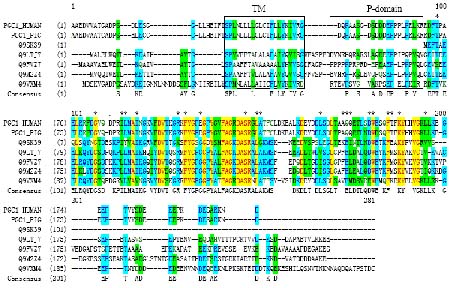
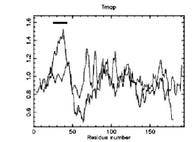
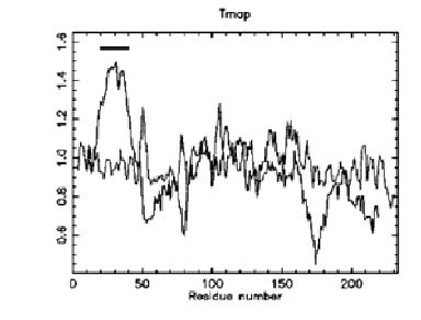
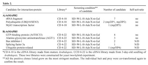
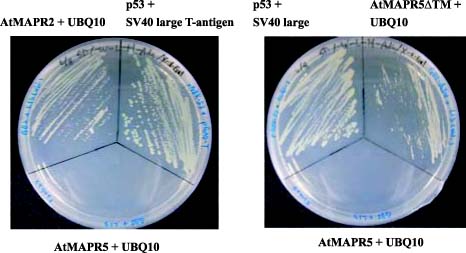
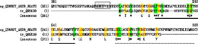
Abstract. Four Arabidopsis genes homologous to the membrane-associated progesterone binding protein (MAPR) were identified. MAPRs have previously been isolated from membrane preparations of porcine liver as proteins binding to progesterone. According to sequence alignment, each of the AtMAPRs, except AtMAPR2, was predicted to contain three domains; the N-terminal domain, except that of AtMAPR2, was predicted to accommodate a single transmembrane alpha-helix. A potentially interesting helical wheel motif SPX10FX2Y was found in the transmembrane domain. Proteins that may interact with AtMAPRs were found using the yeast two-hybrid system. AtMAPR2 and AtMAPR5DTM were found to associate with ubiquitin, where ubiquitination was involved in Aux/IAA modification. Sequence alignment indicated that AtMAPRs are distantly related to the Aux/IAA proteins. AtMAPR2 was also found to bind Myb3, a transcription factor that controls the expression of genes for the biosynthesis of phenylpropanoid. These results imply that AtMAPR may be a part of a plant hormone signaling pathway.

Keywords: AtMAPR; Non-genomic effect; Pant hormone signaling.

Abbreviations: ABA, abscisic acid; GA, gibberellic acid; MAPR, membrane-associated progesterone binding protein; AtMAPR, MAPR homologues in Arabidopsis.

Introduction

The components for signal transduction can be divided into two groups of proteins. These are, first, enzymes that are responsible for the biosynthesis of signaling molecules, or hormones, and second, proteins that are responsible for the perception of hormones, or receptors. The biosynthesis of major plant hormones has become ever clearer in the past 20 years. For example, most of the genes involved in the biosynthesis of gibberellins (GAs) are identified through genetic studies of dwarf cultivars or GA-deficient mutants (Hedden and Phillips, 2000). Many protein components for signal transduction were also determined through the genetic studies of GA-insensitive mutants, e.g. SPY, GAi and RGA (Sun, 2000), which were deficient in the GA signal transduction pathway. However, the receptors that can bind hormones directly, with the exception of the ethylene receptor and one receptor component for brassinosteroid, are still largely unknown. The perception of GA and ABA by membrane receptors has been proposed for nearly a decade, but these receptors still remain elusive (Lovegrove and Hooley, 2000). Data mining of a whole plant genome sequence to find membrane proteins as candidate receptors might offer an alternative approach to the genetic one.

Recently, the membrane receptor, or at least its major part, for brassinosteroids, BRI1, was identified and cloned by Chory's group from a series of BR-insensitive mutants (Wang et al., 2001). Brassinosteroids is similar in chemical structure to the steroid type hormones in animals. Thus, it seems feasible to compare the signaling pathways of steroids in animals and plants. Conventionally, steroid hormones including estrogens, progesterone, and androgens are thought to transmit signals through members of the nuclear receptor protein superfamily, namely estrogen receptor or progesterone receptor. This fact was mainly attributed to the hydrophobic nature of steroids. Steroid hormones diffuse into the cell and bind to the soluble nuclear receptors. In recent years, a different signaling pathway for steroids mediated by membrane-bound receptors has been demonstrated (Falkenstein et al., 2000). This type of signaling is not blocked by transcriptional inhibitors and is therefore described as the "non-genomic actions" of steroids.

A few proteins were proposed to be involved in the "non-genomic actions" of steroids, including MAPR, 25-Dx, IZAg, ratp28 and Hpr6.6, where sequence homology existed in these proteins (Falkenstein et al., 1996; Krebs et al., 2000; Nolte et al., 2000; Raza et al., 2001). Although its function was not fully understood, a membrane-associated progesterone binding protein (MAPR) was discovered from porcine liver cells (Falkenstein et al., 1996) and shown to be membrane-associated (Meyer et al., 1998). MAPR may undergo dimerization upon ligand binding

*Corresponding author. Tel: +886-2-23630231 ext. 3870; Fax: +886-2-23633783; E-mail: ccyang@ntu.edu.tw


Botanical Bulletin of Academia Sinica, Vol. 46, 2005

ed transmembrane helix was displayed using "Pepwheel" in EMBOSS.

Gene Cloning and Recombinant Protein Production

Arabidopsis tissue (7 days seedling, 1 g) was harvested and ground in liquid nitrogen. Total RNA was isolated using TRIZOL reagent (Invitrogen) according to the manufacturer's instructions. In the RT reaction, the cDNA was synthesized from 5 g total RNA using the Superscript II RNase H- Reverse Transcriptase (Invitrogen) following the manufacturer's instructions. 2 ml of cDNA products were used in each PCR reaction in a final volume of 50 l. Primers were designed according to the full-length cDNA sequences available in TAIR together with suitable restriction enzyme sites to facilitate the following construction of plasmids for protein expression. Two primers, 5-GGAATTCCATATGGAA TTCACCGCAGAGCAGC-3 and 5-CGGGATCCCTAAGAGACAACACGGCCAACGAC AGGA-3, were used to amplify the gene AtMAPR2 from first strand cDNA that was reverse transcribed from RNA using the CapFinder PCR cDNA library construction kit (Clontech). A second set of primers, 5-TCCCCCGGGG CAGCGTTCAGCAAATATGGGAGAC-3 and 5-GGAATTCCTTACTCCTTTGCAGCATCATCATCATC-3, were used for the amplification of the AtMAPR3 gene. Primers, 5-TCCCCCGGGGCAGCGCGTTAGAACTATGGC AAAC-3 and 5-GGAATTCCCTACTCCTCCTTC TTCAACACAGTC-3, were used for the amplification of the AtMAPR5 gene. The PCR products were cloned into the pGEMT vector using the T-A cloning kit (Promega). The DNA fragments encoding AtMAPR2, AtMAPR3, and AtMAPR5 were subcloned into pET16, pET21, or pET43, as appropriate. For the production of recombinant wild-type proteins or 6X-His-tagged recombinant proteins, bacterial strain BL21 (DE3) harboring the suitable plasmid was induced by IPTG. The overexpressed target proteins were characterized and identified. The protein N-terminal sequencing was carried out using a Procise 492 Protein Sequencer (Applied Biosystems).

Polyclonal Antibody

A polyclonal antibody for AtMAPR2 was raised against the recombinant wild-type protein AtMAPR2, which was obtained from a preparative SDS-PAGE. A 16.5% Tris-Tricine SDS-PAGE (Schagger and von Jagow, 1987) was used to monitor the overexpression of AtMAPR2. AtMAPR2 was identified and excised directly from a preparative Tricine-PAGE (Hoefer SE600, 16 cm 16 cm). After three immunizations at two-week intervals, ascites was induced and collected as the source for a polyclonal antibody preparation.

Gel Electrophoresis and Immuno-Detection

To detect the presence of AtMAPR2, 1 g of 30-d-old Arabidopsis was homogenized with liquid nitrogen and extracted with 3 mL of RIPA buffer (100 mM Tris-HCl, 150 mM NaCl, 0.1% Triton X-100, 1X protease inhibitor cock

(Falkenstein et al., 2001). Interestingly, 25-Dx, the homolog of MAPR in rat, was regulated by progesterone in brain regions that were involved in female reproductive behavior (Krebs et al., 2000). Except for their putative roles as progesterone receptors as discussed by Krebs et al, a separate experiment by Min et al. demonstrated that IZAg participates the steroidogenesis (Min et al., 2004). The N-terminal sequences of IZAg completely matched with 25-Dx. Furthermore, a hydrophobic motif of 14 amino acids followed by a proline-rich segment in the N-terminal domain of 25-Dx shares 71% sequence homology with the transmembrane domain of the precursor for the interleukin-6 receptor (Selmin et al., 1996). Recently, Dap1p, a protein homologue of MAPR from Saccaromyces cerevisiae, is reported to be involved in the DNA damage response and sterol regulation (Hand et al., 2003). Sequence analysis showed that MAPR possesses a cytochrome b5 domain (Mifsud and Bateman, 2002). From the viewpoint of evolution, Mifsud and Bateman suggested that heme-containing cytochrome b5 domain may serve as a template for a novel ligand-binding pocket, such as a steroid-binding site.

From Arabidopsis, we have found four homologues of animal MAPR, products of at2g24940 (AtMAPR2), at3g48890 (AtMAPR3), at4g14965 (AtMAPR4) and at5g52240 (AtMAPR5). The numbering indicates that the genes encoding AtMAPR2, AtMAPR3, AtMAPR4, and AtMAPR5 are located in the chromosomes 2, 3, 4 and 5 of Arabidopsis, respectively. In this manuscript, we will employ the AtMAPRs as the name for the proteins, AtMAPRs as the genes encoding these proteins. They share approximately 38-44% sequence identity with MAPR/25-Dx/IZAg/Hpr6.6 in 100 or so amino acids in the middle region. Also, a motif (SPX10FX2Y) is revealed in the putative transmembrane domains located at the N-terminus for both animal MAPR and AtMAPRs. Based on these findings, these Arabidopsis proteins would seem worthy of study to investigate any functions related to the hydrophobic signaling molecules in plants.

Materials and Methods

DNA Sequence Analysis

A BLAST (Altschul et al., 1990) search was performed based on the sequence of PGC1 (MAPR/25-Dx/IZAg/ Hpr6.6) against the databases of SwissProt/TrEMBL. Multiple sequence alignment was performed using the GCG package program PileUp (Feng and Doolittle, 1987) and the program AlignX within Vector NTI (Informax). To obtain detailed information on those homologues in Arabidopsis, a BLAST was carried out within TAIR (The Arabidopsis Information Resource, http://www.arabidopsis.org). The definitions for the putative domains were predicted by ProDom (Corpet et al., 1998; Corpet et al., 2000) based on the sequence or structural information. The transmembrane part of the protein was predicted using the "TMAP" (Persson and Argos, 1994, 1997) program within EMBOSS (Rice et al., 2000). The helical presentation of the predict


KAO et al. AtMAPR, a novel protein for signaling

yeast two-hybrid experiment, was kindly provided by ABRC (mature plant, cat. #CD4-10 ; 3-day-old, cat. #CD4-22). The yeast strain AH109 was co-transformed with the bait and cDNA library. Transformation of the bait and prey library into yeast AH109 was carried out according to the manufacturer's instructions (YEASTMAKER yeast transformation system II, CLONTECH). The co-transformation efficiency (growth on SD/-Trp-Leu) in several independent experiments was from 2 104 to 3 105 transformants g-1 of DNA. Total transformants (~3 105) were screened for activation of HIS3, ADE2 and MEL1 on SD/-Trp-Leu-His-Ade/X-a-Gal medium plates. The cDNA library plasmids (pACT) were isolated from positive clones and rescued into E. coli JM109. These library plasmids were identified by digestion analysis with Xho I and DNA sequencing. To eliminate the false positives, the selected library plasmids were retransformed alone into AH109 to test the potential for self-activation. The yeast harboring the candidate plasmid was then transformed with the bait plasmid followed by X-a-Gal analysis and detection of the ability to activate reporter genes.

Results

Sequence Analysis of AtMAPR Family

The sequence of a membrane-associated progesterone binding protein (MAPR) PGC1_Pig (Falkenstein et al., 1996) having 193 amino acids was used for a BLAST search against the EMBL database. PGC1 is the nomenclature currently employed by the SwissProt database, while 25-Dx, IZAg, and Hpr6.6 are utilized by previous reports. More than 30 protein homologues distributed among mammals, yeast, Drosophila, Caenorhabditis elegans, and Arabidopsis were identified. Most of them are annotated through the progesterone binding property established in animal models. In the Arabidopsis genome alone, four protein products AtMAPR2, AtMAPR3, AtMAPR4 and AtMAPR5 had 38-44% sequence identity with PGC1 in the middle domain, comprising more than 100 amino acids (Table 1). A multiple sequence alignment was generated using the two animal PGC1s (PGC1_Pig, PGC1_Human) and selected Arabidopsis homologues (Figure 1). It is clear that the sequence similarity is mostly contributed by the middle region of the putative progesterone binding protein. Using PHD software (Rost et al., 1994), this region was predicted to be a globular domain localized in the cytosol. However, the N-terminal parts of PGC1 and AtMAPR5, for example, share very low primary structural

tail [Roche], pH 8.3) with the addition of 28 mM b-mercaptoethanol before use. The mixture was vortexed for 2 min. The crude extract was centrifuged at 16,000 g for 15 min, and the supernatant was used for immunoadsorption. Various amounts of polyclonal antibody (50 l, prepared as above) were immobilized in 50 l of CNBr-activated Protein A Sepharose CL 4B (Amersham Biosciences, UK), which was equilibrated in RIPA buffer, according to the manufacturer. Crude extract of Arabidopsis (500 l) was then mixed with resin under 4C overnight. The resin was washed several times with PBST and mixed with 25 l of SDS sample buffer and boiled for 5 min. The supernatant was ready for 16.5% Tricine SDS-PAGE and subsequent blotting.

AtMAPRs Gene Expression Studies by Semiquantitative RT-PCR Analysis

Arabidopsis (16-day-old seedlings) were grown on solid MS medium which contains MS salt, 3% sucrose, and 0.8% agar. The seedlings were transferred to MS liquid medium the day before hormone treatment. Different hormones were added to the medium for a further 48 h under normal growth conditions (22C, 16-h light). The plant hormones used in this experiment included 2,4-dichlorophenoxyacetic acid (2,4-D), indole-3-acetic acid (IAA), 6-benzylaminopurine (BA), kinetin, and ()-cis,trans-abscisic acid (ABA).

Arabidopsis tissue was harvested and ground in liquid nitrogen. Total RNA was isolated as above. The PCR reactions consisted of 20 pmol of the gene-specific primers, or with the UBQ10 gene primers, 5-CGACTTGTCATTAGAAAGAAAGAGATAACAGG-3 and 5-GATCTTTGCCGGAAAACAATTGGAGGATGGT-3, used as an internal control.

After 18~25 cycles as appropriately, the PCR products were resolved on a 1.5% agarose gel and visualized by a gel documentation system (AutoChemi automated system, UVP Inc, USA). The density of the PCR bands were normalized with LabWork 4.5 software (UVP Inc., USA) and the expression of UBQ10 was used as a standard.

Yeast Two-Hybrid

The MATCHMAKER GAL4 two-hybrid system 3 (CLONTECH) was used for screening. The ORF of the AtMAPR2 and AtMAPR5 were cloned in the pGBKT7 vector to generate bait in the two-hybrid system. The Arabidopsis cDNA library, which is constructed for a


Botanical Bulletin of Academia Sinica, Vol. 46, 2005

Figure 1. Multiple sequence alignment of MAPRs from animal and their homologues (AtMAPRs) in Arabidopsis. Sequences for PGC1_HUMAN (O00264), PGC1_PIG (Q95250), RMapr (from rice, Q9FVZ7), AtMAPR2 (Q9SK39), AtMAPR3 (Q9M2Z4), AtMAPR5 (Q9XFM6), MAPR homolog from Drosophila (Q9VXM4) are obtained from SwissProt/TrEMBL databases. Those residues of AtMAPR2 which contributed to similarities with Aux/IAA are indicated by asterisks (*) (see Figure 9).

A

similarity. The potential transmembrane parts of PGC1_Pig and AtMAPR5 were predicted using the EMBOSS (Rice et al., 2000) program "TMAP" (Figure 2) (Persson and Argos, 1997). A 14 amino acid peptide fragment for both proteins in similar regions was predicted to be a transmembrane domain. Due to a limited fragment length of less than 20 amino acid residues, the domain most likely folds as a single alpha helix. A striking similarity is observed when the two transmembrane domains (TM) are displayed as helical wheels using the Pepwheel program of EMBOSS. As shown in Figure 3, four amino acids S, P, Y and F are located in the proximal side of the helical wheel. This may lead to the formation of a new motif of SPX10FX2Y, where X represents mostly hydrophobic amino acids. It is not surprising that the transmembrane sequence may vary during evolution as long as it is hydrophobic. In contrast, the conserved helical wheel motif may be indicative of functional conservation between AtMAPR and MAPR.

B

Figure 2. Transmembrane domains of AtMAPR were predicted using the software "TMAP" (described in Materials and methods). A, PGC1_Pig represents the membrane-associated progesterone binding proteins of animals; B, AtMAPR5. Bars are displayed in the plot as predicted to be the regions most likely to form transmembrane segments.


KAO et al. AtMAPR, a novel protein for signaling

A

Cloning and Expression of the AtMAPR2

Appropriate primers were designed to amplify the genes encoding AtMAPR2, AtMAPR3, and AtMAPR5 from Arabidopsis by RT-PCR with Pfu DNA polymerase (Stratagene). The fragments were cloned into the vector pGEM-T (Promega) with Nde I and BamH I or suitable restriction sites flanking the DNA inserts. The DNA sequences of the cloned regions were identical to the corresponding genes published in the database. The fragments were then subcloned into pET21, pET16, or pET43 as appropriate (Novagen). The recombinant plasmids carrying the DNA fragments encoding AtMAPR2, AtMAPR3, and AtMAPR5 were named pET21AM2, pET43AM3, and pET43AM5, respectively. Bacterial strain BL21 (DE3), carrying the recombinant plasmids, was subjected to different induction conditions to optimize protein expression. Expressed AtMAPR2 was observed on a Tricine-PAGE as a distinct band migrating between the 6.5 and 14 kDa markers (Figure 4). This band was found in the soluble fraction under all expression conditions, not surprising since it is the only gene product in the AtMAPR family devoid of the putative transmembrane domain. The protein AtMAPR2 was also expressed with an N-terminal His6 tag to facilitate identification and purification.

Polyclonal Antibody to AtMAPR2

By comparing the total proteins extracted from BL21(DE3) cultures harboring pET21AM2 and pET21, AtMAPR2 at the ~10 kDa position was estimated to be 95% pure (Figure 4). The protein band was transferred to a PVDF membrane and subjected to protein N-terminal sequencing.

B

Figure 3. The predicted transmembrane domains were displayed as helical wheel using "Pepwheel" (see Materials and Methods). A, PGC1_Pig represents the membrane-associated progesterone binding proteins; B, AtMAPR5.

The most conserved region was denoted as the G-domain because it was predicted to be a globular domain. This domain can be easily defined in the full-length AtMAPR2, and is located between residues 73 to 172 of AtMAPR5. This domain has been suggested to be heme-containing and involved in progesterone metabolism (Min et al., 2004). The region between the TM and G-domain is called the P-region due to the presence of multiple proline residues, with at least 4 in animal MAPR (PGC1), 5-7 in the Arabidopsis homologues, and 13 in the rice protein homologues (Q9FVZ7). Proline-rich motifs have been found to interact with many proteins involved in cell signaling, such as the WW and SH3 domains (Kay et al., 2000; Macias et al., 2002). Also interesting to note is that the P-region and other flanking sequences of the G-domain are rich in charged amino acids like D, E, R or K.

Figure 4. Recombinant wild type AtMAPR2 are heterologously expressed in E. coli BL21(DE3) under different conditions. Lanes 1, E. coli BL21(DE3) carrying pET21AM2 were grown at 37C with 0.1 mM of IPTG; lane 2, E. coli BL21(DE3) harboring pET21(b) without insert.


Botanical Bulletin of Academia Sinica, Vol. 46, 2005

A

B

Figure 5. The specificity of Anti-AtMAPR2P polyclonal antibody was examined for interaction with different recombinant proteins. A, Crude extracts of E.coli expressed recombinant wild type AtMAPR2 (lanes 1, 2 and 4), His-Tag-AtMAPR2 (lane 3), AtMAPR3 (from pET43AM3) (lane 5) and Ni-NTA column purified AtMAPR5 (lane 6) were separated using 16.5% Tricine SDS-PAGE and stained with CBR; B, The blot was then stained with the polyclonal anti-AtMAPR2 antibody together with HRP/DAB reagents. The coloring reaction was stopped after 2 min.

The first 10 amino acids were sequenced and confirmed to be identical with those deduced from the gene encoding AtMAPR2. It was excised from a preparative Tricine-PAGE gel and used as the antigen to raise a polyclonal antibody anti-atmp2p. The specificity of the antibody was demonstrated by Western blotting using the recombinant wild-type AtMAPR2 and the recombinant fusion protein AtMAPR2 with an N-terminal His6 tag. The AtMAPR2 and affinity purified His-tagged AtMAPR2 were analyzed in two parallel 16.5% Tricine-PAGE gels; one was stained with CBR and the other subjected to a Western blot procedure. As shown in Figure 5, both proteins could be stained by the polyclonal anti-Atmp2p. The titer of the antiserum was determined to be 3000 to 4000 using Western blot (data not shown). The fusion protein migrated more slowly than the wild-type, presumably due to the extra residues from the His6 tag plus several linker amino acids. In the same gel, recombinant fusion protein derived from pET43AM3 was revealed by CBR staining but not by Western blot, showing that the anti-atmp2p recognized AtMAPR2 but not the AtMAPR3 or AtMAPR5 even though they share homologous domains. The recombinant protein of AtMAPR3 was fused with a ~50 kDa Nus tag and therefore appeared much larger than the recombinant AtMAPR2 protein.

To detect the presence of the native form of AtMAPR2, proteins extracted from 10-day-old Arabidopsis seedlings were analyzed. Detecting AtMAPR2 in a direct Western blot of total proteins using the HRP-linked staining method was difficult. This may reflect the low abundance of the protein in the native host at this growing stage. To harvest enough proteins for detection, immunoprecipitation using anti-Atmp2p was performed. As shown in a 16.5% Tricine SDS-PAGE (Figure 6), a clear band from the immunoprecipitated sample from 30-day-old Arabidopsis co-migrated with the recombinant protein derived from pET21AM2. The heavily stained protein bands represented the IgG heavy

and light chains with Mr of 50 kDa and 25 kDa, respectively. This indicates the presence of AtMAPR2 in vivo, although in a low amount.

Semiquantitative RT-PCR

The AtMAPR2, AtMAPR3, AtMAPR4, and AtMAPR5 transcripts under different hormone treatments were subjected to the semiquantitative RT-PCR analysis. The UBQ10 transcripts, which are believed to be constitutive, were used to verify the uniformity of the various mRNA

Figure 6. Crude extract of 30-day-old Arabidopsis was subjected to immunoprecipitation by 50 mL polyclonal anti-AtMAPR2 antibody (lanes 2, 4), and analyzed by 16.5% Tricine SDS-PAGE. The gel was stained by CBR (3, 4, 5), and a separate blot was immunostained (1, 2); lanes 1 and 3, control (exclude antibody during immunoprecipitation; lane 5, crude extract of E. coli producing recombinant wild type AtMAPR2 protein. The heavily stained polypeptides in lanes 1 and 2 represent the IgG heavy (about 50 kDa) and light (about 25 kDa) chains.


KAO et al. AtMAPR, a novel protein for signaling

samples. As shown in Figure 7, the genes were expressed in most circumstances with some interesting features. In particular, AtMAPR2 expression level was down-regulated by auxin (IAA or 2,4-D) at low concentrations (<10 M) and cytokinin (BA) at 100 M. The RT-PCR results of hormone treatment consisted of microarray analysis data obtained from AFGC (Arabidopsis Functional Genomic Consortium) as described later.

Proteins Interacting with AtMAPR2 and AtMAPR5

To understand the physiological roles played by AtMAPRs, we sought to identify potential proteins interacting with AtMAPR2 and AtMAPR5 using the yeast two-hybrid approach. The intact DNA sequences encoding AtMAPR2 and AtMAPR5 were fused in-frame to the GAL4 DNA-binding domain of pGBKT7 to produce pGBAM2 and pGBAM5, respectively, as baits. The baits were used to

Figure 7. A. Gene expression of AtMAPRs with different hormone treatments detected by semiquantitative RT-PCR method. 16-day-old seedlings were treated with 2,4-dichlorophenoxyacetic acid (2,4-D), indole-3-acetic acid (IAA), 6-benzylaminopurine (BA), Kinetin, gibberellic acid (GA3), and ()-cis, trans-Abscisic acid (ABA), for 48 h. Three kinds of concentration, 1, 10 and 100 M, were used. The PCR bands were normalized with LabWork 4.5 software (UVP Inc., USA) where the expression of UBQ10 was used as standard. The expression levels of the genes without hormone treatment were designated as 1. B. The numbers bigger than 2.0 or smaller than 0.5 obtained above were indicated by and , respectively.


Botanical Bulletin of Academia Sinica, Vol. 46, 2005

screen a mature Arabidopsis cDNA library CD4-10 provided by ABRC (Arabidopsis Biological Resource Center). The CD4-10 library was provided by Dr. Walker (Division of Biological Sciences, University of Missouri) via the distribution of ABRC. Recombinant proteins expressed from the library are fused with the activation domain of GAL4 in their N-terminal. Positive clones were screened by identifying colonies growing on SD (minimal synthetic dropout) medium lacking Trp (bait marker), Leu (prey marker), His and adenine, and also by finding colonies which turned blue when plated on a medium with X-a-Gal. All the positive clones obtained are summarized in Table 2. Among 3105 transformants, ten positive clones were identified from library CD4-10 using AtMAPR2 as the bait. All ten candidates were subjected to DNA sequencing. Seven clones contained variable lengths of rRNA fragment carried by chloroplasts. Two clones (mp2IP1, mp2IP2) were identified which encoded a part (one third) of the UBQ10 (polyubiquitin) gene, which possessed six consecutive ubiquitin genes. One clone (mp2IP3) contained the Myb3 transcription factor gene. Myb3 belongs to the R2R3-Myb gene family, and its expression is correlated with most plant hormones, in particular, auxin and ethylene (Kranz et al., 1998). Ubiquitination has been demonstrated to be as important as the phosphorylation of signaling components in plant signal transduction, particularly in the signal perception of auxin (Reed, 2001; Kepinski and Leyser, 2002).

We screened the ABRC CD4-22 library, which was made from 3-day-old seedlings. Eleven positive clones were identified from only 8 104 transformants. One positive clone (mp5IP4) corresponded to a putative ubiquitin-related protein encoded by at2g17200. The gene contains a full-length 1656 bp CDS coding for 550 amino acids. The N- and C-termini of the at2g17200 protein possess domains similar to ubiquitin (Pfam: PF00240) and a UBA (Pfam: PF00627), respectively (Dieckmann et al., 1998; Whitby et al., 1998; Bateman et al., 2002). This, together with the fact that AtMAPR2 may interact with ubiquitin, caused us to postulate that AtMAPR5 might also interact with UBQ10 pro

tein in the yeast two-hybrid system, but was not detected in the library screening. The plasmids pGBAM5 and pACT-UBQ10 were co-transformed into yeast strain AH109. No colony was found in the SD (-W-L) medium devoid of His and adenine. AtMAPR5 is similar to AtMAPR2 in its middle G domain and possesses extra 72 amino acids in the transmembrane domain-containing N-terminus. A truncated version of AtMAPR5 lacking the first 40 amino acids, AtMAPR5DTM, was constructed in the bait plasmid to test its ability to interact with ubiquitin. Interestingly, the yeast transformed with these two plasmids did grow on SD/-W-L-H-Ade medium and turned light blue in the presence of X-a-Gal (Figure 8). The light blue color indicated the expression of active a-galactosidase. This means that AtMAPR5DTM, but not intact AtMAPR5, interacts with ubiquitin (mp2IP1). This may imply that the presence of the N-terminal TM hampered the interaction with ubiquitin. Alternatively, the presence of TM may have caused the bait protein to become partly trapped in the membrane system, hampering the ability of AtMAPR5 to target into the nucleus and interact with prey proteins.

Additional evidence supported the proposal that AtMAPRs might be modified through ubiquitin-related pathways. We compared the sequences of AtMAPRs and AUX/IAA, a protein family involved in auxin signaling (Abel et al., 1995; Reed, 2001), and found some similarity between them. The sequence similarity mainly falls into two regions corresponding to domains III and IV of the Aux/IAA protein family. AtMAPR2 possesses a motif with ~35 amino acid residues, which is similar (30%) to domain III of AXI9, an Aux/IAA protein (Figure 9). Domains III and IV are responsible for homo- or heterodimerization with ARFs (Reed, 2001), which have an N-terminal DNA binding domain. The 35 amino acids within AtMAPR are predicted to assume a secondary structure arranged in a baa fashion. The corresponding sequences in domain III are also reported to fold into a baa structure (Morgan et al., 1999). Interestingly, the difficulty in the detection of AtMAPR2 protein using Western blot coincided with the


KAO et al. AtMAPR, a novel protein for signaling

Figure 8. Protein-protein interactions are demonstrated by co-transformed the relevant bait and prey plasmids into yeast AH109 followed by plating on the medium SD/-W-L-H-Ade-(X-a-Gal). Light blue color was observed due to the presence X-a-Gal. Positive controls are performed by co-transformation of plasmids encoding p53 and SV40 large T-antigen, respectively.

Figure 9. Sequence comparison of AtMAPR2 (Q9SK39) with an AUX/IAA protein, AXI9 (Q38827). A solid line and a dotted line indicate domain III and domain IV of AXI9, respectively. The consensus sequence motif "VGWPPVRS" in domain II is surrounded by a box.

same side of the helix as revealed by the Pepwheel plot strongly implicates its physiological functionality. Proline residues behave quite differently in soluble protein or in TM, as has been discussed recently (Deber and Therien, 2002). The abundance of proline in membrane transporter proteins suggests that experiments to mutate this residue by the transgenic approach may be worth undertaking. AtMAPR2, unlike other members of the family, does not possess the N-terminal 70 amino acids. If these N-terminal segments do confer the ability to anchor other AtMAPRs in membrane system, AtMAPR2 may play the role of free soluble protein to communicate with other components in a signaling protein complex. However, such a detailed mechanism could only be established through more yeast two-hybrid experiments.

The presence of protein homologues to progesterone-binding protein in plants is intriguing. We have therefore

fact that Aux/IAA was extremely short-lived. If AtMAPRs are not part of the currently accepted model for Aux/IAA, at least, it is likely that AtMAPRs share the similar homo- or heterodimerization mechanism with these proteins.

Discussion

We have presented the results of a study on a novel protein family AtMAPR that was identified from the Arabidopsis genome based on its homology to animal membrane associated progesterone-binding proteins, including IZAg, MAPR, 25-Dx and Hpr6.6. The presence of a common motif SPX10FX2Y showed the feasibility of our hypothesis. Statistical analysis showed that all four of these amino acids are frequently found in the TM (Arkin and Brunger, 1998; Ulmschneider and Sansom, 2001). The fact that these four residues in the motif reside on the


Botanical Bulletin of Academia Sinica, Vol. 46, 2005

inquired into the function of these homologues. Whether signal transduction of steroids, or other hydrophobic signal molecules, in plants and animals diverges from a common ancestor, or to what extent they share the basic structural components is a complex question. Chory has stated that non-genomic action in an animal does not agree with the LRR-RLK type BRI1 receptors which are responsible for the perception of BR in plants (McCarty and Chory, 2000; Friedrichsen and Chory, 2001). However, it is interesting to note that the perception of plant steroid hormones and GA or ABA all require the presence of membrane associated proteins. As MAPR has been proposed to reside in the membrane system and have a role in the steroid-binding or steroid metabolism, it would be interesting to investigate the role played by their homologues in plants. Furthermore, the receptor proteins located in the plasma membrane are often composed of more than one component, and some of them may not be revealed by the genomic approach. It is worth examining the function of the plant homologues of proteins that might be involved in "non-genomic" actions in animals.

The best strategy for obtaining a large quantity of pure protein for antibody production is to construct a fusion protein with a His-tag. However, a His-tag must be removed prior to immunization since it is often a good epitope and occupies a considerable population in a polyclonal antibody preparation. We encountered problems in using proteases to remove the His-tag, and, therefore, we used the recombinant wild-type protein as the antigen. The specificity of the polyclonal antibody was confirmed by two forms of recombinant AtMAPR2 proteins having different chain lengths which could be stained by the antibody. This antibody was employed to show the presence of AtMAPR in Arabidopsis.

Functional expression of AtMAPR was also seen during plant growth at the RNA level. The Stanford Microarray database provides hints about this novel gene family's functions. In particular, the expressions of AtMAPRs are either enhanced or suppressed in response to the treatment of auxin. Using the microarray database available from AFGC (Arabidopsis Functional Genomic Consortium), we searched for experiments that had significant changes (data not shown). Briefly, for the cDNA clone 110K7XP corresponding to the gene encoding AtMAPR2, we found 481 spots in 489 arrays that were open to the public. For the cDNA clone E2A2T7 corresponding to the gene encoding AtMAPR5, 266 spots were found in 274 arrays. Finding that a hormone treatment did induce or repress the expression of these two genes is interesting. We are particularly interested in the expression of these genes in response to the hormone treatment or in hormone insensitive mutants. Auxin treatment in roots reduced the expression of the AtMAPR2 gene up to 50%. As described in Results, AtMAPR2 and AtMAPR5's expressions were responsive to the treatment of auxin and cytokinin. Taking the data from semiquantitative RT-PCR and public microarray database together, we speculated that AtMAPR2 might be subjected to the regulation of auxin.

Yeast two-hybrid experiments reported in this paper revealed that AtMAPRs may be subjected to the modification by ubiquitin or ubiquitin-related proteins. Ubiqitination has been shown to play roles in several plant hormone signaling pathways, including auxin, GA, and ABA (Reed, 2001; Lois et al., 2003; Sasaki et al., 2003). AtMAPRs share at least two features with Aux/IAA, a protein family involved in auxin signaling and regulated by ubiquitination. First, AtMAPR2 is difficult to detect with Western blot, reflecting the low abundance of these proteins, while the Aux/IAA is extremely short-lived. Second, the presence of a common baa motif shared by AtMAPRs and Aux/IAA implies that AtMAPRs might have the ability to dimerize with other signalling components. It is interesting to note that the G-domain is also present in a mammalian HERC2 ubiquitin transferase (Mifsud and Bateman, 2002).

This is the first paper to described the roles of MAPR proteins in plants. Signal transduction is a web of complex cellular information. Conventional genetics and reverse genetics should be combined to explore this complex problem. With yeast two-hybrid and other relevant approaches, we believe that the AtMAPRs represent one starting point to begin exploring this complex web.

Acknowledgements. We thank helpful comments from Dr. Nigel Scrutton and Dr. Martin Mewies during the preparation of this manuscript. We thank Dr. Jian-shing Wu in the O-3B Core lab in preparing the antibodies. The O-3B core lab is headed by Prof. Rong-huay Juang and supported by an "Aiming for Excellence" project of the Ministry of Education. This project was supported by the National Science and Technology Program for Agricultural Biotechnology (NSC90-2317-B-002-006).

Literature Cited

Abel, S., M.D. Nguyen, and A. Theologis. 1995. The PS-IAA4/5-like family of early auxin-inducible mRNAs in Arabidopsis thaliana. J. Mol. Biol. 251: 533-549.

Altschul, S.F., W. Gish, W. Miller, E.W. Myers, and D.J. Lipman. 1990. Basic local alignment search tool. J. Mol. Biol. 215: 403-410.

Arkin, I.T. and A.T. Brunger. 1998. Statistical analysis of predicted transmembrane alpha-helices. Biochim. Biophys. Acta 1429: 113-128.

Bateman, A., E. Birney, L. Cerruti, R. Durbin, L. Etwiller, S.R. Eddy, S. Griffiths-Jones, K.L. Howe, M. Marshall, and E.L.L. Sonnhammer. 2002. The Pfam Protein Families Database. Nucl. Acids. Res. 30: 276-280.

Corpet, F., J. Gouzy, and D. Kahn. 1998. The ProDom database of protein domain families. Nucleic Acids Res. 26: 323-326.

Corpet, F., F. Servant, J. Gouzy, and D. Kahn. 2000. ProDom and ProDom-CG: tools for protein domain analysis and whole genome comparisons. Nucleic Acids Res. 28: 267-269.

Deber, C.M. and A.G. Therien. 2002. Putting the beta-breaks on membrane protein misfolding. Nat. Struct. Biol. 9: 318-319.

Dieckmann, T., E.S. Withers-Ward, M.A. Jarosinski, C.F. Liu,


KAO et al. AtMAPR, a novel protein for signaling

Mifsud, W. and A. Bateman. 2002. Membrane-bound progesterone receptors contain a cytochrome b5-like ligand-binding domain. Genome Biol. 3: RESEARCH0068.

Min, L., H. Takemori, Y. Nonaka, Y. Katoh, J. Doi, N. Horike, H. Osamu, F.S. Raza, G.P. Vinson, and M. Okamoto. 2004. Characterization of the adrenal-specific antigen IZA (inner zone antigen) and its role in the steroidogenesis. Mol. Cell. Endocrinol. 215: 143-148.

Morgan, K.E., T.I. Zarembinski, A. Theologis, and S. Abel. 1999. Biochemical characterization of recombinant polypeptides corresponding to the predicted betaalphaalpha fold in Aux/IAA proteins. FEBS Lett. 454: 283-287.

Nolte, I., D. Jeckel, F.T. Wieland, and K. Sohn. 2000. Localization and topology of ratp28, a member of a novel family of putative steroid-binding proteins. Biochim. Biophys. Acta 1543: 123-130.

Persson, B. and P. Argos. 1994. Prediction of transmembrane segments in proteins utilising multiple sequence alignments. J. Mol. Biol. 237: 182-192.

Persson, B. and P. Argos. 1997. Prediction of membrane protein topology utilizing multiple sequence alignments. J. Protein. Chem. 16: 453-457.

Raza, F.S., H. Takemori, H. Tojo, M. Okamoto, and G.P. Vinson. 2001. Identification of the rat adrenal zona fasciculata/reticularis specific protein, inner zone antigen (IZAg), as the putative membrane progesterone receptor. Eur. J. Biochem. 268: 2141-2147.

Reed, J.W. 2001. Roles and activities of Aux/IAA proteins in Arabidopsis. Trends Plant Sci. 6: 420-425.

Rice, P., I. Longden, and A. Bleasby. 2000. EMBOSS: the European Molecular Biology Open Software Suite. Trends Genet. 16: 276-277.

Rost, B., C. Sander, and R. Schneider. 1994. Redefining the goals of protein secondary structure prediction. J. Mol. Biol. 235: 13-26.

Sasaki, A., H. Itoh, K. Gomi, M. Ueguchi-Tanaka, K. Ishiyama, M. Kobayashi, D.H. Jeong, G. An, H. Kitano, M. Ashikari, and M. Matsuoka. 2003. Accumulation of phosphorylated repressor for gibberellin signaling in an F-box mutant. Science 299: 1896-1898.

Schagger, H. and G. von Jagow. 1987. Tricine-sodium dodecyl sulfate-polyacrylamide gel electrophoresis for the separation of proteins in the range from 1 to 100 kDa. Anal. Biochem. 166: 368-379.

Selmin, O., G.W. Lucier, G.C. Clark, A.M. Tritscher, J.P. Vanden Heuvel, J.A. Gastel, N.J. Walker, T.R. Sutter, and D.A. Bell. 1996. Isolation and characterization of a novel gene induced by 2,3,7,8-tetrachlorodibenzo-p-dioxin in rat liver. Carcinogenesis 17: 2609-2615.

Sun, T. 2000. Gibberellin signal transduction. Curr. Opin. Plant Biol. 3: 374-380.

Ulmschneider, M.B. and M.S. Sansom. 2001. Amino acid distributions in integral membrane protein structures. Biochim. Biophys. Acta 1512: 1-14.

Wang, Z.Y., H. Seto, S. Fujioka, S. Yoshida, and J. Chory. 2001. BRI1 is a critical component of a plasma-membrane receptor for plant steroids. Nature 410: 380-383.

Whitby, F.G., G. Xia, C.M. Pickart, and C.P. Hill. 1998. Crystal structure of the human ubiquitin-like protein NEDD8 and interactions with ubiquitin pathway enzymes. J. Biol. Chem. 273: 34983-34991.

I.S. Chen, and J. Feigon. 1998. Structure of a human DNA repair protein UBA domain that interacts with HIV-1 Vpr. Nat. Struct. Biol. 5: 1042-1047.

Falkenstein, E., C. Meyer, C. Eisen, P.C. Scriba, and M. Wehling. 1996. Full-length cDNA sequence of a progesterone membrane-binding protein from porcine vascular smooth muscle cells. Biochem. Biophys. Res. Commun. 229: 86-89.

Falkenstein, E., H.C. Tillmann, M. Christ, M. Feuring, and M. Wehling. 2000. Multiple actions of steroid hormonesa focus on rapid, nongenomic effects. Pharmacol. Rev. 52: 513-556.

Falkenstein, E., C. Eisen, K. Schmieding, M. Krautkramer, C. Stein, R. Losel, and M. Wehling. 2001. Chemical modification and structural analysis of the progesterone membrane binding protein from porcine liver membranes. Mol. Cell. Biochem. 218: 71-79.

Feng, D.F. and R.F. Doolittle. 1987. Progressive sequence alignment as a prerequisite to correct phylogenetic trees. J. Mol. Evol. 25: 351-360.

Friedrichsen, D. and J. Chory. 2001. Steroid signaling in plants: from the cell surface to the nucleus. Bioessays 23: 1028-1036.

Hand, R.A., N. Jia, M. Bard, and R.J. Craven. 2003. Saccharomyces cerevisiae Dap1p, a Novel DNA Damage Response Protein Related to the Mammalian Membrane-Associated Progesterone Receptor. Eukaryotic. Cell 2: 306-317.

Hedden, P. and A.L. Phillips. 2000. Gibberellin metabolism: new insights revealed by the genes. Trends Plant Sci. 5: 523-530.

Kay, B.K., M.P. Williamson, and M. Sudol. 2000. The importance of being proline: the interaction of proline-rich motifs in signaling proteins with their cognate domains. Faseb. J. 14: 231-241.

Kepinski, S. and O. Leyser. 2002. Ubiquitination and auxin signaling: a degrading story. Plant Cell 14(Suppl): S81-95.

Kranz, H.D., M. Denekamp, R. Greco, H. Jin, A. Leyva, R.C. Meissner, K. Petroni, A. Urzainqui, M. Bevan, C. Martin, S. Smeekens, C. Tonelli, J. Paz-Ares, and B. Weisshaar. 1998. Towards functional characterisation of the members of theR2R3-MYB gene family from Arabidopsis thaliana. Plant J. 16: 263-276.

Krebs, C.J., E.D. Jarvis, J. Chan, J.P. Lydon, S. Ogawa, and D.W. Pfaff. 2000. A membrane-associated progesterone-binding protein, 25-Dx, is regulated by progesterone in brain regions involved in female reproductive behaviors. Proc. Natl. Acad. Sci. USA 97: 12816-12821.

Lois, L.M., C.D. Lima, and N.H. Chua. 2003. Small ubiquitin-like modifier modulates abscisic Acid signaling in Arabidopsis. Plant Cell 15: 1347-1359.

Lovegrove, A. and R. Hooley. 2000. Gibberellin and abscisic acid signalling in aleurone. Trends Plant Sci. 5: 102-110.

Macias, M.J., S. Wiesner, and M. Sudol. 2002. WW and SH3 domains, two different scaffolds to recognize proline-rich ligands. FEBS Lett. 513: 30-37.

McCarty, D.R. and J. Chory. 2000. Conservation and innovation in plant signaling pathways. Cell 103: 201-209.

Meyer, C., R. Schmid, K. Schmieding, E. Falkenstein, and M. Wehling. 1998. Characterization of high affinity progesterone-binding membrane proteins by anti-peptide antiserum. Steroids 63: 111-116.


Botanical Bulletin of Academia Sinica, Vol. 46, 2005