Bot. Bull. Acad. Sin. (2005) 46: 169-173

CHANG and WANG — A new species of Podospora

A new species of Podospora from Taiwan

Jong-How CHANG1 and Yei-Zeng WANG2,*

1Department of Life Science, National Chung-Hsing University, 250 Kuokuang Rd., Taichung 402, Taiwan

2National Museum of Natural Science, 1 Kuan-Chien Rd. Taichung 404, Taiwan

(Received August 9, 2004; Accepted December 14, 2004)

Abstract. A new species of Podospora, P. multipilosa is described and illustrated. It is characterized by large tufted hairs composed of swollen cells and ascospores with a slender pedicel. A key to the 22 Podospora species recorded thus far from Taiwan is also provided.

Keywords: Fungi; Podospora multipilosa; Schizothecium; Sordariales; Taiwan.

Introduction

Twenty-one species of Podospora have been reported in Taiwan (Wang, 1992, 1994, 2000; Chang and Wang, 2003). A new species, P. multipilosa is described and illustrated in this paper. Specimens were collected from dung samples incubated in moist chambers and examined in fresh condition. Microscopic structures were studied by light microscopy, and measurements were made in distilled water mounts. Terms used for the description follow Lundqvist (1972). Voucher specimens are deposited at the herbarium of the National Museum of Natural Science, Taichung, Taiwan (TNM).

Podospora multipilosa J.H. Chang & Y.Z. Wang, sp. nov. (Figures 1, 2)

Etymology. Latin, multi = multiple and pilosa = pilose, referring to the multiple tufted hairs of the perithecium.

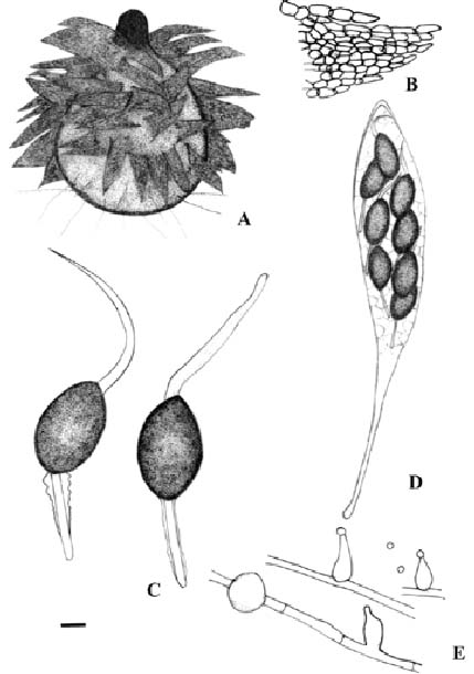
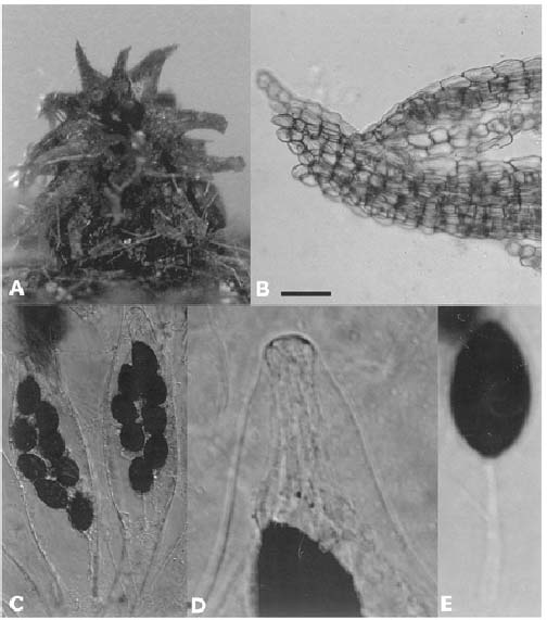
Perithecia dispersa vel gregaria, superficialia, pyriformia, 900-1700 × 675-1060 µm, pili cristata, brunnea, agglutinata, fasciculate ex toto, 400-500 × 160-170 µm. Colli atra, 130-200 µm alta. Peridium pseudoparenchymatosum, membranaceum, semitranslucidum, viridulum ad olivacibrunneum. Asci octospori, clavati ac stipitati longi, 295-400 × 32-38.5 µm, exannulati apicales. Ascosporae ellipsoideae, fuscae, 18-26 × 12-15 µm, pedicelli angusti, recti, baculiformes, 19.5-24.5 × 2-2.5 µm; cauda superae flagelliformes, eccentricae 28.5-35 × 2.5-4.5 µm, cauda basalis quasi gelatinosis vaginatis circumdare pedicellis, inaequalis an undatis aliquando. Poro germinali apicali, 2-2.5 µm in diametro.

Perithecia scattered or gregarious on substrate, superficial, pyriform, 900-1700 × 675-1060 µm, almost

entirely covered by brown, agglutinated, fasciculate, tufted hairs, 400-500 × 160-170 µm. Necks dark, 130-200 µm high. Peridium greenish to olivaceous brown, pseudoparen-chymatous, membranous, semi-transparent. Asci 8-spored, clavate and long-stalked, 295-400 × 32-38.5 µm, lacking apical ring. Ascospores dark brown, ellipsoid, 18-26 × 12-15 µm, with narrow, erect, baculiform pedicel, 19.5-24.5 × 2-2.5 µm; upper cauda lash-like, eccentric, 28.5 -35 × 2.5- 4.5 µm, sometimes inconspicuously striate under oil microscope; basal cauda as a gelatinous sheath surrounding pedicel, irregular or sometimes undulate. Germ pore apical, 2-2.5 µm in diam.

Colonies on 2% Difco malt extract agar spreading slowly, reaching 1.1-1.75 cm in diam. in 3 weeks at room temperature, grayish olivaceous to dark olivaceous, mostly submerged; yellowish olivaceous to olivaceous on reverse. Hyphae occasionally swollen, inflations globose, 6-12 µm in diam. Phialides borne singly and sparsely from aerial hyphae; hyaline, small, flask-shaped, 7-11 × 3.5-5 µm. Conidia produced at the apex of phialides; globose to ovoid, hyaline, 2-3 µm in diam.

Holotype. Kaohsiung, Mailan forestry industrial road, on Cervus unicolor swinhoei (Formosan sambar) dung, J.H. Chang; Jong 23, Oct. 24, 2003 (TNM F16503).

Additional specimens examined. Kaohsiung, Mailan forestry industrial road, on Cervus unicolor swinhoei (Formosan sambar) dung, J.H. Chang; Jong 24, Nov. 20, 2003 (TNM F16504). Mailan forestry industrial road, J.H. Chang; Jong 32, Feb. 25, 2004 (TNM F16505). Mailan forestry industrial road, on Muntiacus reevesi micrurus (Formosan Reeve's muntjac) dung, J.H. Chang; Jong 27, Feb. 25, 2004 (TNM F16506).

This species is characterized by large tufted hairs composed of swollen cells and ascospores with a slender pedicel. It is a member of the P. conica group (=Schizothecium) (Lundqvist, 1972; Bell and Mahoney, 1995), but the olivaceous peridial color is unusual among

*Corresponding author. E-mail: yzwang@mail.nmns.edu.tw


Botanical Bulletin of Academia Sinica, Vol. 46, 2005

Figure 1. Podospora multipilosa. A, perithecium; B, large tufted hair of swollen cells; C, ascospores; D, ascus containing eight ascospores; E, phialides and a globose inflation formed on the hypha. Scale bar: A = 150 µm, B, D = 14 µm, C = 5 µm, and E = 8 µm.


CHANG and WANG — A new species of Podospora

Figure 2. Podospora multipilosa. A, perithecium on substrate; B, large tufted hairs; C, asci; D, upper part of an ascus; E, ascospore. Scale bar: A = 300 µm, B = 30 µm, C = 25 µm, and D, E = 8 µm.

the species in this group. Podospora aloides (Fuckel) J. H. Mirza & Cain appears to be closely related to this species, but it has larger ascospores (31-37 × 16-20 µm) with a shorter pedicel (12-14 × 2 µm), a single and lash-like basal cauda, and perithecial hairs (ca. 230 × 70 µm)

mainly covering the neck portion (Mirza and Cain, 1969; Bell and Mahoney, 1995). Podospora conica (Fuckel) A.E. Bell & Mahoney differs in having larger ascospores (22-29 × 13-18 µm) and shorter perithecial hairs (30-110 µm) (Bell and Mahoney, 1995).


Botanical Bulletin of Academia Sinica, Vol. 46, 2005

Key to species of Podospora from Taiwan (inspired by Mirza and Cain, 1969; Wang, 2000).

1. Peridium thin, consisting of swollen cells; hairs agglutinated 2

Peridium consisting of angular cells; hairs if present, not agglutinated 6

2. Asci 8-spored 3

Asci 32- or 64-spored 5

3. Agglutinated hairs up to 400-500 µm long P. multipilosa J. H. Chang & Y. Z. Wang

Agglutinated hairs shorter 4

4. Ascospores more than 30 µm long; upper cauda grooved P. curvuloides Cain

Ascospores 25-29.5 × 17-20 µm; upper cauda not seen P. aff. conica (Fuckel) A. E. Bell & Mahoney

5. Asci 32-spored; ascospores 20-24 × 10-13 µm P. dakotensis (Griffiths) J. H. Mirza & Cain

Asci 64-spored; ascospores 21-24 × 13-15 µm P. formosana Y. Z. Wang

6. Hairs on the neck scattered, short, straight and erect 7

Hairs not as above 10

7. Hairs inflated at the apex; ascospores 24-30 × 4-6 µm P. inflatula Cain

Hairs not inflated at the apex 8

8. Asci 8-spored; ascospores 45-57 × 22-35 µm P. fimiseda (Ces. & De Not.) Niessl

Asci containing more than 8 spores 9

9. Asci 128-spored; ascospores 17-19 × 10-12 µm P. setosa (G. Winter) Niessl

Asci 256-spored; ascospores 11-17.5 × 7.5-10 µm P. araneosa (Cain) Cain

10. Hairs tuberculate; upper cauda lyre-shaped 11

Hairs not tuberculate; upper cauda not lyre-shaped 14

11. Asci 8-spored 12

Asci 32-spored 13

12. Ascospores 25-34 × 14-16 µm; with a cauda attached at the end of pedicel P. argentinensis (Speg.) J. H. Mirza & Cain

Ascospores 34-39 × 20-22.5 µm; without a cauda attached at the end of pedicel

P. decipiens (G. Winter ex Fuckel) Niessl

13. Two to four caudae attached to the base of the pedicel, 10-16 × 4-8 µm P. myriaspora (H. & P. Crouan) Niessl

Five caudae attached to the base of the pedicel, 8-11.5 × 1.5-2 µm P. pleiospora (Winter) Niessl

14. Hairs in tufts 15

Hairs not in tufts, flexuous or lacking 18

15. Asci 4-spored P. anserina (Ces. ex Rabenh.) Niessl

Asci containing more than 4 spores 16

16. Asci 256-spored; ascospores 14-18 × 10-11 µm. P. curvicolla (G. Winter) Niessl

Asci 8-spored 17

17. Ascospores 24.5-32 × 15-17.5 µm; pedicel swollen in middle.................................. ........................... P. prethopodalis Cain

Ascospores 32.5-50 × 18.75-25 µm; covered by short filaments................................................. P. immersa (Stratton) Cain

18. Perithecia glabrous; cauda absent......................................................................... P. globosa (Massee & E. S. Salmon) Cain

Perithecia not glabrous; ascospores with at least 1 cauda 19

19. Ascospores with 4 upper caudae 20

Ascospores with 1 upper cauda 21

20. Ascospores with 4 basal caudae..................................................................................................... P. communis (Speg.) Niessl

Ascospores with 1 basal cauda............................................................................................ P. hyalopilosa (R. Stratton) Cain

21. Upper and basal caudae composed of numerous filaments, and enveloping ascospores P. longicaudata (Griffiths) Cain

Caudae not as above; pedicels 44-64 µm long............................................................P. dolichopodalis J. H. Mirza & Cain


CHANG and WANG — A new species of Podospora

Chang, J.H. and Y.Z. Wang. 2003. New records of coprophilous Pyrenomycetes from Taiwan. (III). Fung. Sci. 18: 145-150.

Lundqvist, N. 1972. Nordic Sordariaceae s. lat. Symb. Bot. Upsal. 20: 1-374.

Mirza, J.H. and R.F. Cain. 1969. Revision of the genus Podospora. Can. J. Bot. 47: 1999-2048.

Wang, Y.Z. 1992. New records of coprophilous Pyrenomycetes from Taiwan. (I). Trans. Mycol. Soc. R.O.C. 7: 18-22.

Wang, Y.Z. 1994. New records of coprophilous Pyrenomycetes from Taiwan. (II). Trans. Mycol. Soc. R.O.C. 9: 185-194.

Wang, Y.Z. 2000. The genus Podospora in Taiwan. Mycotaxon 76: 373-391.

Acknowledgments. The authors thank Mr. Y.H. Leong of National Pingtung University of Science and Technology for providing dung samples, Dr. N. Lundqvist for providing critical comments on the description, Mr. Wen-Neng Chou for Latin description, and Mr. Dan Chamberling for revising English writing of the manuscript.

Literature Cited

Bell, A. and D.P. Mahoney. 1995. Coprophilous fungi in New Zealand I. Podospora species with swollen agglutinated perithecial hairs. Mycologia 87: 375-396.