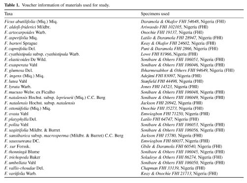
Bot. Bull. Acad. Sin. (2005) 46: 231-238

SONIBARE et al. — Scanning electron microscopy studies of Ficus

A survey of epidermal morphology in Ficus Linn. (Moraceae) of Nigeria

Mubo A. SONIBARE1,*, Adeniyi A. JAYEOLA2, Adeyemi EGUNYOMI2, and Jin MURATA1

1Koishikawa Botanical Gardens, Graduate School of Science, Faculty of Science, University of Tokyo, Japan

2Department of Botany and Microbiology, University of Ibadan, Nigeria

(Received July 14, 2004; Accepted January 14, 2005)

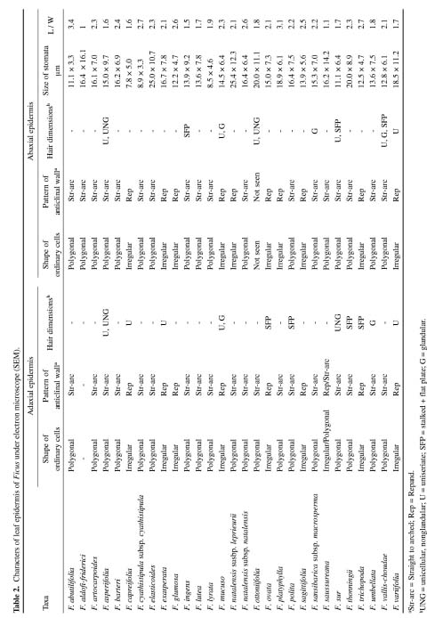
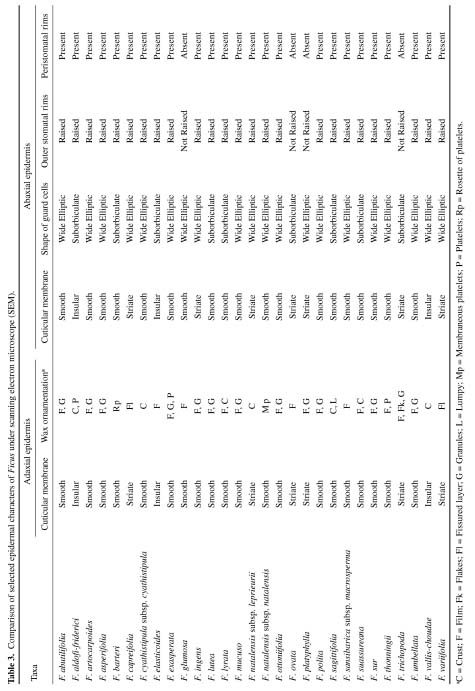
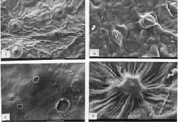
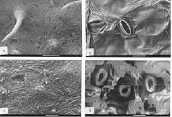
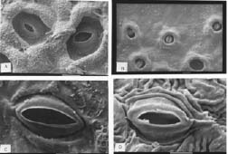
Abstract. A detailed morphological study of the leaf epidermis of the tropical genus Ficus Linn. in Nigeria is presented. The study revealed several interesting epidermal features that have not previously been reported in the genus. Leaf epidermal characters such as pattern of epidermal cells, type of stomata, shape of guard cell pairs, and cuticular ornamentation are constant in some species and variable in others and thus of great significance in understanding the relationships between and within species. Leaves are hypostomatic in all species except in F. vallis-choudae Del., which has amphistomatic leaves. Poral epidermal walls of the outer stomatal ledge are distinctly higher than the peristomatal rims in F. cyathistipula subsp. cyathistipula Warb., F. lyrata Warb., and F. sagittifolia Mildbr. & Burret.

Keywords: Epidermal characters; Ficus; Morphological survey; Scanning electron microscopy (SEM); Taxonomy.

Introduction

Literature on the epidermal morphology of Ficus is relatively rare though the taxonomic value of epidermal morphology is well documented in botanical literature for several other groups of angiosperms (Stace, 1965). As an example, epidermal characters of Euphorbiaceae have been studied by Amelunxen et al. (1967), Kakkar and Paliwal (1974), and Raju and Rao (1977). Several reports were published on the epidermal structures and stomatal ontogeny by Karatela and Gill (1984a, b; 1985) who stressed their usefulness in plant taxonomy.

In his taxonomic study of Jatropha L. (Euphorbiaceae), Dehgan (1980) reported that all species, with the exception of J. fremontioides Standley, have paracytic stomata that are with few exceptions restricted to the abaxial surface. Wax morphology has been used extensively in taxonomic work. The classic work of Hallam and Chambers (1970) on SEM studies of the leaf of Eucalyptus L'Heritier has demonstrated the potential of epicuticular wax studies to aid the classification of a complex genus. Although morphological studies of the epidermis may not themselves be sufficient as taxonomic evidence, they could in conjunction with anatomical features serve as good taxonomic tools for delimiting taxa.

Stomata and associated epidermal cells are providing an increasingly important source of taxonomic characters. The taxonomic significance of stomata distribution and mor

phology in the Epacridaceae was surveyed by Watson (1962), who found that members of the tribe Styphelieae typically possess only adaxial stomata (on the sepal) while in the Epacrideae they are usually abaxial. Trichomes and hydathodes are other characters found on the epidermis and other organs of plants that can serve as good taxonomic tools. They have been employed both for classification and identification purposes by many systematists (Theobald et al., 1979; Rollins, 1993; Potgieter and van Wyk, 1999; Dickison, 2000; Batterman and Lammers, 2004). De Bary (1884) and Renner (1907) have also discussed hydathodes in Ficus.

Preliminary field observation of Nigerian Ficus populations revealed conspicuous differences in vegetative and floral features among the taxa. Previous classifications of the genus have been largely based on the systematics of pollinating wasps (Corner, 1958, 1965; Ramirez, 1977). It is imperative therefore to attempt a search for epidermal characters that may be of taxonomic utility. The main objective of this work was to survey the leaf epidermis of Ficus species in Nigeria in search of taxonomic characters that might assist in identification and understanding of the variations among the species.

Materials and Methods

Plant samples were obtained from herbarium specimens or personal collections. Small pieces (c. 7 mm2) of leaf from herbarium materials were dehydrated in an absolute ethanol:acetone series (90% ethanol, 30 min; absolute ethanol 30 min; 50:50 absolute ethanol:acetone, 10 min; and finally two steps of acetone, 10 min each), according to Luckow and Grimmes (1997). Dehydrated material was then critical point dried using an Oriental 100V drier and sputter coated with

*Corresponding author. Present address: Department of Biological Sciences, Olabisi Onabanjo University, P.M.B. 2002, Ago-Iwoye, Nigeria. Tel: 234-803-365-9517; Fax: 234-2-8103043; E-mail: sonibaredeola@yahoo.com


Botanical Bulletin of Academia Sinica, Vol. 46, 2005

gold in a fine coat ion sputter JC 1100. Observation was done with a Jeol JSM 5200 LV scanning electron microscope with a camera (Jeol 45042) attachment. Measurements of lengths and width of guard cell pairs were made and recorded. Voucher specimens were deposited in Forest Herbarium Ibadan (FHI), Nigeria. Voucher information of the specimens examined is presented in Table 1.

Results

Leaf Surface

Microcharacters of taxonomic significance obtained from selected features of the leaf surfaces, using SEM, are presented in Tables 2 and 3.

Hairs

Based on the presence or absence of hairs on the leaf epidermal surfaces, the taxa studied may be divided broadly into two groups, a glabrous group consisting of taxa without hairs on either their adaxial or abaxial surfaces and a pubescent group comprising taxa with one or more hair types on their leaf surfaces.

Glabrous group: This group comprises taxa such as F. abutilifolia (Miq.) Miq., F. aldofi-friderici Mildbr., F. artocarpoides Warb., F. cyathistipula subsp. cyathistipula

Warb., F. elasticoides De Wild., F. glumosa Del., F. lutea Vahl, F. lyrata Warb., F. natalensis subsp. leprieurii (Miq.) C.C. Berg, F. natalensis Hochst subsp. natalensis, F. platyphylla Del., F. sagittifolia Mildbr. & Burret, and F. saussureana DC.

Pubescent group: This consists of taxa in which hairs of different dimensions occur on either the adaxial / abaxial or both surfaces. Hairs are found on the adaxial surfaces but not on the abaxial surfaces of F. capreifolia Del., F. exasperata Vahl, F. ovata Vahl, F. polita Vahl, F. thonningii Blume, F. trichopoda Baker, and F. umbellata Vahl. Hairs occur on the abaxial leaf surfaces but not on the adaxial leaf surfaces of F. ingens Del., F. ottoniifolia (Miq.) Miq., F. sansibarica subsp. macrosperma (Mildbr. & Burret) C.C. Berg, F. vallis-choudae Del. Hairs are present on both the adaxial and abaxial leaf surfaces in F. asperifolia Miq., F. mucuso Welw. ex Ficalho, F. sur Forssk, and F. variifolia Warb. While only one type of hair is present in F. capreifolia, F. exasperata, F. ovata, F. polita, F. sansibarica subsp. macrosperma, F. thonningii, F. trichopoda, F. umbellata, F. variifolia, two or more types of hairs are found in F. asperifolia, F. mucuso, F. ottoniifolia, F. sur, and F. vallis-choudae. Unicellular, non-glandular, simple hairs with narrow apiculate tips were found on the adaxial and abaxial surfaces of F. asperifolia. In F. sur, they were only observed on the adaxial surface.


SONIBARE et al. — Scanning electron microscopy studies of Ficus


Botanical Bulletin of Academia Sinica, Vol. 46, 2005


SONIBARE et al. — Scanning electron microscopy studies of Ficus

The adaxial and abaxial surfaces of F. mucuso had unicellular glandular hairs that were observed only on the abaxial surface of F. vallis-choudae. Stalked hairs with flat plates were observed on the abaxial surfaces of F. ingens and F. sur, and also on the adaxial surfaces of F. ovata, F. polita, F. thonningii, and F. trichopoda (Table 2).

Epidermal Cells

Anticlinal walls are repand in F. capreifolia, F. exasperata, F. glumosa, F. ovata, F. sagittifolia, F. trichopoda, F. variifolia, but straight to arched in all other species. Cell shape is irregular in F. capreifollia, F. exasperata, F. glumosa, F. mucuso, F. ovata, F. sagittifolia, F. trichopoda, and F. variifolia while it is polygonal in other species, except in F. saussureana, in which both irregular and polygonal shapes are represented.

Stomatal Apparatus

The stomata are restricted to the abaxial surface of lamina (hypostomatic) except in F. vallis-choudae (Figure 1B), in which leaves are amphistomatic, but stomata more abundant on abaxial surface. Stomata are dense or evenly scattered on the abaxial leaf surfaces in most species, but they are few in F. abutilifolia, F. capreifolia, F. ottoniifolia, F. platyphylla, F. sansibarica subsp. macrosperma. Stomata are mostly of a paracytic type. The outlines of the pair of guard cells are usually suborbiculate to wide elliptical as seen in the surface view (Figures 1-3), as summarized in Table 3. The highest and lowest stomata length/width (L/W) ratios of 1 and 3.4 are found among the glabrous species. The outer stomatal rims are raised in all species except in F. glumosa, F. ovata, F. platyphylla, and F. trichopoda, in which the outer stomatal rims are flat.

In F. cyathistipula subsp. cyathistipula (Figure 2D), F. lyrata (3A) and F. sagittifolia (3B), the poral epidermal walls of the outer stomatal ledge are distinctly higher than the peristomatal rims while in both F. barteri and F. elasticoides the peristomatal rims overlap the outer ledge.

Cuticular and Wax Ornamentation

The outer surface of the cuticle is smooth to weakly undulate in most species (Table 2), striate to wrinkled in some as in F. capreifolia, F. ingens, F. natalensis subsp. leprieurii, F. ovata, F. platyphylla, and F. variifolia. The striations are irregular in orientation and height or occasionally radiating from the guard cells. In F. aldofi-friderici, F. elasticoides and F. vallis-choudae, cuticle is divided into numerous small regions by the deep folds between them. Epicuticular waxes on adaxial epidermis as indicated in Table 3 exhibit great micromorphological diversity in all the taxa studied. Film type in F. elasticoides, F. glumosa, F. ovata and F. sansibarica subsp. macrosperma; Film and platelets in F. exasperata, film and granular waxes were observed in F. abutilifolia, F. artocarpoides, F. asperifolia, F. ingens, F. lutea, F. mucuso, F. ottoniifolia, F. platyphylla, F. polita, F. sur, F. trichopoda, F. umbellata (Table 2). Crust and platelets were observed in F. aldofi-friderici. Ficus saussureana had a mixture of crust and film wax ornamentation. Membraneous platelets were observed in F. natalensis subsp. natalensis. Rossettes of platelets were found in F. barteri Sprague while F. capreifolia exhibited a fissured layer (Figure 1A).

Hydathodes

Hydathodes were observed on the adaxial leaf surfaces of F. lutea (Figure 1C), F. sansibarica subsp. macrosperma

Figure 1. Characteristics of adaxial epidermal cells of Ficus (SEM). A, Punt & Daramola 78407 (FHI) F. capreifolia (Scale bar: 100 µm), wax ornamentation showing fissured layer. B, Wit & Daramola 79243 (FHI) F. vallis-choudae (Scale bar: 10 µm), note the presence of adaxial stomata. C, Sonibare & Others 22254 (UIH) F. lutea (Scale bar: 50 µm), note the presence of hydathodes on adaxial epidermis. D, Domen 32431 (FHI) F. ovata (Scale bar: 10 µm), showing stalked hair with flat plate.

Figure 2. Characteristics of abaxial epidermis of Ficus (SEM). A, Latilo & Daramola 28947 (FHI) F. asperifolia (Scale bar: 50 µm), showing long trichomes with apiculate tips and dense stomata. B, Okeke 24695 (FHI) F. mucuso (Scale bar: 10 µm), with more elliptic guard cell outline. C, Wit & Daramola 79243 (FHI) F. vallis-choudae (Scale bar: 100 µm), note the presence of hydathodes. D, Lowe 81966 (FHI) F. cyathistipula subsp. cyathistipula (Scale bar: 5 µm), with poral epidermal wall higher than peristomatal rim.


Botanical Bulletin of Academia Sinica, Vol. 46, 2005

The absence of sufficient overlap among the character values of the taxa considered provides support for the consideration of this genus as a very diverse one. One such character is the lamina hyadathode. Metcalfe and Chalk (1950) have recorded hydathodes from thirty-four families of dicotyledons, and De Bary (1884) noted water pores in adaxial epidermal depressions in at least six species of Ficus: F. neriifolia, F. deltoidea, F. callosa, F. granatum, F. saussureana, and F. septica. In the present study, hydathodes were not observed in the leaves of Ficus species such as F. asperifolia, F. capreifolia, F. exasperata and F. saussureana. Their absence from the leaves of F. saussureana is in particular at variance with the observation of de Bary in 1884, who observed hydathodes on its abaxial epidermis. However, hydathodes observed on the adaxial leaf surfaces of F. lutea, F. sansibarica subsp. macrosperma, F. sur, and F. vallis-choudae fit the observations made by Lersten and Peterson (1974) on the hydathodes in leaves of F. deltoidea, in which hydathodes were scattered in a seemingly random pattern in the leaf, each opening on a shallow circular depression on the adaxial surface. The hydathodes may be said to fit the passive type according to the classification of Haberlandt (1914). This inference is drawn because of prominent intercellular spaces in the epithem (internal portion of the hydathodes).

Although the ecological significance of the surface features revealed in this study are not yet fully understood (Juniper and Jeffree, 1983), Jayeola (1998) opined that certain epidermal features might help to reduce wind speed on the leaf surface, thereby curtailing excessive water loss from the stomata.

Since Ficus species are widely distributed in Nigeria, occurring from the coastal to dry sahelian savanna, it is possible that each species responds to its environment in specific ways by modifying the basic plan of certain features to improve its adaptation.

Acknowledgements. Mr. Akitoshi Iwamoto of the University of Tokyo Museum is gratefully acknowledged for technical assistance in the use of the scanning electron microscope.

Literature Cited

Amelunxen, F., K. Morgenroth, and T. Picksak. 1967. Untersuchungen an der Epidermis mit dem stereoscan-Elektronenmikroskop. Z. Pflanzenphysiol. 57: 79-95.

Batterman, M.R.W. and T.G. Lammers. 2004. Branched foliar trichomes of Lobeliodeae (Campanulaceae) and the infrageneric classification of Centropogon. Syst. Bot. 29(2): 448-458.

Corner, E.J.H. 1958. An introduction to the distribution of Ficus. Reinwardtia 4: 425-355.

Corner, E.J.H. 1965. Checklist of Ficus in Asia and Australasia with keys to identification. Gard. Bull. Singapore 21: 1-186.

De-Bary, A. 1884. Comparative Anatomy of Vegetative Organs of the Phanerogams and Ferns. English Translation by

Figure 3. Abaxial stomatal morphology of Ficus (SEM). A, Jones 14523 (FHI) F. lyrata (Scale bar: 10 µm), note the raised poral epidermal wall. B, Adejimi 78819 (FHI) F. sagittifolia (Scale bar: 10 µm), with film type of wax ornamentation and raised poral epidermal wall. C, Brenan 39370 (FHI) F. artocarpoides (Scale bar: 5 µm). D, Keay 37245 (FHI) F. sansibarica subsp. macrosperma (Scale bar: 5 µm), note the folding.

(Figure 2C), F. sur and F. vallis-choudae, while hydathodes were absent in the other species.

Discussion

The study of the epidermal surfaces of Ficus revealed a number of important micromorphological characters, and these characters exhibit interesting interspecific variations that are of significance for identification. Much variation is found in the glabrous and glabrescent species. For example, the highest and lowest stomata length / width (L/W) ratios were found among the glabrous species, indicating little or no correlation between stomata dimensions and the presence or absence of trichome.

The taxonomic value of trichomes in angiosperm is well documented in the botanical literature (Theobald et al., 1979; Batterman and Lammers, 2004). In the present study, their presence or absence on the epidermal surfaces of Ficus species were found to be less informative taxonomically. The nature of the trichomes in the genus seems to be more reliable than their mere presence or absence. Three basic types of trichomes prominent in the species studied are uniseriate, stalked with flat plate, and glandular. The taxonomic value of trichomes in this work was greatly limited by their absence in 22 out of the 28 species of Ficus studied. Trichome type has been found to be of little diagnostic value in this genus.

The presence of a stomatal ledge in about 80% of the taxa studied is of special note. This high percentage of occurrence may be due to ecological factors in such species as F. atocarpoides, F. elasticoides, F. lyrata and F. sagittifolia, but further investigation is required. Another feature worthy of note is wax ornamentation. Again, the presence of waxes of various types on the leaf surfaces of Ficus could be related to adaptation to drought.


SONIBARE et al. — Scanning electron microscopy studies of Ficus

Bower F.O. and Scott D.H. Oxford. Oxford University Press.

Dehgan, B. 1980. Application of epidermal morphology to taxonomic delimitations in the genus Jatropha L. (Euphobiaceae). J. Linn. Soc. Bot. 80(3): 257-278.

Dickison, W.C. 2000. Integrative Plant Anatomy. San Diego: Harcourt Academic Press.

Haberlandt, G. 1914. Physiological Plant Anatomy, 4th edn. translated by M.Drummond. Macmillan, London.

Hallam, N.D. and T.C. Chambers. 1970. The leaf waxes of the genus Eucalyptus L'Heritier. Austral. J. Bot. 18: 335-386.

Jayeola, A.A. 1998. Surface sculpturing of Alstonia boonei De Wild. and A. congensis Engl. (Apocynaceae) and its importance on their taxonomy. Feddes Repert. 109(5-6): 429-433.

Juniper, B.E. and C.E. Jeffree. 1983. Epidermal Surfaces. In Plant Surfaces. Paris, London.

Kakkar, L. and G.S. Paliwal. 1974. Studies of the leaf anatomy of Euphorbia V. Epidermis. Proc. Indian Natl. Sci. Acad., B 40: 55-67.

Karatela Y.Y. and S.L. Gill. 1985. Epidermal Morphology and stomatal ontogeny in some West African Convolvulaceae species. Herba Hung. 24(2-3):11-12.

Karatela Y.Y. and S.L. Gill. 1984a. Epidermal structures and stomatal ontogeny in some Gymnosperms from Nigeria. Feddes Rep. 95(5-6): 351-354.

Karatela Y.Y. and S.L. Gill. 1984b. Structure and ontogeny of some Nigerian ferns. Nova Hedwigia 40: 477-486.

Lersten, H.R. and W.H. Peterson. 1974. Anatomy of hydathodes and pigment disks in leaves of Ficus diversifolia (Moraceae). Bot. J. Linn. Soc. 68: 109-113.

Luckow, M. and J. Grimmes. 1997. A survey of anther glands in the Mimosoid legumes, tribe Parkiaea and Mimosaceae. Amer. J. Bot. 84: 285-298.

Metcalfe, C.R. and L. Chalk. 1950. Anatomy of the Dicotyledons. Oxford Clarendon Press.

Potgieter, M.J. and A.I. van Wyk. 1999. Leaf anatomy of the South African Icacinaceae and its taxonomic significance. S. African J. Bot. 65: 153-163.

Raju, V.S. and P.N. Rao. 1977. Variation in the structure and development of foliar stomata in the Euphorbiaceae. Bot. J. Linn. Soc. 75: 69-97.

Ramirez, W.B. 1977. A new classification of Ficus. Ann. Missouri Bot. Gard. 64: 296-310.

Renner, O. 1907. Beitrage zur anatomie und systematic der Artocarpeen und Conocephalen, insbesondere der Gattung Ficus. Bot. Jahrb. 39: 319-448.

Rollins, R.C. 1993. The Cruciferae of Continental North America. Stanford University Press.

Stace, C.A. 1965. The significance of leaf epidermis in the taxonomy of Combretaceae I. A general review of the tribal, generic and specific characters. Bot. J. Linn. Soc. 59: 229-250.

Theobald, W.L., J.L. Krahulik, and R.C. Rollins. 1979. Trichome description and classification. In C.R. Metcalfe and L. Chalk (eds.), Anatomy of the Dicotlydons, 2nd edition, Volume 1, Clarendon Press, Oxford, pp. 40-53.

Watson, L. 1962. The taxonomic significance of stomatal distribution and morphology I. Epacridaceae. New Phytol. 61: 36-40.


Botanical Bulletin of Academia Sinica, Vol. 46, 2005